
Asia-Pacific Chiropractic Journal
Issue 7-2 Published 1 September 2026;
Next issue: 1 Nov 2026
Proudly written and produced by Human Intelligence
Read in English, Maori, Hindi, Amharic, Sinhala, Tamil, Vietnamese and more
In this issue ...
In memoriamMarion Todres-Masarsky: ab imo pectoraEditorialCommentaryHilary Taglio: The language our hands speakDialogueResearchHypothesesSuhorukovs & Kirdoglo: Long-term consequences of Herpesvirus infection and chronic inflammation of connective tissue
Charles Masarsky: Long COVID Olfactory dysfunction can be exacerbated by Dysautonomia generated by Vertebral Subluxation
Professional identity
Clinical practice IndexHenry Pollard: Joint Hypermobility in Chiropractic Practice: Part 2: Manipulation safety and evidence-based rehabilitationClinical Case reportsAlexandra Zoda et al [ASRF]: Improvements in emotional regulation, motor coordination and executive function in an 8-year-old male with ADHD and dyslexia concomitant with Chiropractic care: A case report
Jacey Pyrjma et al [ASRF]: Improvements in mood, latching, range of motion and gastrointestinal function in 3-month-old twins: A Chiropractic case report
Shaan Rai et al [ASRF]: Chiropractic management of a 49-year-old female with Arnold-Chiari Malformation and associated neurological sequelae: a case report
Dudum & Gatterman: Rapid MRI confirmed reduction of lumbar disc herniation following an accelerated nonsurgical spinal decompression protocol in an 18y female: A case report
Quick-take
Charles Blum: Nano- and Microplastics and Maternal–Fetal Health
In memoriam

Editorial
Commentary
AU: Hilary Taglio BSc, DC. Gonstead Diplomate. Private practice of Chiropractic, Roseville CA. w: https://tagliochiropractic.com/
Narrative: Long before an adjustment is delivered, our patients are already reading our hands. Patients cannot explain why they trust one doctor immediately and another only after months of care, yet we have all observed this. We may introduce ourselves to our patients, but our hands are truly what acquaint us.
Indexing terms: Chiropractic; touch; technique; hands.
Cite: Taglio H. The language our hands speak. Asia-Pac Chiropr J. 2026;7.2. https://www.apcj.site/TaglioHands.pdf
Take-away: 'Gonstead’s hands no longer adjust, yet the language they spoke has never fallen silent’
Introduction


Dialogue
AU1: Beau Woods B.Sc, B.Chiro, B.Com, MPH, PhD(c). Private Practice of Chiropractic, Western Australia. e: s4143439@student.rmit.edu.au
AU2: Joshua Tymms BHealthSci, MClinChiropr. Private practice of Chiropractic, Duncraig, Western Australia.
AU3: Harriet Walker BHSc, BAppSc(Chiro). Private practice of Chiropractic, Kingswood, South Australia.
AU4: Ben Coupe BChiroSc, MChiro. Private practice of Chiropractic, Melbourne, Australia.
Narrative: Advanced Bio-Structural Correction (ABC™) is a methodology used internationally for the assessment and correction of subluxation, yet it remains sparsely represented in the peer-reviewed literature, where its evidence base is presently confined to single case reports and small case series. (1–22)
Indexing terms: Chiropractic; functional neurology; neuroplasticity; clinical intervention; nutrients.
Introduction
Advanced Bio-Structural Correction (ABC™) is a technique used by Chiropractors and other manual practitioners that is directed at the assessment and correction of subluxation. The ABC™ protocol combines spinal, pelvic and lower-limb corrections with meningeal releases and, as an adjunct for advanced practitioners, Endonasal Cranial Correction™. ABC™ has been adopted by growing numbers of practitioners across North America, Europe, Southern Africa and Australasia
In Australasia it is supported by Advanced Bio-structural Correction Australasia (ABCA), a not-for-profit association formed in 2011 to advance the awareness, training and responsible utilisation of the method. Despite this international uptake, ABC™ has, in the words of one of the first reports to describe it in the peer-reviewed literature, ‘not been well described in the literature’. (3)
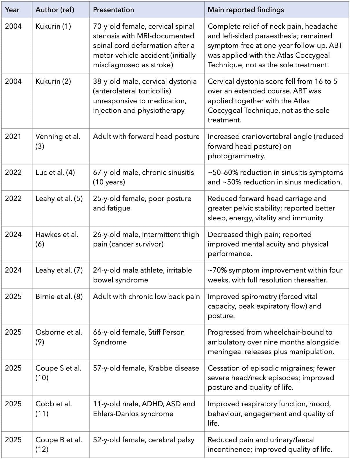
The peer-reviewed footprint of ABC™ is small but has grown significantly since 2021. The earliest reports appeared in 2004 under the technique’s earlier name, Advanced Bio-structural Therapy (ABT), and described its use, in each instance as one component of a multimodal course of care rather than the sole intervention. (1, 2)
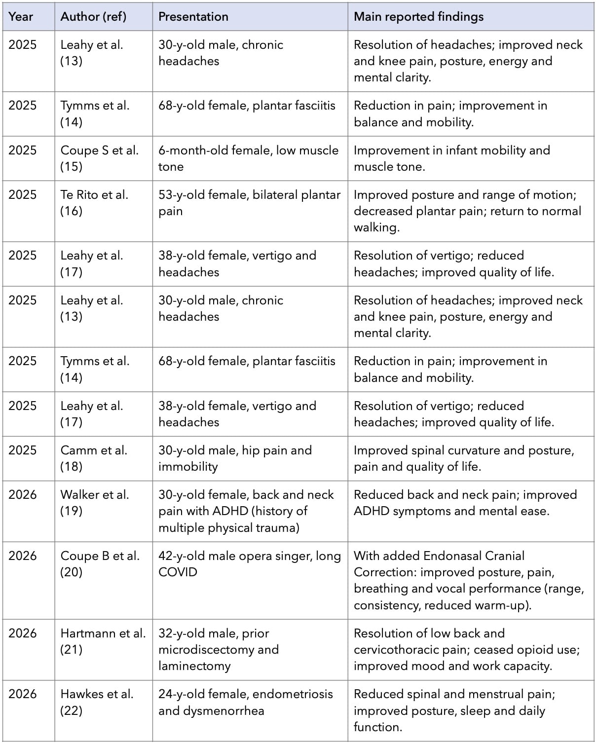
The contemporary literature consists predominantly of single case reports and small case series describing changes observed during a course of care. (3 – 22) These reports document objective postural measures such as the craniovertebral angle (3) and spirometric indices, (8) alongside a wide range of patient-reported outcomes across diverse and sometimes serious presentations, including neurological and neurodevelopmental conditions, chronic and post-surgical pain, and various non-musculoskeletal complaints. (4 – 7, 9 - 22) Case reports of this kind are valuable for documenting clinical experience and generating hypotheses, but by design they cannot establish efficacy: they describe single patients, lack control groups, and cannot exclude natural history, concurrent care or placebo effects. Case studies are insufficient to determine cause and effect.
It is also relevant that the existing reports derive from a small number of contributing centres, and that proponents themselves acknowledge an absence of high-quality research substantiating the technique’s broader claims, claims that have attracted published criticism from perennial critics. (25) The published reports identified to date are summarised in Table 1.
None of this is a reason to avoid research; it is the reason research is needed.
Table 1 shows published case reports describing care using Advanced Bio-Structural Correction (ABC™), including its earlier designation Advanced Biostructural Therapy (ABT), in chronological order.
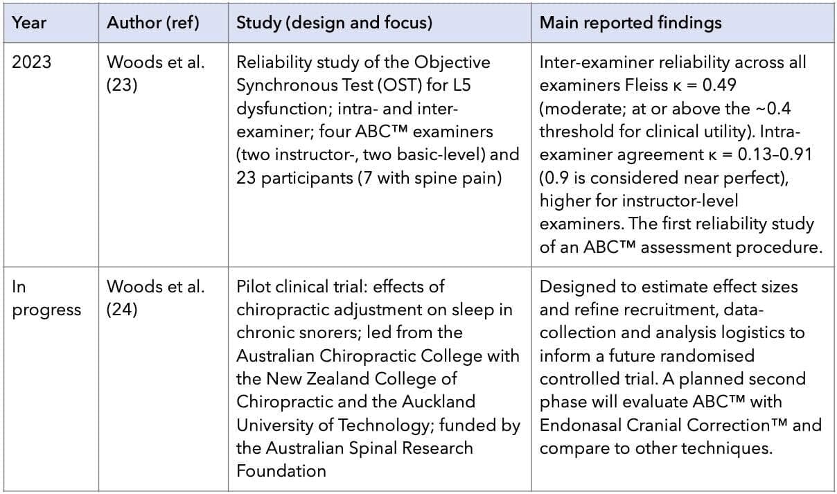
The literature has, however, begun to extend beyond case description. The first study to evaluate the reliability of an ABC™ assessment procedure has been published: an examination of the intra- and inter-examiner Reliability of the Objective Synchronous Test (ROOSTA-L5), the procedure used to indicate correction of the fifth lumbar vertebra, which reported inter-examiner agreement (Fleiss κ = 0.49) at or above the threshold commonly accepted for clinical utility in physical medicine. (23) In parallel, a funded pilot clinical trial is in progress (see Discussion: Early progress). These studies are summarised in Table 2.
Table 1: Published case reports describing care using Advanced Bio-Structural Correction (ABC™), including its earlier designation Advanced Biostructural Therapy (ABT), in chronological order. Findings are as reported by the authors and reflect changes observed concurrently with care; as uncontrolled single cases — and, in some instances, with ABC/ABT forming only one part of a multimodal course — they do not establish efficacy.
Part 1 -
Part 2-
The wider chiropractic profession has confronted comparable evidence gaps (to varying degrees) by developing formal recommendations and research agendas to coordinate effort and channel limited resources. In the United States, successive reviews set out priorities for health-services, clinical, basic-science and outcomes research and for the research infrastructure the profession would require, (26, 27) and these were revisited and renewed a decade later. (28) More recently, the research priorities of the Australian chiropractic profession have been articulated through a structured survey of academics and practitioners. (29) Against this backdrop it is both timely and appropriate for ABCA to set out a coordinated research strategy of its own. This paper presents that strategy, describes how it was developed and ratified, and discusses its strengths, limitations and the safeguards required for it to be credible.
Development of the strategy
The strategy was drafted by the ABCA Research Committee, a standing committee constituted to oversee and coordinate research activity within the association. The committee developed the document iteratively, drawing on the structure of established chiropractic research recommendations (26 - 29) and on the wider literature concerning research capacity building in allied health. (30, 31) The draft was then presented to the ABCA Board and to the membership and was ratified in 2025. Ratification by both governance and membership is a deliberate feature; it is intended to give the strategy a mandate, to align practitioner expectations with the realities and timelines of research, and to encourage broad participation. The strategy is intended to be a living document, with scheduled review every two years (see Implementation).
The strategy
Vision
To advance the understanding and recognition of ABC™ by investigating the clinical outcomes observed in practice, fostering research that tests its effectiveness, and integrating findings to support evidence-informed application within the broader healthcare community. The framing is deliberately investigative rather than promotional: the aim is to determine, through rigorous scientific enquiry, where, for whom and to what extent the technique is effective.
Objectives
Expand the evidence base: investigate and substantiate the clinical outcomes observed in ABC™ practice, progressing from descriptive reports toward higher-level designs.
Build practitioner research capacity: equip practitioners with the skills, tools and opportunities to document outcomes and contribute to research by making the first step as ‘clinician-authors’.
Engage in postgraduate and doctoral studies: support practitioners pursuing Masters and PhD study on topics relevant to ABC™.
Promote research literacy: foster a culture of informed decision-making by developing practitioners’ ability to critically appraise and apply research.
Facilitate collaboration: build partnerships with academic institutions, independent researchers and funding bodies.
Enhance dissemination: publish and present findings to chiropractic and multidisciplinary audiences through both traditional and contemporary channels.
Strategic initiatives
The objectives are operationalised through seven interlinked initiatives. They are presented separately for clarity, but the evidence on research capacity building emphasises that such strategies are interdependent and most effective when implemented as a coordinated, whole-of-system approach with sustained leadership support. (30, 31)
- Research development and capacity building: Methodology workshops, training in case-report writing, study design and basic statistics, and a mentorship programme pairing novice researchers with experienced supervisors. Scholarships and small grants will support relevant research education, including training that is not ABC™-specific but that builds transferable research skills. This reflects evidence that skill development, mentorship, protected time and funded opportunity are core, mutually reinforcing components of building research capability in allied-health settings. (30,31)
- Establishing a research network: Formalising the research committee, partnering with universities and research organisations, and encouraging co-authorship with interdisciplinary and international teams. A practice-based research network (PBRN) is a natural vehicle for this work: PBRNs are sustained collaborations between clinicians and academics that collect data within everyday practice, improving the relevance and generalisability of findings relative to laboratory settings. The components required of a chiropractic PBRN — practitioner–researcher partnership, centralised data management, standardised quality assurance, and funding from multiple sources — have been described in the literature and would provide the governance that distinguishes a credible network from ad hoc reporting. (32)
- Research projects: A graded programme is proposed, beginning with larger and better-reported case series and progressing toward controlled designs. Reporting of all case material will follow the CARE guidelines, the consensus standard for clinical case reports, which improve rigour and allow individual cases to be aggregated and compared. (33) Subsequent work includes: controlled clinical studies of ABC™ in defined musculoskeletal and, where justified, non-musculoskeletal presentations — a stream already begun with a funded pilot trial of the effects of chiropractic adjustment on sleep in chronic snorers, with a planned second phase evaluating ABC™ and Endonasal Cranial Correction™ (see Early progress); (24) evaluation of supportive products (for example pillows, mattresses, seat-wedges and chairs); evaluation of assessment technologies (postural photography and three-dimensional analysis, radiographic and gait assessment, spirometry and heart-rate variability); reliability and validity studies of ABC™ assessment procedures, extending the first such study of the Objective Synchronous Test; (23) and anatomical and biomechanical investigation of the proposed mechanisms, including meningeal and dural tension. The methodology is grounded in the foundational work of Breig on adverse mechanical tension in the central nervous system, (34) and of Yamada and colleagues on the stretch-induced pathophysiology of the spinal cord, (35) while contemporary cadaveric work on suboccipital soft-tissue connections to the dura mater offers one model for rigorous further enquiry. (36) The strategy also explicitly values qualitative and mixed-methods research, recognising that patient experience and meaning are not fully captured by trial designs alone.
- Engagement and dissemination: Encouraging conference presentation, targeting peer-reviewed and open-access journals, communicating progress to the community through webinars, newsletters and social media, and recognising practitioners who publish, with particular encouragement for first-time authors. Dissemination is treated as a core obligation of publicly accountable research rather than as promotion.
- Postgraduate and education support: Resources and pathways for practitioners entering research training (for example Master of Health Research, Master of Public Health or Master of Research methods) and, in time, doctoral study. An education–practice–research feedback loop is proposed whereby research findings inform how the technique is taught and practised, supporting iterative refinement. This addresses a recognised need to build people, not only fund projects, within the profession.
- Funding and resources: A blended model of external grant applications, an in-house research fund for pilot studies and scholarships, and member fundraising. Diversified funding from multiple sources is a recognised requirement for sustaining a research network. (32) This model is already producing results: approximately AUD 40,000 has been committed to the programme to date, including a competitive grant from the Australian Spinal Research Foundation and the case study project (see Early progress). (24)
- Ethical and rigorous research standards: All research will comply with ethical requirements and obtain approval from a relevant Human Research Ethics Committee, and quality-assurance protocols will be implemented to support reproducible, best-practice research. Given the governance considerations discussed below, these standards are central rather than peripheral to the strategy.
Implementation
The strategy is sequenced across five years, with an ongoing phase to sustain activity thereafter.
Table 3: Phased implementation of the ABCA research strategy.
Key performance indicators
Progress will be monitored against the following indicators:
- Number of ABC™-related papers published annually.
- Number of practitioners engaged in research projects.
- Attendance at research workshops and training programmes.
- Volume of research funding secured and funds raised annually.
- Postgraduate enrolments and completions related to ABC™.
- Citations and recognition in academic and professional circles.
- Number of partnerships formed with academic or research institutions.
- Number of ABC™-specific research training modules and resources created.
Discussion
This strategy aligns ABCA with a well-established approach in the profession, in which organisations articulate research priorities explicitly so that scarce resources are directed coherently and collaboratively. (26 – 29) Its emphasis on practitioner engagement, mentorship and a practice-based network is consistent with the best available evidence on building research capacity in allied health, which finds that capacity-building strategies are interdependent and require sustained organisational and leadership commitment. (30 – 32) Its insistence on standardised case reporting (33) and on ethics approval and quality assurance reflects the methodological and governance expectations of contemporary health research.
Early progress
Implementation is already under way. The case study project has thus far precipitated the publication of 20 case studies by clinician-authors.
The first reliability study of an ABC™ assessment procedure, ROOSTA-L5, has been published, (23) and an initial pilot clinical trial has been funded and is in progress: a study of the effects of Chiropractic adjustment on sleep in chronic snorers, in collaboration with the New Zealand College of Chiropractic and the Auckland University of Technology. (24) The trial is designed to estimate effect sizes and refine the logistics of recruitment, data collection and analysis to inform a future randomised controlled trial, with a planned second phase evaluating Endonasal Cranial Correction™. Approximately AUD 40,000 has been committed to the programme to date, including a competitive grant of AUD 19,500 from the Australian Spinal Research Foundation, AUD 10,000 from ABCA and AUD $10,000 from SOT-Australasia. (24) That this early work is being conducted with independent university-based researchers is itself consistent with the governance safeguards set out below.
The current state of the evidence
Candour about the present evidence base is essential. The published ABC™ literature is at an early, descriptive stage, dominated by single case reports and small series drawn from a limited number of contributors. (1 – 22) Such reports describe associations, changes observed concurrently with care, and cannot, by design, demonstrate that the intervention caused the observed change, control for natural history or placebo effects, or support generalisation. This limitation is most acute for the serious and rare conditions that appear in some reports, (9, 10, 12) where enthusiastic interpretation could foreseeably mislead patients or delay other care. The reliability study and pilot trial noted above mark the beginning of a move beyond description, (23, 24) and the strategy commits to continuing that graded progression toward controlled and reliability studies, and to reporting standards that allow cases to be appraised and aggregated rather than read as proof. (33) Framing the work this way is not a concession; it is what will make the resulting evidence persuasive to readers outside the technique’s existing community of practice. (25)
Challenges and safeguards
Three challenges deserve particular attention:
First, resourcing and time: surveys using the Evidence-Based Practice Attitude and Utilisation Survey consistently report that Chiropractors hold favourable attitudes toward evidence-based practice and wish to improve their skills, while identifying lack of time and lack of accessible clinical evidence as the principal barriers, and access to free online databases and full-text articles as key facilitators. (37) The strategy’s investment in protected opportunity, funding and accessible tools responds directly to these findings.
Second, methodological rigour and the risk of bias: an early-stage field investigating outcomes it expects to find is vulnerable to confirmation bias, selective reporting and outcome-measure drift. Pre-specified protocols, registration of studies where applicable, validated and objective outcome measures, blinded assessment where feasible, and the inclusion of independent researchers are the appropriate countermeasures, and are embedded in the strategy’s commitment to best-practice quality assurance and external collaboration.
Third, and most importantly, independence and conflict of interest: ABCA both promotes and trains practitioners in the technique and proposes to fund and conduct research into it. This dual role is not unusual for a professional association, but it must be managed transparently if findings are to carry weight. Genuine partnership with universities and independent investigators, already evident in the collaborations underpinning the current pilot trial (32), transparent declaration of funding and interests, ethics-committee oversight, and a willingness to publish negative or null results are the mechanisms by which the strategy can convert advocacy into credible enquiry. The explicit valuing of qualitative and mixed-methods research is a strength in this regard, provided it complements rather than substitutes for the controlled designs needed to test effectiveness claims.
Strengths
The strategy’s principal strengths are its breadth, its sequencing and its mandate. It addresses people, infrastructure, projects, funding and ethics together rather than in isolation, consistent with whole-of-system capacity-building models. (30) It sequences activity realistically, beginning where the field is, case material, and building toward controlled studies, a progression already begun. And, having been ratified by both Board and membership, it carries an organisational mandate that capacity-building initiatives often lack.
Conclusion
ABCA has set out a structured, time-bound research strategy intended to move ABC™ from clinical observation toward formally tested knowledge. The strategy is consistent with how the wider profession has approached research prioritisation and capacity building, (26 – 31) and it is realistic about the early state of the ABC™ evidence base. Its success should be judged not by the volume of supportive findings it produces but by the rigour, transparency and independence of the enquiry it enables. Pursued in that spirit, with ethical oversight, methodological discipline and genuine external collaboration, the strategy can make a meaningful contribution to the evidence base and, ultimately, to the care of patients.
Funding, conflicts of interest and declarations
No funding was received for the writing of this work. Beau Woods is an instructor with ABCA. Ben Coupe is an Advanced Level instructor with ABCA. All authors primarily practice ABC™. During the preparation of this manuscript the authors used Claude (Anthropic; Opus 4.8, accessed June 2026) to assist with literature searching, drafting and editing of text, and preparation of the manuscript and its reference list and tables. All authors reviewed and edited the content, independently verified the accuracy and source of every reference, and take full responsibility for the integrity and accuracy of the entire work.
References
- Kukurin GW. The amelioration of symptoms in cervical spinal stenosis with spinal cord deformation through specific chiropractic manipulation: a case report with long-term follow-up. J Manipulative Physiol Ther. 2004;27(5):366. https://doi.org/10.1016/j.jmpt.2004.04.009
- Kukurin GW. Reduction of cervical dystonia after an extended course of chiropractic manipulation: a case report. J Manipulative Physiol Ther. 2004;27(6):421–426. https://doi.org/10.1016/j.jmpt.2004.05.008
- Venning G, Vivier T, Doyle M. Improvement in craniovertebral angle in a patient with anterior head posture using Advanced Biostructural Correction™: a case report. Asia-Pac Chiropr J. 2021;2.1. https://www.apcj.net/papers-issue-2-1/#Venningetal
- Luc A, Postlethwaite R, McIvor C. Chiropractic management of chronic sinusitis and breathing difficulty in a 67-year-old male: a case report. Asia-Pac Chiropr J. 2022;2.5. https://www.apcj.net/papers-issue-2-5/#LucSinusitis
- Leahy A, Postlethwaite R, McIvor C. Improvement in posture, sleep and energy in a 25-year-old female under chiropractic care: a case report. Asia-Pac Chiropr J. 2022;2.5. https://www.apcj.net/papers-issue-2-5/#LeahyPostureSleep
- Hawkes D, Postlethwaite R, McIvor C. Decreased thigh pain and increased mental acuity and physical performance in a 26-year-old male under chiropractic care: a case report. Asia-Pac Chiropr J. 2024;4.4. https://www.apcj.net/papers-issue-4-4/#HawkesMentalAcuity
- Leahy AE, Postlethwaite R, McIvor C. Improvement in digestive health in a 24-year-old male under chiropractic care: a case report. Asia-Pac Chiropr J. 2024;4.4. https://www.apcj.net/papers-issue-4-4/#LeahyDigestiveHealth
- Birnie L, Woods B, Chu E. Advanced Biostructural Correction™ and its impact on pulmonary function: a case report. Asia-Pac Chiropr J. 2025;5.3. https://www.apcj.net/papers-issue-5-3/#BirniePulmonaryFunction
- Osborne M, Woods B, Chu E. Wheelchair to walking in 9 months: ABC™ meningeal releases and Stiff Person Syndrome: a case report. Asia-Pac Chiropr J. 2025;5.3. https://www.apcj.net/papers-issue-5-3/#OsborneWheelchairWalking
- Coupe S, Postlethwaite R, McIvor C. Improvement in migraines, neck pain and balance in a 57-year-old female with Krabbe disease: a case report. Asia-Pac Chiropr J. 2025;6.1. https://www.apcj.net/papers-issue-6-1/#CoupeKrabbeDs
- Cobb J, Postlethwaite R, McIvor C. Improvement in balance, behaviour, respiratory function and quality of life in an 11-year-old male with ADHD, Autism Spectrum Disorder and Ehlers-Danlos Syndrome: a case report. Asia-Pac Chiropr J. 2025;6.1. https://www.apcj.net/papers-issue-6-1/#CobbQoL
- Coupe B, Postlethwaite R, McIvor C. Improvement in muscle spasticity, pain and continence in a 52-year-old female with cerebral palsy: a case report. Asia-Pac Chiropr J. 2025;6.1. https://www.apcj.net/papers-issue-6-1/#CoupeCerebralPalsy
- Leahy A, Postlethwaite R, McIvor C. Resolution of headaches, and improvement in energy and mental clarity in a 30-year-old male: a case report. Asia-Pac Chiropr J. 2025;6.1. https://www.apcj.net/papers-issue-6-1/#LeahyMentalClarity
- Tymms J, Postlethwaite R, McIvor C. Improvement in balance and mobility in a 68-year-old female with plantar fasciitis: a case report. Asia-Pac Chiropr J. 2025;6.2. https://www.apcj.net/papers-issue-6-2/#TymmsPlantarFasciitis
- Coupe S, Postlethwaite R, McIvor C. Improvement in mobility and tone in a 6-month-old female under chiropractic care: a case report. Asia-Pac Chiropr J. 2025;6.2. https://www.apcj.net/papers-issue-6-2/#CoupeMobilityTone
- Te Rito V, Postlethwaite R, McIvor C. Improvement in bi-lateral plantar pain, posture and movement in a 53-year-old female: a case report. Asia-Pac Chiropr J. 2025;6.2. https://www.apcj.net/papers-issue-6-2/#TeRitoPlantarPain
- Leahy A, Postlethwaite R, McIvor C. Resolution of vertigo and headaches in a 38-year-old female: a case report. Asia-Pac Chiropr J. 2025;6.2. https://www.apcj.net/papers-issue-6-2/#LeahyVertigo
- Camm J, Postlethwaite R, McIvor C. Improvement in spinal curvature and quality of life in a 30-year-old male with hip pain and immobility: a case report. Asia-Pac Chiropr J. 2025;6.2. https://www.apcj.net/papers-issue-6-2/#CammHipPain
- Walker H, Postlethwaite R, McIvor C. Improvement in ADHD symptoms and mental ease in a 30-year-old female with multiple physical trauma: a case report. Asia-Pac Chiropr J. 2026;6.3. https://www.apcj.net/papers-issue-6-3/#WalkerMultipleTrauma
- Coupe B, Postlethwaite R, McIvor C. Improvement in vocal performance and musculoskeletal function following subluxation-based chiropractic care in a professional singer: a case report. Asia-Pac Chiropr J. 2026;6.4. https://www.apcj.net/papers-issue-6-4/#CoupeVocalPerformance
- Hartmann R, Postlethwaite R, McIvor C. Improvement in cervicothoracic pain, mood, work capacity and quality of life in a post-surgical patient with a history of microdiscectomy and laminectomy: a case report. Asia-Pac Chiropr J. 2026;6.4. https://www.apcj.net/papers-issue-6-4/#HartmannPostSurgery
- Hawkes D, Postlethwaite R, McIvor C. Improvements in endometriosis, dysmenorrhea and broad impacts on quality of life in a 24-year-old female: a case report. Asia-Pac Chiropr J. 2026;6.4. https://www.apcj.net/papers-issue-6-4/#HawkesFemaleQoL
- Woods B, Thomas N, Stanners M, Holt K. Reliability of the Objective Synchronous Test as used in Advanced BioStructural Correction™ to assess for L5 dysfunction (ROOSTA-L5). J Contemp Chiropr. 2023;6(1):1–7. https://journal.parker.edu/article/78094-reliability-of-the-objective-synchronous-test-as-used-in-advanced-biostructural-correction-to-assess-for-l5-dysfunction-roosta-l5
- Woods B (Chief Investigator), Kumari N, Niazi I, Haavik H, White D. Effects of chiropractic adjustments on sleep in chronic snorers [funded research project, in progress]. Australian Spinal Research Foundation. https://spinalresearch.com.au/funded_research_projects/effects-chiro-adjustments-on-sleep-in-chronic-snorers/
- Barrett S. Advanced BioStructural Correction. Quackwatch. https://quackwatch.org/chiropractic/dd/abc/
- Mootz RD, Coulter ID, Hansen DT. Health services research related to chiropractic: review and recommendations for research prioritization by the chiropractic profession. J Manipulative Physiol Ther. 1997;20(3):201–217. https://pubmed.ncbi.nlm.nih.gov/9127258/
- Haas M, Bronfort G, Evans RL. Chiropractic clinical research: progress and recommendations. J Manipulative Physiol Ther. 2006;29(9):695–706. https://doi.org/10.1016/j.jmpt.2006.09.005
- Lawrence DJ, Meeker WC. Commentary: the National Workshop to Develop the Chiropractic Research Agenda — 10 years on, a new set of white papers. J Manipulative Physiol Ther. 2006;29(9):690–694. https://doi.org/10.1016/j.jmpt.2006.09.004
- Research priorities of the Australian chiropractic profession: a cross-sectional survey of academics and practitioners. J Manipulative Physiol Ther. 2022. https://www.sciencedirect.com/science/article/abs/pii/S0161475422000331
- Matus J, Wenke R, Mickan S. Research capacity building frameworks for allied health professionals — a systematic review. BMC Health Serv Res. 2018;18:716. https://doi.org/10.1186/s12913-018-3518-7
- Matus J, Wenke R, Mickan S. A practical toolkit of strategies for building research capacity in allied health. Asia Pac J Health Manag. 2019;14(2):i261. https://journal.achsm.org.au/index.php/achsm/article/view/261
- Bussières A, Côté P, French S, et al. Creating a chiropractic practice-based research network (PBRN): enhancing the management of musculoskeletal care. J Can Chiropr Assoc. 2014;58(1):8–15. https://www.ncbi.nlm.nih.gov/pmc/articles/PMC3924502/
- Gagnier JJ, Kienle G, Altman DG, Moher D, Sox H, Riley D; CARE Group. The CARE guidelines: consensus-based clinical case report guideline development. Glob Adv Health Med. 2013;2(5):38–43. https://doi.org/10.7453/gahmj.2013.008
- Breig A. Adverse Mechanical Tension in the Central Nervous System: An Analysis of Cause and Effect; Relief by Functional Neurosurgery. Stockholm: Almqvist & Wiksell International; New York: Wiley; 1978. ISBN 9780471041375. https://search.worldcat.org/oclc/3950425
- Yamada S, Zinke DE, Sanders D. Pathophysiology of “tethered cord syndrome”. J Neurosurg. 1981;54(4):494–503. https://doi.org/10.3171/jns.1981.54.4.0494
- Sillevis R, Hogg R. Anatomy and clinical relevance of sub occipital soft tiss ue connections with the dura mater in the upper cervical spine. PeerJ. 2020;8:e9716. https://doi.org/10.7717/peerj.9716
- Alcantara J, Leach MJ. Chiropractic attitudes and utilization of evidence-based practice: the use of the EBASE questionnaire. Explore (NY). 2015;11(5):367–376. https://doi.org/10.1016/j.explore.2015.06.002

Beau Woods, MPH, is a Perth-based clinician-author with more than 20 years of clinical experience. He is the director of a group of eight chiropractic practices.
Beau is an instructor and Research Committee member with Advanced Biostructural Correction Australasia and a PhD candidate at RMIT University, where he is researching musculoskeletal health and rehabilitation systems in Timor-Leste.
Dr Woods has received more than A$200,000 in competitive grant funding from multiple sources, including the Australian Spinal Research Foundation.
Google Scholar: https://scholar.google.com/citations?hl=en&user=2_81v70AAAAJ
Perth Chiro: https://www.perthchiro.com.au
'The strategy is offered as a transparent, accountable framework intended to move ABC™ from clinical observation
toward formally tested knowledge, in the service of patients and the wider profession.’
All sites are under construction ...
Clinical practice
AU: Henry Pollard, BSc, Grad Dip Chiro, Grad Dip App Sc, M Sport Sc, PhD, ICSSD, FICC, FAICE. Adjunct Professor, Durban University of Technology. e: drhenrypollard@gmail.com
Cite: Pollard H. Joint Hypermobility in Chiropractic Practice: Part 2: Manipulation safety and evidence-based rehabilitation. Asia-Pac Chiropr J. 2026;7.2. www.apcj.site/PollardHypermobilityPart2
Take-away: 'Chiropractors must consider whether the symptomatic region is functioning as an unstable cost centre requiring active control rather than additional mobility ...'
Introduction
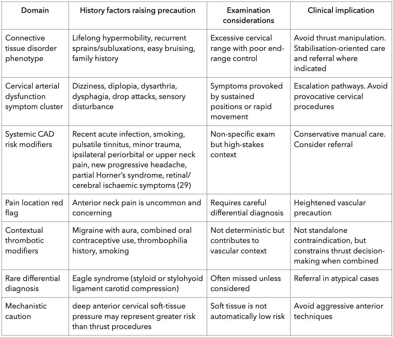
Table note: These factors represent precautionary constraints rather than deterministic predictors. Associations do not confirm causality. In hypermobility phenotypes, excessive cervical motion combined with autonomic symptoms or connective tissue disorder features should heighten precaution, as instability and vascular differentials may be more difficult to exclude clinically (19). For example, a young hypermobile patient presenting with new unilateral headache, anterior neck pain, pulsatile tinnitus, or syncope requesting cervical manipulation represents a context where cervical arterial dysfunction and craniocervical instability must be carefully considered before any thrust intervention (18).
Evidence-based rehabilitation approaches
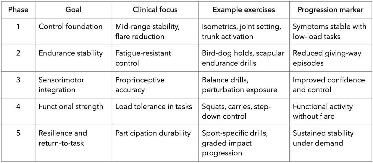
The strongest evidence for managing symptomatic hypermobility supports rehabilitation strategies emphasising stability, neuromuscular control, endurance, and graded exposure. (2) Hypermobility is therefore rarely a disorder requiring increased mobility. It is more accurately a stability and load tolerance disorder, where excessive motion exists alongside reduced control and poor proprioceptive integration. (20) Contemporary clinical classifications of hypermobility spectrum disorders similarly emphasise impaired connective tissue restraint and neuromuscular control. (1)
Table 2: Rehabilitation Progression Framework
Progressive strengthening
Progressive resistance training improves active joint stability by enhancing muscular restraint and increasing tolerance to mechanical load. (21) Randomised trials in children with joint hypermobility syndrome have demonstrated meaningful reductions in knee pain and improvements in function following strengthening programmes. (21)
Structured rehabilitation programmes have also been shown to improve physical capacity and reduce symptom burden in hypermobility populations. (22) Importantly, the therapeutic effect arises not from restricting mobility but from improving the capacity of surrounding musculature to compensate for reduced ligament stiffness.
Engelbert et al. emphasised that strengthening programmes in hypermobility syndromes should prioritise neuromuscular endurance and dynamic joint support rather than maximal force production. (2) Clinically, programmes should begin with low-load mid-range control tasks before progressing toward higher demand functional movements. Early aggressive end-range loading may provoke symptom flares in irritable unstable joints. (9)
Proprioceptive and sensorimotor training
Proprioceptive deficits are well documented in individuals with hypermobility and contribute directly to instability risk. (20) Impaired joint position sense has been demonstrated in patients with hypermobile Ehlers-Danlos syndrome compared with healthy controls. (20) Reduced proprioceptive acuity limits the neuromuscular system’s ability to regulate joint position and maintain mechanical stability during functional tasks (23).
Rehabilitation approaches targeting proprioception have demonstrated improvements in symptoms and functional stability. (23) Proprioceptive training programmes can improve joint position awareness and reduce symptom severity in patients with joint hypermobility syndrome. (23)
Clinically, sensorimotor rehabilitation may include balance training, perturbation exposure, closed-chain stability tasks, and coordination exercises that challenge joint control under gradually increasing load. (2)
Motor control and spinal stabilisation
Spinal symptoms in hypermobile patients often reflect impaired stabilisation strategies rather than structural pathology alone. Delayed activation of the transversus abdominis muscle has been demonstrated in individuals with chronic low back pain, highlighting motor control impairment as a central mechanism in spinal dysfunction. (24)
Exercise therapy has strong evidence for improving outcomes in chronic non-specific low back pain, particularly when programmes focus on control, endurance, and progressive functional loading. (25) Spinal stability depends on interaction between passive restraints, muscular control, and neural coordination. (26)
When passive ligamentous support is compromised, as in hypermobility syndromes, neuromuscular control becomes increasingly important in maintaining functional stability. Rehabilitation programmes therefore prioritise endurance-based stabilisation exercises, trunk control training, and progressive functional loading tasks designed to improve the capacity of the active stabilising system. (11)
Kinetic chain load-sharing and the symptomatic cost centre
A clinically useful construct in symptomatic hypermobility is that painful segments may represent the cost centre of task demand rather than the sole driver of dysfunction. In kinetic chain models of human movement, mechanical load is distributed across multiple segments and tissues during functional tasks. (27)
When one link in the system under-contributes because of stiffness, motor control deficit, or fatigue, other regions may compensate by absorbing greater mechanical demand. (28)
In hypermobility syndromes, excessive motion may therefore occur in regions that are repeatedly recruited to compensate for reduced contribution elsewhere in the chain. This phenomenon can increase eccentric braking requirements in vulnerable tissues and may contribute to symptom recurrence. (28)
Kinetic chain reasoning should be applied as a clinically testable hypothesis rather than a deterministic rule. Functional reassessment following targeted rehabilitation of adjacent regions can help determine whether a symptomatic joint is the primary driver of dysfunction or the site where load failure becomes clinically apparent. Durable improvement depends on graded capacity building across the entire movement system rather than repeated passive care directed at a single segment.
Clinical instability vignettes
Vignette 1:
Multidirectional shoulder instability presenting as rotator cuff tendinopathy
A 19-year-old overhead athlete presents with lateral shoulder pain during serving. She reports slipping sensations rather than traumatic dislocation. History reveals lifelong flexibility and recurrent sprains, raising suspicion of a generalised hypermobility phenotype.
Examination demonstrates excessive external rotation, translation in multiple directions, scapular dyskinesis, and pain on resisted cuff testing. The dominant impairment is instability-driven load failure rather than primary tendinopathy.
Rehabilitation should prioritise scapular control, rotator cuff endurance, proprioceptive retraining, and graded exposure rather than further mobilisation. (28) These rehabilitation priorities are consistent with stability-focused exercise approaches recommended for hypermobility syndromes. (2)
Vignette 2:
Low lumbar instability with short-lived response to manipulation
A 42-year-old man reports episodic low back pain provoked by sustained standing and bending. Manipulation provides only transient relief with rapid recurrence.
Examination demonstrates excessive mid-lumbar motion, reduced trunk endurance, and hip sequencing deficits, with early lumbar substitution during extension tasks.
This pattern aligns with instability-dominant degenerative trajectories described in biomechanical models of spinal stability. (26) Repeated thrust into a lax segment may reinforce symptom cycling, whereas endurance-based motor control rehabilitation is more consistent with evidence-based management of chronic spinal pain. (25)
Vignette 3:
Connective tissue disorder phenotype with cervical neurovascular constraint
A 28-year-old woman presents with chronic neck pain, episodic dizziness, fatigue, recurrent sprains, easy bruising, and orthostatic intolerance. She requests cervical manipulation due to brief prior relief.
Examination reveals marked cervical range with poor restraint rather than restriction. History raises concern for symptomatic connective tissue disorder phenotype with possible craniocervical instability risk.
In this context, HVLA thrust is inappropriate. Contemporary safety framing emphasises cervical arterial dysfunction constraints rather than outdated provocation testing. (18) International manual therapy safety frameworks emphasise structured history screening and escalation pathways when vascular symptoms are present. (19)
Conclusion
Symptomatic hypermobility is not primarily a mobility disorder. It is a stability and load tolerance disorder. Chiropractors contribute most effectively when care is aligned with evidence-based principles: strengthening, proprioceptive retraining, motor control rehabilitation, pacing, and cautious tool selection whether the chiropractor provides it or refers for it. Symptomatic hypermobility is not primarily a mobility problem. It is a stability and load tolerance disorder requiring active restraint, neuromuscular control, and graded capacity building.
References
- Castori M, Tinkle B, Levy H, Grahame R, Malfait F, Hakim A. A framework for the classification of joint hypermobility and related conditions. Am J Med Genet C Semin Med Genet. 2017;175:148-157.
- Engelbert RHH, Juul-Kristensen B, Pacey V, Smeenk S, Woinarosky N, Sabo S, et al. Evidence-based rationale for physical therapy treatment of hypermobile Ehlers–Danlos syndrome. Am J Med Genet C Semin Med Genet. 2017;175:158-167.
- Malfait F, Francomano C, Byers P, Belmont J, Berglund B, Black J, et al. The 2017 international classification of the Ehlers-Danlos syndromes. Am J Med Genet C Semin Med Genet. 2017;175:8-26.
- Bialosky JE, Bishop MD, Price DD, Robinson ME, George SZ. The mechanisms of manual therapy in the treatment of musculoskeletal pain: a comprehensive model. Man Ther. 2009 Oct;14(5):531-8.
- Pickar JG. Neurophysiological effects of spinal manipulation. Spine J. 2002;2:357-371.
- Triano JJ. Biomechanics of spinal manipulative therapy. Spine J. 2001;1:121-130.
- Herzog W. The biomechanics of spinal manipulation. J Bodyw Mov Ther. 2010;14:280-286.
- Vernon H, Humphreys BK, Hagino C. Chronic mechanical neck pain treated by manual therapy: systematic review. J Manipulative Physiol Ther. 2007;30:215-227.
- Simmonds JV, Keer RJ. Hypermobility and the hypermobility syndrome. Man Ther. 2007;12:298-309.
- Bronfort G, Haas M, Evans R, Leininger B, Triano J. Effectiveness of manual therapies: the UK evidence report. Chiropr Osteopat. 2010;18:3.
- Globe G, Farabaugh RJ, Hawk C, Morris CE, Baker G, Whalen WM, et al. Chiropractic clinical practice guideline for low back pain. J Manipulative Physiol Ther. 2016;39:1-22.
- Bussières AE, Stewart G, Al-Zoubi F, Decina P, Descarreaux M, Haskett D, et al. The treatment of neck pain-associated disorders. J Manipulative Physiol Ther. 2016;39:523-564.
- Henderson FC, Austin C, Benzel E, Bolognese P, Ellenbogen R, Francomano CA. Neurological and spinal manifestations of Ehlers–Danlos syndromes. Neurosurg Rev. 2017;40:495-513.
- Cassidy JD, Boyle E, Côté P, He Y, Hogg-Johnson S, Silver FL, et al. Risk of vertebrobasilar stroke and chiropractic care. Spine. 2008;33:S176-S183.
- Haldeman S, Carey P, Townsend M, Papadopoulos C. Arterial dissections following cervical manipulation. CMAJ. 2001;165:905-906.
- Church EW, Sieg EP, Zalatimo O, Hussain NS, Glantz M, Jabbour P. Chiropractic care and cervical artery dissection. Cureus. 2016;8:e498.
- Ernst E. Vascular accidents after neck manipulation. Int J Clin Pract. 2000;54:554-556.
- Rushton A, Rivett D, Carlesso L, Flynn T, Hing W, Kerry R, et al. International framework for cervical region examination. Man Ther. 2014;19:222-228.
- IFOMPT. International framework for examination of the cervical region for vascular pathologies. Auckland: IFOMPT; 2020.
- Rombaut L, Malfait F, De Paepe A, Rimbaut S, Verbruggen G, De Wandele I, Calders P. Impairment and impact of pain in female patients with Ehlers-Danlos syndrome: a comparative study with fibromyalgia and rheumatoid arthritis. Arthritis Rheum. 2011;63(7):1979-87.
- Pacey V, Tofts L, Adams RD, Munns CF, Nicholson LL. Exercise in children with joint hypermobility syndrome and knee pain: a randomised controlled trial comparing exercise into hypermobile versus neutral knee extension. Pediatr Rheumatol Online J. 2013;11(1):30.
- Corrado B, Ciardi G. Hypermobile Ehlers-Danlos syndrome and rehabilitation: taking stock of evidence based medicine: a systematic review of the literature. J Phys Ther Sci. 2018 Jun;30(6):843-847.
- Ferrell WR, Tennant N, Sturrock RD, Ashton L, Creed G, Brydson G, et al. Enhancement of proprioception in patients with joint hypermobility syndrome. Rheumatology. 2004;50(10):3323-8.
- Hodges PW, Richardson CA. Inefficient muscular stabilisation of the lumbar spine associated with low back pain. Spine. 1996;21:2640-2650.
- Hayden JA, van Tulder MW, Malmivaara A, Koes BW. Exercise therapy for treatment of non-specific low back pain. Ann Intern Med. 2005;142:765-775.
- Panjabi MM. The stabilizing system of the spine. Spine. 1992;17:362-371.
- Almansoof HS, Nuhmani S, Muaidi Q. Role of kinetic chain in sports performance and injury risk: a narrative review. J Med Life. 2023 Nov;16(11):1591-1596.
- Kibler WB, Press J, Sciascia A. The role of core stability in athletic function. Sports Med. 2006;36:189-198.
- Pollard H. Inappropriate use of the title chiropractor and profession of chiropractic when reporting an adverse vascular event. Chiropractic Journal of Australia. 2021;48:57-65.
Research
AU: Jagadeesh A, MOT (Advanced OT in Neurology), MS. (Counselling Psychology), CKTP – KTAI (USA). Program Head, Department of Occupational Therapy, Akkara Foundation, Kasaragod, Kerala. e: ajagadeeshmars@gmail.com ORCID: https://orcid.org/0009-0000-6401-0513
AU2: Pratiksha Mohanty. Faculty of Health and Wellness, Sri Sri University, Cuttack.
AU3: Tirthankar Ghosh MSc (Human Physiology), MPH, PhD (Physiology & Ergonomics). Dean, Ergonomics and Exercise Physiology Laboratory, Faculty of Health and Wellness, Sri Sri University, Cuttack, Odisha.
Abstract: Industrial workers, including cashew workers, often face work-related issues, including Pain, Musculoskeletal Discomfort (MSD) affecting muscles, joints, tendons, ligaments, and nerves. Work-related MSD (WMSD) & pain usually develop over time due to the nature of the work, environment, or machinery used. This study aimed to address that gap by identifying the intensity of pain and MSD among cashew workers, examining gender-based and task-specific differences, and highlighting the importance of joint protection, work simplification, and energy conservation techniques as preventive measures.
Methods: A cross-sectional survey was conducted among 40 cashew workers from Boovikanam and nearby areas in Kasaragod district, Kerala, who met the inclusion criteria. Data were collected using a convenient sampling technique. Standardised assessment tools, namely Rapid Entire Body Assessment (REBA) and Visual Analogue Scale (VAS), were used to evaluate WMSD and pain intensity. The data were analysed using IBM SPSS Statistics Version 26.
Results: Findings revealed a significant risk of WMSD among cashew workers, particularly during shelling and peeling activities. Most shelling workers demonstrated medium risk, while peeling workers demonstrated higher, very high-risk scores on REBA. Moderate pain intensity was common in both groups on the VAS. A significant positive correlation was observed between pain intensity and WMSD among male workers (r = 0.725, p < 0.001) and female workers (r = 0.538, p = 0.014), indicating that higher pain levels were associated with increased WMSD risk.
Conclusion: The findings highlight the need for ergonomic interventions, especially for workers with mild to severe pain. Gender differences were also observed; males commonly reported moderate pain, whereas females showed a wider range of pain intensities.
Indexing Terms: Cashew Workers; Ergonomics; Pain; Survey Study; Work-Related Musculoskeletal Discomfort.
Musculoskeletal Discomfort (MSD) is a broad group of inflammatory and degenerative conditions affecting muscles, tendons, joints, ligaments, nerves, and other soft tissues that support movement. Work-related musculoskeletal discomfort (WMSD) has emerged as a major concern in both developed and developing countries, often caused by repetitive tasks, awkward postures, forceful exertions, and suboptimal ergonomic environments. This discomfort not only compromises the physical well-being of workers but also leads to reduced productivity, increased absenteeism, and higher compensation and healthcare costs. (1)
In India, the cashew industry is a major export sector, with Kerala alone accounting for 35% of processing units. Cashew processing is a labour-intensive, multistage process involving drying, roasting, shelling, peeling, grading, and packing, most of which are manually performed and physically demanding. Studies have consistently reported low back pain as the most prevalent symptom across various occupations. (2, 3)
A cross-sectional study among 246 cashew factory workers in Udupi district, Karnataka, found that 28.5% of workers experienced musculoskeletal pain, most frequently in the knees (32.4%), lower back (30.9%), and shoulders (11.8%). Cutting tasks were associated with the highest pain prevalence, and over 70% of affected workers had over five years of experience. (4, 5)
Despite the long-standing operation of cashew factories since the 17th century in South India, limited research has addressed the ergonomic risks and musculoskeletal implications for workers in this sector. Other regional studies report WMSD prevalence ranging from 28.5% to over 70% among various cashew processing tasks.6 Kerala-specific surveys highlight female gender and lower education levels as significant risk factors for MSD in this workforce. (7)
While ergonomic assessments and WMSD studies exist in other industrial settings, cashew workers remain underrepresented in the literature. This study aims to address that gap by identifying the intensity of musculoskeletal discomfort and pain among cashew factory workers, examining gender-based and task-specific differences, and highlighting the importance of joint protection, work simplification, and energy conservation techniques as preventive measures. The findings are expected to contribute to occupational health literature and inform ergonomic interventions and policies for this underserved workforce.
Methods
This study used a quantitative cross-sectional design to assess the intensity of work-related musculoskeletal discomfort (WMSD) among cashew workers. It was conducted in villages around Boovikanam, Kasaragod district, Kerala, approximately 10 - 15 km from Kasaragod town. Data collection aimed to provide various postures adopted during cashew work. A total of 40 cashew workers (20 males, 20 females), aged 35 - 55 years, were selected through convenient sampling due to accessibility and willingness to participate, and data were collected over a period of two months, from May to June 2024. This age group was targeted as they are commonly engaged in physically demanding tasks in the cashew industry.
Inclusion criteria comprised workers involved in post-harvest cashew processing activities. Although cashew processing includes drying, roasting, shelling, peeling, and grading, the present study specifically focused on workers engaged in shelling and peeling tasks, as these activities are predominantly manual and ergonomically demanding. Drying, roasting, and grading processes were not included because these activities were largely mechanised in the selected processing units. Both literate and illiterate individuals were included.
Exclusion criteria covered those involved in other stages like cultivation or marketing, individuals recently ill, part-time workers, those with major neurological or orthopaedic issues, psychosocial distress, or unwillingness to participate. Data were collected using the standardised tools like Rapid Entire Body Assessment (REBA) and the Visual Analogue Scale (VAS).
The Rapid Entire Body Assessment (REBA) tool has demonstrated excellent intra-rater reliability (ICC = 0.925) and moderate inter-rater reliability (Fleiss’ κ = 0.54), supporting its use as a reliable ergonomic assessment instrument for evaluating musculoskeletal disorder risk in occupational settings. (8)
The Visual Analogue Scale (VAS) is a highly reliable and valid instrument for subjective pain assessment, demonstrating excellent test–retest reliability with ICC values commonly reported above 0.90. (9)
REBA, developed by Hignett and McAtamney, is an ergonomic tool used to evaluate posture-related risk without needing specialised training or equipment. (10, 11) Scores were assigned to body regions (neck, trunk, upper limbs, legs), and final risk scores were calculated from task observations.
The REBA scoring system categorises ergonomic risk into negligible (score = 1), low (2 – 3), medium (4 – 7), high (8 – 10), and very high risk levels (11 – 15), with increasing scores indicating greater urgency for corrective action. (10)
VAS, a validated subjective scale, was used to assess pain intensity. It features a 10 – 15 cm line ranging from 0 (no pain) to 10 (worst pain), with visual cues to assist illiterate participants. Pain levels were categorised as no (0 – 1), mild (1 – 3), moderate (4 – 6), or severe. (12)
Descriptive statistics were used to determine WMSD and intensity, and inferential statistics assessed differences across gender and task categories. All methodological details were set before data collection; results are presented separately.
Results
A survey was conducted among 40 cashew workers in Kerala using a convenient sampling method. The data collected underwent extensive analysis using Statistical Product and Service Solutions version 22 (SPSS 22). Work-related musculoskeletal discomfort (WMSD) was assessed using the Rapid Entire Body Assessment (REBA) tool, and pain perception was measured through the Visual Analogue Scale (VAS). A questionnaire, interviews and observational methods were used to gather further information.
Descriptive statistics were used to analyse the frequency and percentage values of REBA and VAS scores. To examine the correlation between REBA and VAS among male and female cashew workers, the Pearson Correlation Coefficient (2-tailed) was applied. Since the data were normally distributed, the Pearson correlation was considered appropriate for analysis. (13)
- Table 1 shows the mean and standard deviation of demographic data, such as age (43.50 + 5.43), REBA and VAS scores for Cashew workers, such as shelling and peeling workers.
Table 1: Description of Demographic Data
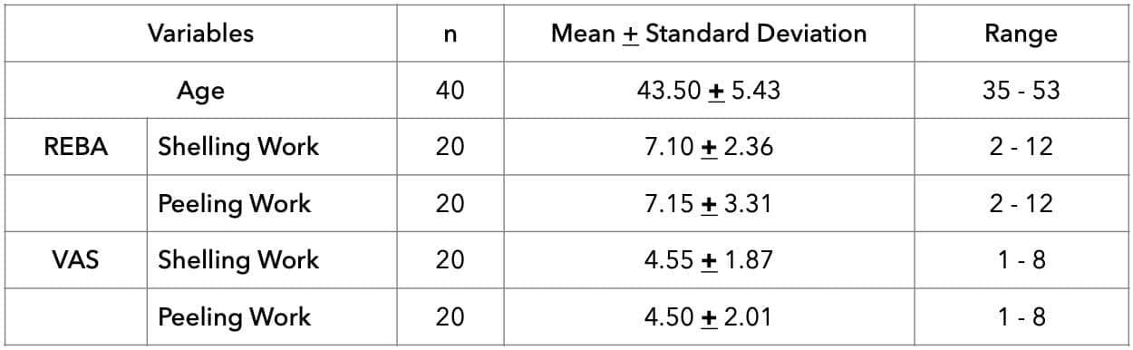
Table 2 shows the frequency and percentage of risk levels of Work-related Musculoskeletal discomfort through the REBA score for the cashew workers involved in shelling and peeling work. Results denote that the majority of the workers involved in shelling and peeling work had medium risk (i.e., 60% and 40%, respectively).
Table 2: Frequency and Percentage of Levels of Risk of WMSD as scored by Rapid Entire Body Assessment (REBA)
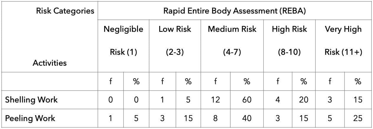
Table 3 shows the frequency and percentage of levels of pain through the VAS score for the cashew workers. Both in shelling workers and peeling workers found moderate pain, i.e., 50% and 55% respectively.
Table 3: Frequency and Percentage of Pain of WMSD as scored by Visual Analog Scale (VAS)
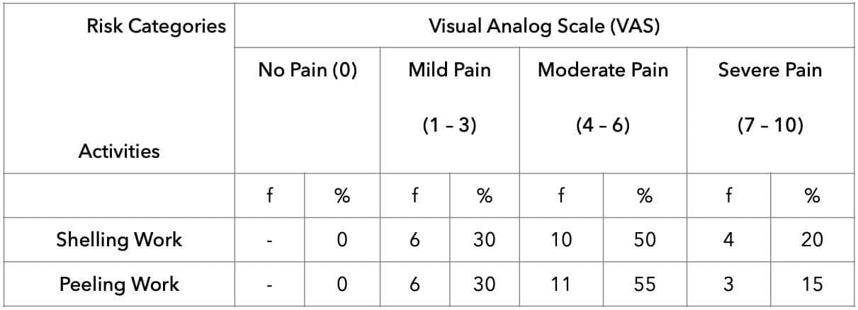
Table 4 shows the ergonomic risk factors / WMSD among 40 participants (20 males and 20 females). None of the participants had a negligible risk. Among males, 3 participants had low risk, 8 participants had medium risk, 2 participants had high risk, and 7 participants had very high risk. Among females, 2 participants had low risk, 12 participants had medium risk, 5 participants had high risk, and only one participant had very high risk. In total, 5 participants had low risk, 20 participants had medium risk, 7 participants had high risk, and 8 participants had very high risk.
Table 4: Comparison of REBA scores between male and female workers
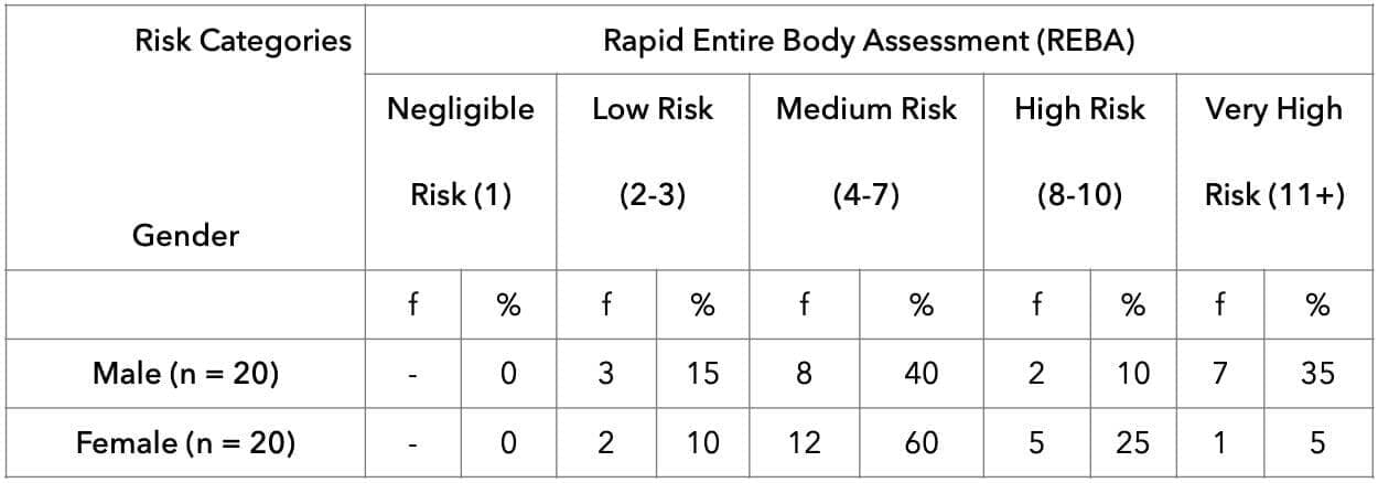
Table 5 shows the VAS pain level distribution among 40 participants (20 males, 20 females). None of the participants reported no pain (i.e., score: 0). Among males, 4 participants had reported mild pain (score: 1 - 3), 12 participants had reported moderate pain (score: 4 - 6), and 4 participants had reported severe pain (score: 7 - 10). Among females, 8 participants had reported mild pain, 9 participants had reported moderate pain, and 3 participants had reported severe pain. In total, 12 participants had reported mild pain, 21 participants had reported moderate pain, and 7 participants had reported severe pain.
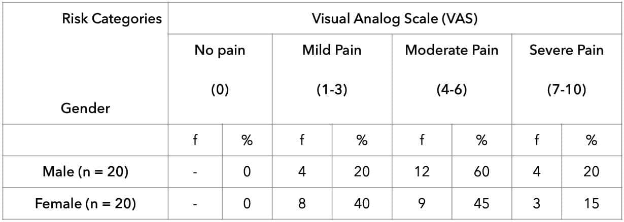
Table 6 shows the correlation between VAS and REBA scores among 20 male cashew workers. A strong positive correlation (r = 0.725) was found, significant at p = 0.0001, indicating that higher pain levels (VAS) are linked to higher ergonomic risk (REBA) for male cashew workers.
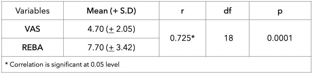
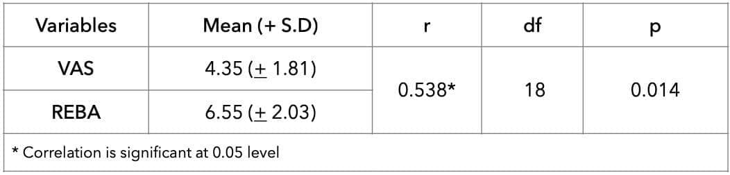
Table 7 shows the correlation between VAS and REBA scores among 20 female cashew workers. A moderate positive correlation (r = 0.538) was observed, statistically significant at p = 0.014, indicating that higher pain levels (VAS) are moderately linked to greater ergonomic risk (REBA) for female cashew workers. As expected, both VAS and REBA show perfect self-correlation (r = 1).
Table 7: Correlation of REBA and VAS scores between Female Cashew Workers. (n = 20)
Discussion
The present study evaluated ergonomic risk factors, work-related musculoskeletal discomfort (WMSD), and pain intensity among cashew workers engaged in shelling and peeling activities. The findings demonstrated a considerable ergonomic risk of WMSD and pain among the workers, with significant correlations between REBA and VAS scores for both male and female participants. These findings indicate that increased ergonomic risk of WMSD is associated with greater pain intensity among cashew workers.
The present findings are consistent with previous studies conducted among cashew-processing workers and other manual labour populations exposed to repetitive movements, awkward postures, and prolonged static work. Girish et al. reported that musculoskeletal pain was highly prevalent among cashew factory workers, particularly affecting the knees, lower back, and shoulders, with repetitive cutting and shelling activities identified as major contributing factors.4 Similarly, Chaitanya et al. observed that female cashew shelling workers experienced substantial musculoskeletal discomfort due to repetitive upper-limb movements and poorly designed workstations. (3) The current study further supports these findings, as most shelling workers demonstrated medium ergonomic risk, while peeling workers exhibited higher proportions of very high-risk REBA scores.
The elevated ergonomic risk of WMSD observed among peeling workers may be attributed to sustained neck flexion, repetitive wrist movements, prolonged sitting posture, and fine motor hand activities performed during peeling tasks. Similar ergonomic concerns were highlighted by Mallampalli et al., who identified awkward postures and repetitive hand-intensive activities as major predictors of WMSD among cashew-processing workers. (2) Another ergonomic intervention study by Mallampalli et al. demonstrated that workstation modification and ergonomic redesign significantly reduced REBA and RULA scores among cashew shellers, emphasising the importance of preventive ergonomic strategies in this sector. (14)
Pain intensity findings from the present study revealed that moderate pain was the most frequently reported category among both shelling and peeling workers. These findings correspond with Satheeshkumar’s study among cashew industry workers in Kerala, which documented a high prevalence of musculoskeletal discomfort associated with repetitive occupational exposure and prolonged working duration. (7) Similar findings have also been reported in manufacturing and agricultural occupations, where repetitive manual tasks and sustained postures contribute significantly to musculoskeletal symptoms and pain development. (15)
Gender-based differences identified in this study are also supported by previous literature. Male workers predominantly reported moderate pain, whereas female workers demonstrated a broader distribution of pain intensity levels. Previous epidemiological studies have consistently shown that women report greater WMSD and severity of musculoskeletal pain than men. Picavet and Schouten found that musculoskeletal pain prevalence was systematically higher among women across multiple body regions. (16) Likewise, Treaster and Burr reported increased susceptibility of women to upper-extremity musculoskeletal disorders due to differences in biomechanics, muscle capacity, task allocation, and occupational exposure patterns. (17) The wider variation in pain intensity among female workers observed in the current study may therefore reflect biological, ergonomic, and psychosocial differences in occupational workload and pain perception.
A significant positive correlation was observed between REBA and VAS scores among both male (r = 0.725, p < 0.001) and female workers (r = 0.538, p = 0.014), indicating that higher ergonomic risk was associated with increased pain intensity. This finding reinforces the clinical relevance of ergonomic assessment tools such as REBA in identifying workers at greater risk of developing WMSDs. Comparable findings have been reported in occupational ergonomic studies where increased postural risk scores were strongly associated with musculoskeletal pain severity and functional limitations. (1, 14)
The present study contributes important evidence to occupational health literature, particularly in the context of the cashew-processing industry, which remains relatively underrepresented in ergonomic research. Unlike many previous studies focusing only on prevalence, the current study combined observational ergonomic assessment with subjective pain evaluation and gender-based comparison, thereby providing a more comprehensive understanding of occupational risk among cashew workers.
Despite these strengths, certain limitations should be considered. The cross-sectional design limits the ability to establish causal relationships between ergonomic exposure and WMSD. The relatively small sample size and convenient sampling technique may also restrict generalisability to larger populations of cashew workers. Additionally, psychosocial and organisational factors such as workload, job stress, rest breaks, and work environment were not assessed and may have influenced pain perception and ergonomic risk.
Overall, the findings emphasise the urgent need for ergonomic interventions within the cashew-processing industry. Strategies such as workstation redesign, posture correction training, scheduled rest breaks, task rotation, and education regarding joint protection and energy conservation techniques may help reduce WMSD risk and improve worker well-being. Gender-sensitive ergonomic approaches may also be beneficial considering the observed differences in pain distribution and ergonomic exposure between male and female workers.
Conclusion
In conclusion, the findings suggest that there is a high risk of WMSD among cashew workers. Also, the study findings indicate a clear trend where higher pain levels are associated with higher ergonomic risk of WMSD for both male and female cashew workers, also emphasising the need for ergonomic interventions, particularly for those experiencing mild to severe pain. This study's findings suggest gender-related variations in pain perception or reporting, with males more frequently reporting moderate levels of pain compared to females, who displayed a broader range of pain experiences.
Limitations
This study focused solely on physical factors contributing to WMSD risk. Psychosocial aspects (e.g., workplace conditions, autonomy, monotony, work-rest cycle, task demands, social support) and individual factors (e.g., alcohol/tobacco use, stress) were not included. The study was limited to a single industrial worker group, limiting generalizability. Future research should include diverse farm settings to enhance applicability. Additionally, this study assessed only WMSD risk; further studies identifying actual MSD cases are recommended for effective intervention planning.
Ethical consideration
Informed and written consent was obtained from all participants. Ethical clearance was also obtained from Sri Sri University.
Acknowledgement
We express our sincere gratitude to the craftsmen who rendered immense cooperation during the completion of this study.
References
- PunnettL, Wegman DH. Work-related musculoskeletal disorders: The epidemiologic evidence and the debate. J Electromyogr Kinesiol. 2004;14(1):13–23. DOI: 10.1016/j.jelekin.2003.09.015
- MallampalliKC, Dhar D, PalS. Ergonomic assessment of postures and risk factors associated with work-related musculoskeletal disorders among cashew workers in Eastern India. In: Adv Phys, Social & Occup Ergonomics (AHFE 2020). Berlin, Heidelberg: Springer; 2020:79–86. DOI 10.1007/978-3-030-51549-2_11
- ChaitanyaK, Dhar D, PalS. Prevalence of musculoskeletal disorders among female cashew nut shelling workers in India. In: Convergence of Ergonomics and Design (ACEDSEANES 2020). Cham: Springer; 2021:281–291. DOI 10.1007/978-3-030-63335-6_29
- GirishN, Ramachandra K, ArunGM, Asha K. Prevalence of musculoskeletal disorders among cashew factory workers. Arch Environ Occup Health. 2012;67(1):37–42. DOI 10.1080/19338244.2011.573020
- NarsiaRH, Joseph OR. Participatory ergonomics: Work-related musculoskeletal disorders among cashew nut factory workers in Karkala taluka. Acta Sci Orthop. 2020;3(6):36–40.
- ChaitanyaK, Dhar D, PalS. Ergonomic assessment and workstation design for women workers in cashew processing. In: Smart Innovation, Systems and Technologies (ICORD 2023). Cham: Springer; 2023:37–47.
- SatheeshkumarM. Work-related musculoskeletal disorders among cashew industry workers in Kerala. Int J Adv Res Eng Technol. 2018;9(4):162–173.
- Schwartz, A. H., Albin, T. J., & Gerberich, S. G. (2019). Intra-rater and inter-rater reliability of the Rapid Entire Body Assessment (REBA) tool. International Journal of Industrial Ergonomics, 71, 111–116. https://doi.org/10.1016/j.ergon.2019.02.005
- Bijur, P. E., Silver, W., & Gallagher, E. J. (2001). Reliability of the visual analog scale for measurement of acute pain. Academic Emergency Medicine, 8(12), 1153–1157. https://doi.org/10.1111/j.1553-2712.2001.tb01132.x
- HignettS, McAtamney L. Rapid Entire Body Assessment (REBA). Appl Ergon. 2000 Apr;31(2):201–205. DOI 10.1016/S0003-6870(99)00039-3
- HignettS, McAtamney L. REBA: a rapid entire body assessment method for investigating work-related musculoskeletal disorders. In: Proc Ergonomics Society Aust, Adelaide 1995:45–51.
- GouldD, Kelly D, GoldstoneL. Visual Analogue Scale (VAS). J Clin Nurs. 2001;10:697–706.
- Dancey CP, Reidy J. Statistics Without Maths for Psychology. 7th ed. Harlow (UK): Pearson Education; 2017.
- MallampalliKC, Dhar D, PalS. Ergonomic assessment of cashew nut sheller reduces REBA and RULA scores. Work J. 2023; Usercentered design study. DOI 10.1007/9783030633356_29 pubmed.ncbi.nlm.nih.gov+4researchgate.net+4researchgate.net+4
- SilversteinBA, et al. Occupational exposures and WMSDs in manufacturing sectors. Am J Ind Med. 1996;29(3):234–248. doi:10.1002/(SICI)10970274(199603)29:3<234::AIDAJIM2 3.0.CO;2T
- PicavetHS, Schouten JS. Musculoskeletal pain is systematically higher in women than in men. J Clin Epidemiol. 2003;56(7):522–528. DOI 10.1016/S08954356(03)00040X
- TreasterDE, Burr D. Gender differences in prevalence of upper extremity musculoskeletal disorders. Ergonomics. 2004;47(5):495–526. At pubmed.ncbi.nlm.nih.gov
Professional identity
AU: Jayul Doshi MBA (Eco), PT, MPT, PhD (Hon), PhD (c, scoliosis). Associate Editor (Diagnostic Imaging). Private practice of Chiropractic. India. e: ais@drspine.in
Narrative: This paper is a short, descriptive report of conventional Chiropractic practice in India. It is the first data reported to describe the Case-Mix of Chiropractic practice in India. It is noted that the data sets are drawn from 21,386 patients and are not analyses of ‘patient visits’, but of n="21,396" individual patients. This renders the number of patients documented in this report to be a very large sample.
In brief the most numerous presentation was LBP with male predominance. The male dominance held for all other conditions to a varying and lesser degree, and only reached parity with females at 50/50 for ‘knee pain’.
An interesting finding is a greatly increased number of presentations due to seasonal conditions, peaking in June.
This report sets the baseline for future reports of Chiropractic practice throughout India and carries value to inform the distribution of learning objects in India’s first formal program of Chiropractic education, commencing 2026.
Indexing Terms: Chiropractic; India; Case-Mix; Conventional Chiropractic; education; regulation.
Cite: Doshi J. An initial report of Chiropractic practice in India: The case-mix of 21,386 patients. Asia-Pac Chiropr J. 2026;7-2.
https://www.apcj.site/DoshiCasemixIndia26.pdf
Take-away: 'This is the first descriptive report of chiropractic practice in India. It shows the case-mix is similar to that reported for other locations but with a higher than expected degree of presentations for LBP ...'
Introduction
This paper presents the first report of the case-mix of conventional subluxation-inclusive Chiropractic in the nation on India. At the time of writing there is no Chiropractic-specific regulation, although common business practices must be adhered to.
There are 12 Chiropractors formally recognised across the nation on India yet evidence exists to show there are some 2,200 imposter clinics claiming to provide chiropractic services without any trained Chiropractor on-site. This is similar, but vastly larger in scale and potential damage, to the early situation in Australia where individuals could call themselves a ‘chiropractor’ regardless of not having any training. (1) The negative outcome of this was that on the eventual enactment of registration these folk became registered as Chiropractors under grandfathering provisions.
This must not be allowed to happen in India and the Government is urged to act immediately to protect the public in India.
In contrast, the dominant provider of Chiropractic services with recognised Chiropractors in India is a commercial operation called Dr Spine, and this report is drawn from the clinical data of that group.
Regulation
There is no regulation of Chiropractic in India. The country cannot continue to let untrained and unsupervised hands learn physical techniques on the spines of its citizens. This paper calls for government to do what every serious health system before it has done: set a standard, protect a title, and build a profession in India, for India, by Indian-trained chiropractors.
The education is being built and the evidence is in. What remains is the law and the will to enact it as an urgent matter of public safety the State alone can deliver. This Journal will report updates on the development of the required legal framework in India as it happens.
The author recognises accredited, university-anchored Chiropractic education as the only legitimate pathway into the profession. To this end, Sri Sri University is starting the Post Graduate Diploma in Chiropractic Sciences & Masters Fellowship programs, to be followed by a 5½ year ‘full’ program of Chiropractic education for school-leavers.
Work is also under way to constitute a Council on Chiropractic in India to set standards, register practitioners, and shield patients from unqualified practice. The NGO Chiropractic India has formulated this to be handed over to the govt to further legitimise and regulate the profession.
About case-mix studies
Case-mix studies are the first approach to understanding the nature of Chiropractic practice in a particular environment and are specifically useful for identifying the variety of clinical presentations. First-level case-mix studies such as this report the broad range of clinical conditions for which patients seek care. At this point it is not known in the evidential sense whether the 12 Chiropractors in India manage patients with low-back pain or headache, for example. Neither anecdotal evidence nor assumptions drawn from Chiropractic practice in other countries constitute sufficient evidence to inform the profession’s development in India.
Case-mix studies report essential foundational knowledge for informing further studies perhaps of the characteristics of a particular demographic with a particular clinical condition. Case-mix studies are also prime informants of the curriculum for Chiropractic education, ensuing that the real health needs of the nation will be met by Chiropractors specifically trained to identify and manage such conditions within the national health system.
In this manner they differ to demographic studies which report socio-economic information and express it statistically. (2) They also differ to descriptive studies, of which there are several for North America (3, 4, 5) and Europe (6, 7, 8) in that they represent exploratory research with a focus on clinical conditions and are readily undertaken by clinicians.
Case-mix studies also have value in informing public health decisions in developing countries by reporting a related group of patients; an example being a group of patients with tuberculosis. (9) Although common to medicine, case-mix studies are infrequently reported in the Chiropractic literature with the exception of the case-mix in teaching clinics, such as that of an Australian institution by Walsh in 1992. (10) Subsequent reports have been published for teaching clinics (11) in North America (12) and its West Coast region, (13) New Zealand, (14) Mexico (15) and Britain. (16) While not including statistical analysis they typically report a case-mix thought to be representative of private practice in each country. (17)
This paper also indicates future approaches for scholarly inquiry in India as the profession develops. The generation and publication of peer-reviewed descriptive studies and statistical reports is an important process for an emerging profession. It speaks to transparency in its practices and an accountability to the public.
Methods
This study reports prospective documentation of individual patients Chiropractic care provided by one clinic group in India. Providers included 5 Doctors of Chiropractic, 10 clinical technicians, and 3 physiotherapists.
The 1993 Case-Mix studies (18, 19) of Ebrall formed the seed template for data collection. The database of Dr Spine was interrogated to collate reports on case-mix and demographics of all patients registered over a 45-month-(August 2022 to April 2026)-year period. The top 13 reasons for patient presentation are given in Table 1. The percentage is the frequency of each category of all 13 categories. Categorisation by gender is given in Table 2.
Table 1: Distribution of presenting complaints
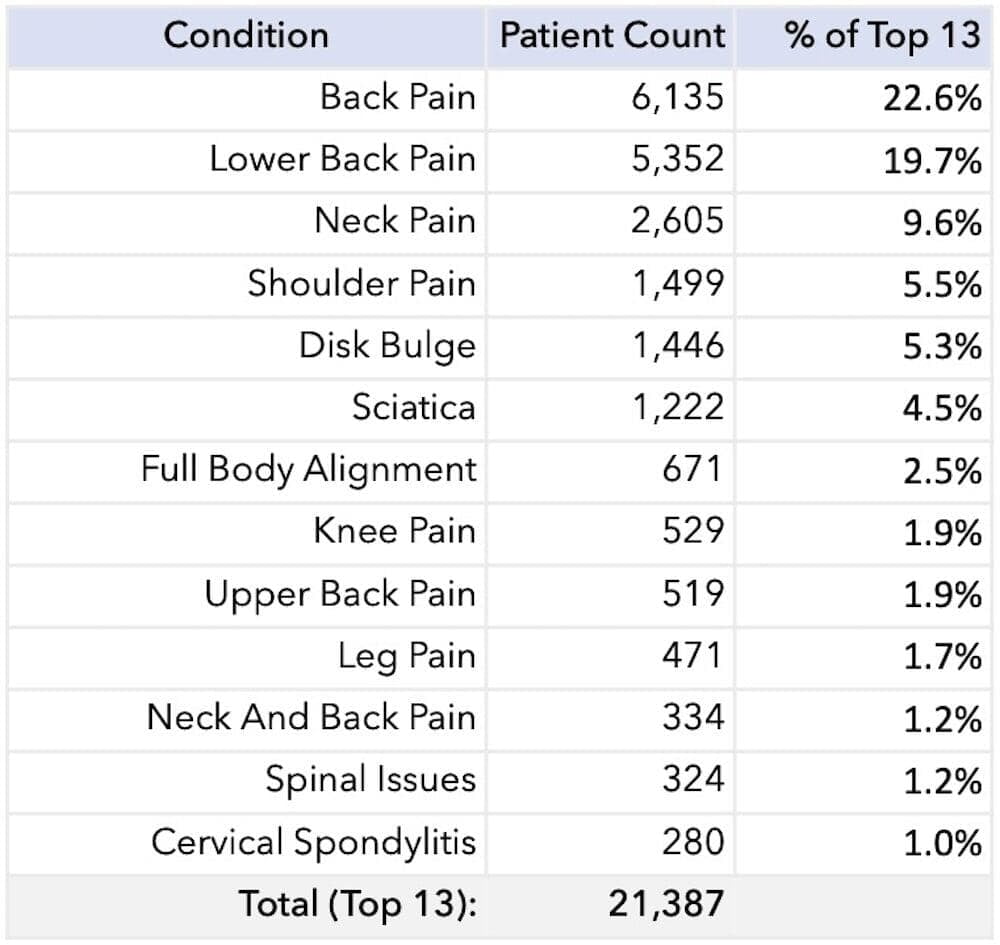
Table 2: Categories of presenting complaints
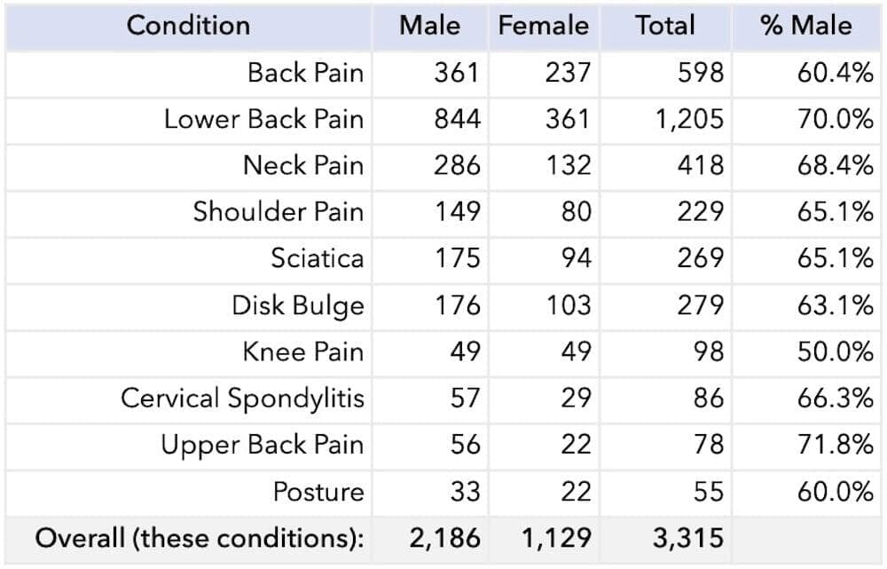
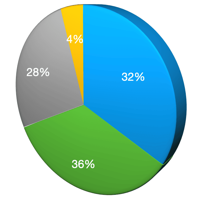
Figure 1: Percent distribution grouped complaints
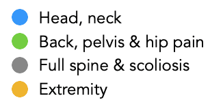
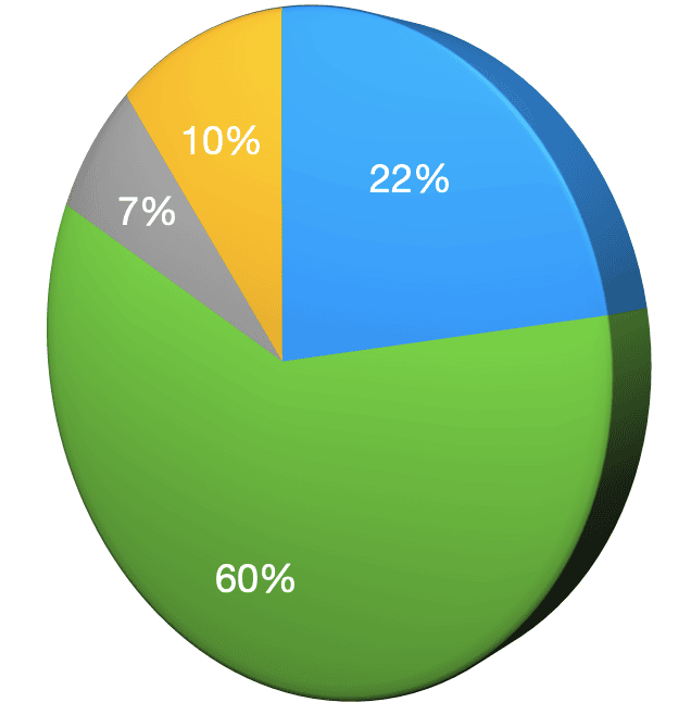
Figure 2: Comparison to the Philippines
Results
Distribution of typical Chiropractic presentations
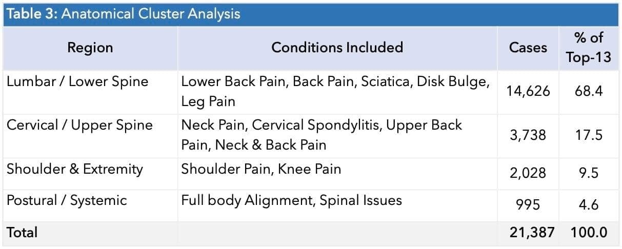
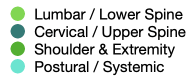
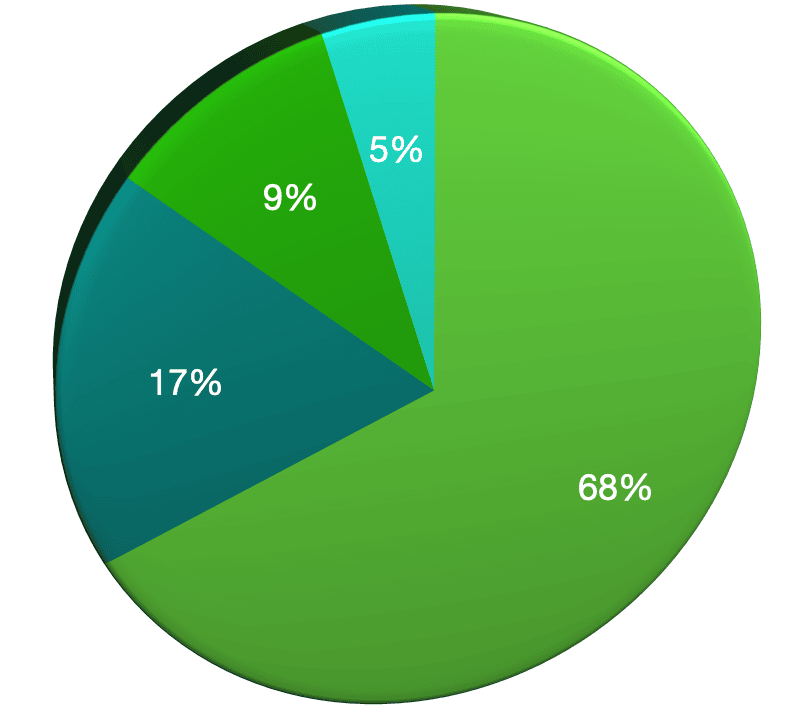
Figure 3: Anatomical cluster analysis
A total of 21,387 patient-files were accepted from a 45-month window. The clinics were located in three Dr Spine centres:
- Bengaluru, Karnataka, New BEL Road (No. 385, opp. Starbucks, RMV 2nd Stage, 2nd Block, Geddalahalli, Sanjayanagara 560094;
- Whitefield (ThePlanet #302, 54 Whitefield Main Rd, Brooke Bond First Cross, Narayanappa Garden, Varthur 560066;
- Indiranagar (947, 12th Main Rd, above ICICI Bank, Sodepur, Appareddipalya 560008).The CRM system remained constant. and recorded all details with consistency for case classification. Some variance is noted as clinicians refined their diagnostic statements.
Of all consecutive visits 65.9% were by males and 34.1% by females. India does not record transgender information. The mean age of female patients was 39.5y and of male patients, 39.7y. The all-patient age range was 8y to 72y.
Case-Mix analysis
Lower-back complaints dominate
Back Pain (n=6,135) + Lower Back Pain (n=5,352) n = 11,487 cases: This clinical category represents ~54% of all top-13 complaints. When we include Sciatica, Disk Bulge, Upper/Mid back and the lumbar region, these presentations drive the majority of visits.
Cervical (neck) issues are the #2 cluster
Neck Pain (n=2,605) + Cervical Spondylitis (n=280) + Shoulder Pain (n=1,499) n ≈ 4,384 cases. This is consistent with screen-heavy lifestyles and poor desk posture.
Skew toward males
Across the monthly samples, ~66% of patients are male. Lower Back Pain skews most male (70%); Knee Pain is the only category that is 50/50.
Nerve-root and spinal disc problems are of concern
Disk Bulge (n=1,446) + Sciatica (n=1,222) + ‘Slip’ Disk + Herniated disc n~2,750+ cases. These are structural issues, not just muscular and typically needed imaging-led care.
Lifestyle / posture conditions are growing
Full-body alignment, posture, scoliosis, frozen shoulder, TMJ and headache/migraine together represent a meaningful tail. We see this as evidence of sedentary, desk-bound presentations.
Seasonal pattern
Lower Back Pain peaks in Sep (n=216) and Aug (n=193); Back Pain peaks in Jun (n=233). Monsoon/post-monsoon months show the highest spine load, perhaps due to cold/damp weather and reduced individual activity.
Long tail of rare conditions
We recorded over 80 distinct complaints but the top 6 conditions accounted for over 85% of volume, a classic Pareto distribution. Care pathways can be standardised for the top 6 yet Chiropractors must remain vigilant for the presentation of serious and complex cases. The emerging education program is cognisant of these findings and its syllabus is appropriately tailored to the presentation profiles, including rare conditions, given in this report.
Critical insights from our findings
Insight 1: Disc disease is the fastest-growing spine condition in India
Disk bulge cases are up 126% and Sciatica up 93% comparing the first 12 months to the latest 12 months.
Together they are the single biggest growth story in our 4-year patient base. Indians are not just getting more back pain, they are presenting with more structural disc disease.
Insight 2: The diagnosis is sharpening, not the disease disappearing
Generic ‘Back Pain’ presentations dropped 46% over 4 years, while specific diagnoses (Disk Bulge, Sciatica, Lower Back Pain) rose.
The spine is not getting healthier, clinicians and patients are getting better at naming the actual condition. This is a maturity signal for Indian spine care.
Insight 3: Total spine volume is down 12%, but severity is up
Monthly visit volume fell from 549/mo (first year) to 481/mo (latest year), a 12.4% decline. But the mix has shifted decisively toward harder, imaging-grade cases (disc, sciatica).
This translates to fewer patients but with sicker spines: the case for specialist care has never been stronger.
Insight 4: Lower Back Pain is still the #1 condition and is growing
Lower Back Pain remains the single largest condition across the entire 45-month window and grew 14% comparing first-year to latest-year volumes.
LBP is the most addressable, most preventable, and most under-managed condition in Indian primary spine care.
Insight 5: Peak demand: Jun-23 hit 1,029 complaints in a single month
The clinic's highest-ever monthly volume was June 2023 at n="1,029" patient complaints. This number is 10x the lowest month (Sep 2022, n="102)."
We conclude that the demand for spine care is highly seasonal and event-driven. Future capacity planning must assume swings of this magnitude.
Insight 6: Neck and Shoulder cases are declining, and this may be the surprise
Neck Pain is down 12% and Shoulder Pain down 13% in the latest year vs the first year. Despite the screen-and-WFH narrative, our data shows cervical / upper-extremity complaints are actually flattening or contracting in this patient base.
We consider this finding merits investigation against national posture and ergonomics studies
.
Insight 7: The cumulative spine burden: 24,099 patient complaints over 4 years
Across 45 months and 8 tracked conditions, the clinic has logged n="24,099" spine-related patient complaints, an average of n="535" every month. This is the largest continuously-tracked single-clinic spine dataset in the country we are aware of, and forms the empirical basis of the India Spine Report.
Insight 8: Knee and Upper Back are flat. The spine is the real story
Non-spine conditions (Knee +7%, Upper Back -24%) show no meaningful directional growth. The growth in patient demand is concentrated in the spinal column, lumbar disc and sciatic disease specifically, and that is where the next decade of clinical investment, training, and policy must focus.
Discussion
The purpose of a case-mix study is to take a snap shot of practice within a short time period, in this case 45 weeks from one location. This window is sufficiently long to allow data to reflect the influence of seasonal factors, a weakness with case-max reports over a shorter period. Also, political fluctuations were relatively stable during this longer period and there was no electioneering at the time of data collection.
Figure 1 showing Indian data is compared with Figure 2, showing data from the first Philippine case -mix study. Although just 6y apart the variances are striking. While the categories are not precise matches, the broad groupings are, and these revel a startling doubling of the presentations of ‘back, pelvis and hip pain’ (India:Philippines). As noted in our insights above, it could be said that India is experiencing an epidemic of LBP and the effects on this on national productivity deserve serious attention from Government.
The practice style of the clinics
The practice style of the clinic from which these data are reported can be described as a conventional, subluxation-inclusive, multimodal model of care. Spinal assessment and segment- specific adjustment form the core, drawing principally on the Gonstead (21) system integrated with structural posture rehabilitation, (22) therapeutic spinal decompression, whole-body vibration, and supervised physiotherapy. Imaging-led diagnosis informs care planning for structural disc and nerve-root presentations.
We consider this to be conventional subluxation-based Chiropractic care even though it is multi-modal and inclusive of physical therapies and other putative therapeutic interventions. This pattern of care delivery seems appropriate to the Indian community while maintaining a focus on the constitutional premise of Chiropractic that subluxation may be identified and corrected for the good of the patient’s well-being.
This style of conventional subluxation-based Chiropractic practice typically prescribes a course of care based on known responses by other patients with similar presentations. On the other hand the extreme of concessional Chiropractic only provides care that is based on published evidence, usually relating to back pain and neck pain where the intervention is generic spinal manipulation instead of segment-specific adjustment. Given the lack of data pertinent to the Indian population it is not practical to fall-back onto a demand for specific case-based clinical evidence.
The underlying premise in this conventional style of Chiropractic is the primacy of individualised or patient-centred care and to this end we are now appearing to validate standard approaches in conventional Chiropractic such as maintenance care. (23) This is a topic for a future report.
Conclusion
This paper is the first known report of chiropractic practice in India. It is a descriptive paper reporting pragmatic clinical data and categorising them as broadly similar to those reported elsewhere with some notable outliers we have addressed as Insights
The paper tells us that known patient presentations in Chiropractic practice in India approximate the patient presentations reported from other countries.
This paper allows the statements ‘chiropractors in India largely manage male patients presenting with low-back pain with or without hip and pelvic pain’. The beneficial outcomes as reported by these patients deserves further exploration to which we are attending.
It is considered important to understand that the outcomes reported in this study represent the only formal practice in India and we acknowledge a pollution of the idea of Chiropractic from unscrupulous people proving service in Chiropractic but without any training in Chiropractic. This is a matter of some urgency for Government to address in the interests of protecting India patients from perilous amateurish manipulation.
We intend to provide future reports examining matters such as cost:benefit ratios, patient health outcomes, and community health benefits. This knowledge is critical to the development of an appropriate education curriculum for Chiropractic in India.
A final matter warranting our comment is that the practice of Chiropractic institutions in other countries sending student outreach activities to India must cease immediately. Chiropractic in India has reached the stage where is is a rapidly growing professional discipline now delivering education at and beyond current international standards driven by a nascent professional association and impending National Law.
References
- Ebrall PS. Finding the professional identity of chiropractic in Australasia: A pragmatic narrative of the Formative Period to 1960. Chiropr Hist. 2020:40(1):49-71
- Chappelow J. Investopedia / Economics/ Demographics accessed 10 December 2019 https://www.investopedia.com/terms/d/demographics.asp
- Coulter ID, Shekelle PG. Chiropractic in North America: a descriptive analysis. J Manipulative Physiol Ther 2005;28:83-9
- Mootz RD, Cherkin DC, Odegard CE, Eisenberg DM, Barassi JP, et al. Characteristics of chiropractic practitioners, patients, and encounters in Massachusetts and Arizona. J Manipulative Physiol Ther 2005;28:645-53
- Hurwitz EL, Chiang L. A comparative analysis of chiropractic and general practitioner patients in North America: findings from the joint Canada/United states survey of health 2002–03. BMC Health Service Research 2006;6:49-10
- Pedersen P. A survey of chiropractic practice in Europe. Euro J Chiropr 1994;42:3-28
- Rubinstein S, Pfeifle CE, van Tulder MW, Assendelft WJJ. Chiropractic patients in the Netherlands: A descriptive study. J Manipulative Physiol Ther 2000;23:557-63
- Hartvigsen J, Sorensen LP, Graesborg K, Grunnet-Nilsson N. Chiropractic patients in Denmark: A short description of basic characteristics. J Manipulative Physiol Ther 2002;25:162-67.
- Peabody JW, Taguiwalo MM, Robalino DA, Frenk J. Improving the Quality of Care in Developing Countries. World Bank Group e Library / Disease control priorities in developing countries 2e accessed 10 December 2019. https://mpra.ub.uni-muenchen.de/12252
- Walsh MJ. A study of patients and patients complaints at chiropractic teaching clinics. Chiropr J Aust 1992;22(2):61-4
- Sawyer CE, Stewart LA. Demographic, clinical, and utilization characteristics of chiropractic teaching clinic patients: implications for clinical and epidemiological investigation J Chiropr 1984;18:58-66
- Nyiendo J, Phillips BR, Meeker WC, Konsler G, Jansen R, et al. A comparison of patients and patient complaints at six chiropractic college teaching clinics. J Manipulative Physiol Ther 1989;12:79-85
- Phillips RB, Mootz RD, Nyiendo J, Cooperstein R, Konsler J, et al. The description of low back pain patients of field practicing chiropractors contrasted with those treated in the clinics of west coast chiropractic colleges. J Manipulative Physiol Ther 1992;15:512-17
- Beck RW, Holt KR. Chiropractic patients presenting to the New Zealand College of Chiropractic teaching clinic: a short description of patients and patient complaints. Chiropr J Aust 2005;35(4):122-4
- Martinez DA, Rupert RL, Ndetan HT. A demographic and epidemiological study of a Mexican chiropractic college public clinic. Chiropr & Osteop 2009;17(1): DOI 10.1186/1746-1340-17-4
- Miller J. Demographic survey of pediatric patients presenting to a chiropractic teaching clinic. Chiropr & Osteop 2010;18(33): http://www.ncbi.nlm.nih.gov/pmc/articles/PMC3014954/
- Jamison JR, Walsh MJ. A comparison of patients and patient complaints at chiropractic teaching clinics and private clinics. Chiropr J Aust 1992;22(3):87-91
- Ebrall PS. A descriptive report of the case-mix within Australian Chiropractic practice, 1992. Chiropr J Aust 1993; 23:92-7
- Ebrall PS. A classification system for use within the case-mix context of Chiropractic practice. Chiropr J Aust 1993; 23:122-6
- Tetrault MY. An initial case-mix report of chiropractic practice in the Philippines. Asia-Pac Chiropr J. 2020;1:006.
- Plaugher G, Lopes MA, eds. Textbook of Clinical Chiropractic: A Specific Biomechanical Approach. Baltimore: Williams & Wilkins; 1993.
- Oakley PA, Harrison DD, Harrison DE, Haas JW. Evidence-based protocol for structural rehabilitation of the spine and posture: review of clinical biomechanics of posture (CBP®) publications. J Can Chiropr Assoc. 2005;49(4):270–296.
- Eklund A, Hagberg J, Jensen I, Leboeuf-Yde C, Kongsted A, et al. The Nordic maintenance care program: maintenance care reduces the number of days with pain in acute episodes and increases the length of pain free periods for dysfunctional patients with recurrent and persistent low back pain - a secondary analysis of a pragmatic randomized controlled trial. Chiropr Man Therap. 2020;28:19 https://doi.org/10.1186/s12998-020-00309-6
Hypotheses
AU1: Oleg Suhorukovs MD, DIBAK, ICAK. Orthopedic Traumatologist Spinal Surgeon. Chief Physician, VOKS Medical and Educational Center, Riga, Latvia. e: olegs.suhorukovs@icloud.com
AU2: Gleb K Kirdoglo MD, PhD. Orthopedic Traumatologist Rehabilitation Specialist, Applied Kinesiologist. Varashava Rivne Oblast, Ukraine www.kinesiohub.pl
Narrative: Cyst-like formations in various tissues of the body may reflect a common mechanism of their development: uniform pressure of the contents (blood, cerebrospinal fluid, synovial fluid, glandular secretions, etc.) under conditions of connective tissue weakness leads to the formation of spherical structures. The association between these viruses and connective tissue may serve as a unifying factor linking diverse clinical conditions and structural abnormalities.
Given the widespread distribution of connective tissue and its derivatives in the body, structural abnormalities potentially associated with herpesvirus infection may be expected to occur almost ubiquitously. The author has not identified references to this in the available literature. Further studies aimed at a more detailed investigation of this phenomenon may allow prediction of cyst development and assessment of connective tissue status, and in many cases may support a shift from surgical treatment toward more conservative and less invasive therapeutic approaches.
Indexing Terms: Herpesvirus; cyst; fibrosis; viral burden; immune-mediated inflammation; connective tissue; Tarlov cyst.
Cite: Suhorukovs O, Kirdoglo GK. Long-term consequences of Herpesvirus infection and chronic inflammation of connective tissue. Asia-Pac Chiropr J. 2026-7-2. At https://www.apcj.site/SuhorukovsHerpes.pdf
Take-away: 'The clinical diversity of manifestations associated with chronic consequences of exposure to these viruses suggests a profound level of viral impact at the level of fibroblasts and during the formation of different tissues'.
However, clinical practice demonstrates that surgical and manual interventions do not lead to sustained improvement in all patients. A proportion of patients return after surgical treatment with recurrent symptoms and persistent complaints, despite technically successful procedures.
This suggests that there is still an incomplete understanding of the causes, contributing factors, and underlying mechanisms involved in the development of chronic inflammation and fibrosis.
One of such factors may be persistent herpesvirus infection (including cytomegalovirus [CMV], Epstein–Barr virus [EBV], varicella-zoster virus [VZV], human herpesvirus 6 [HHV-6], herpes simplex virus [HSV], among others), which can maintain immune response imbalance even in the absence of active viral replication and exert a destructive effect on connective tissue.
Observations
The authors are the first to suggest a potential role of CMV and EBV in the development of long-term destructive changes in connective tissue. Using the example of facet joint cysts, a conceptual framework for the aetiology and mechanisms of cyst-like formations is proposed, along with therapeutic approaches for their correction.
Based on empirical observations, the authors noted the presence of IgG antibodies to CMV and EBV in a group of patients presenting with cysts, hemangiomas, connective tissue weakness, lipomas, and related conditions. Diseases traditionally associated with CMV infection include lymphomas, inflammatory bowel diseases, nasopharyngeal carcinoma, gastric cancer, and multiple sclerosis.
To date, the authors have not identified reports in the available literature describing a relationship between the sequelae of this viral infection and cystic formations.
Epstein–Barr virus (EBV) (Fig. 1), cytomegalovirus (CMV) (Fig. 2), and varicella-zoster virus (VZV) are herpesviruses typically acquired in childhood that establish persistent, latent infection and may influence the development of the immune system.
EBV, in particular, is an extremely prevalent herpesvirus: by adulthood, more than 90% of individuals demonstrate serological evidence of prior infection (IgG antibodies), and in many populations, seroprevalence reaches 90–95%. Infection usually occurs during childhood or adolescence and results in lifelong latency.
EBV should therefore be regarded as a near-universal infectious agent in the adult population, with its clinical significance determined not by the mere presence of infection, but by the phase of viral activity and the host immune status. (1 - 3)
Fig 1: Epstein–Barr virus (EBV). Schematic representation.
Fig 2: Cytomegalovirus (CMV). Schematic representation.
However, insufficient attention has been given to the latent and subclinical consequences of exposure to this group of viruses. Clinically significant lesions and conditions, such as hemangiomas, lipomas, cystic changes, and connective tissue weakness at various anatomical sites, are typically interpreted as degenerative processes. However, in our opinion, they may also have a causal relationship with herpesvirus infection.
Observations of patients with such changes suggest this association, supported both by laboratory findings and by the positive effects of targeted antihomotoxic therapy. Note: Homotoxicology is a branch of biological (regulatory) medicine developed in the 1950s by the German physician Hans-Heinrich Reckeweg. It is based on the concept that disease represents a manifestation of the body's response to homotoxins, endogenous and exogenous toxins, inflammatory mediators, infectious agents, and metabolic byproducts. The primary aim of therapy is not to suppress symptoms, but to support the body’s self-regulatory capacity, enhance detoxification and regenerative mechanisms, and facilitate the restoration of functional equilibrium.
The primary aim of therapy is not to suppress symptoms, but to support the body’s self-regulatory capacity, enhance detoxification and regenerative mechanisms, and facilitate the restoration of functional equilibrium.
Synovial and facet joint cysts of the spine are generally regarded as degenerative formations. In some cases, histological examination reveals pseudocystic degenerative changes of the ligamentum flavum, accompanied by characteristic microcalcifications and a foreign body-type giant cell reaction. According to the literature, these cystic formations are most often not associated with an infectious process and are attributed to degenerative changes and increased segmental mobility of the spine. (4 - 8)
Tarlov cysts
Perineural cysts (Tarlov cysts) are described in clinical and morphological literature as meningeal dilatations of the spinal nerve root sheaths filled with cerebrospinal fluid (CSF). The cyst wall is typically composed of fibrous tissue with elements of the leptomeningeal (arachnoid) membrane, often including nerve fibres within its structure (Fig. 3).
Fig 3: Magnetic resonance imaging (MRI): 1 = facet joint cyst; 2 = Tarlov cyst
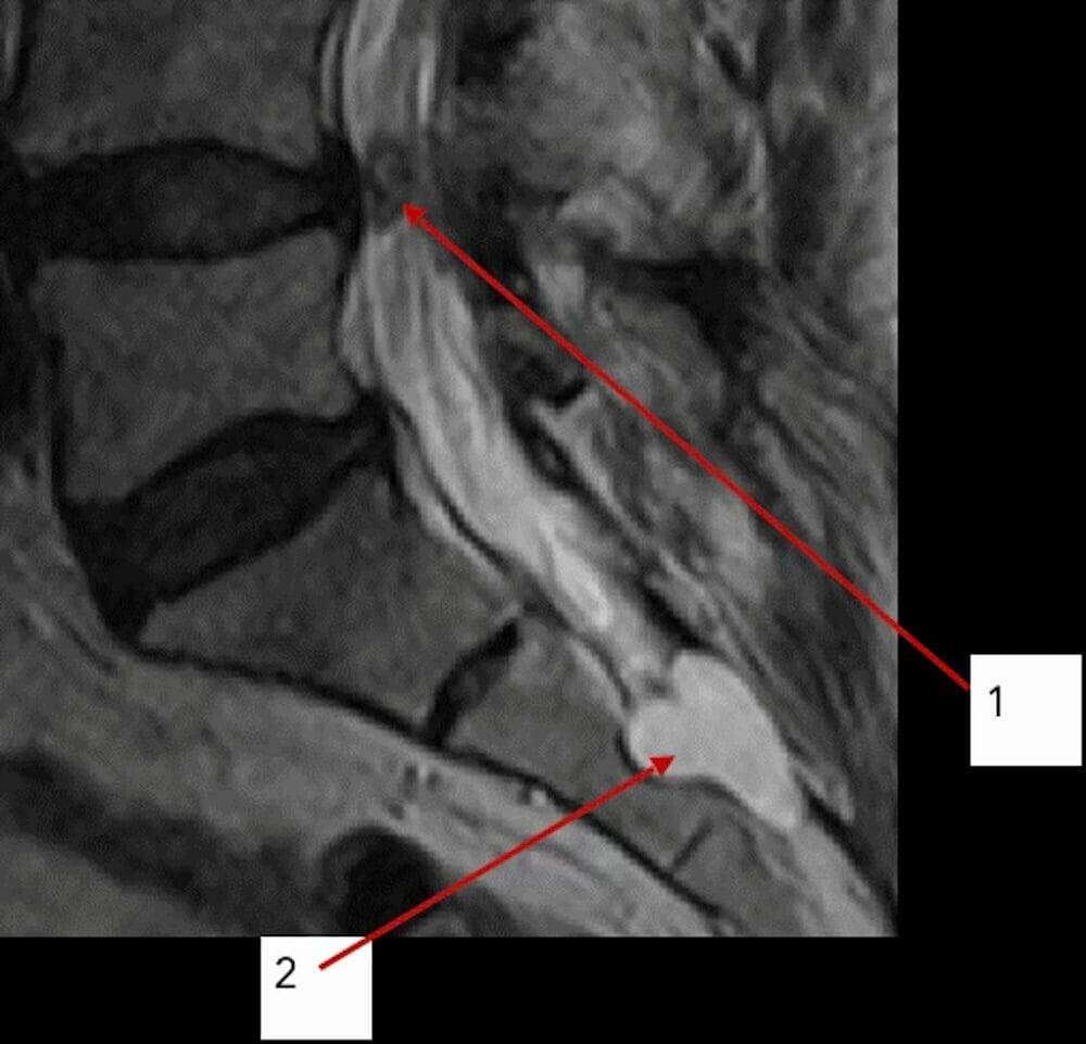
Their origin has been associated with congenital factors, disturbances of cerebrospinal fluid (CSF) dynamics, a valve-like mechanism, or post-traumatic changes. Although their aetiology remains incompletely understood, unlike a number of other pathological processes, perineural cysts do not demonstrate features of infectious or viral involvement. (9 - 13)
A rare complication following microdiscectomy includes discal, annular, and hemorrhagic cysts of the facet joints (Fig. 3, 1), as well as fibrocystic cavities lacking a true synovial lining. (14 - 20) Morphological examination has not revealed specific cytomegalovirus inclusion bodies or viral cytopathic effects.
Synovial cysts are abnormal fluid-filled sacs within the spinal joints and are relatively common in patients with spinal spondylosis. These cysts are benign and resemble cystic formations in other anatomical locations, such as wrist ganglion cysts. They are generally considered to develop as a result of age-related degenerative changes and are most frequently observed in patients over 65 years of age. Although they may occur throughout the spine, they are most commonly found in the lumbar region. For spine surgeons, the presence of a facet joint cyst is indicative of segmental instability at the corresponding spinal level. (41 - 45)
Various conservative treatment approaches for these cysts have been described; however, in cases of chronic pain or neurological deficit, surgical intervention is required. (46, 47)
Clinical manifestations in patients with synovial cysts requiring surgical treatment are typically associated with nerve root compression and spinal instability at the level of the cysts (Figs. 4, 5).
Figs 4, 5: Magnetic resonance imaging (MRI): (1) facet joint synovial cyst causing nerve compression and requiring surgical removal
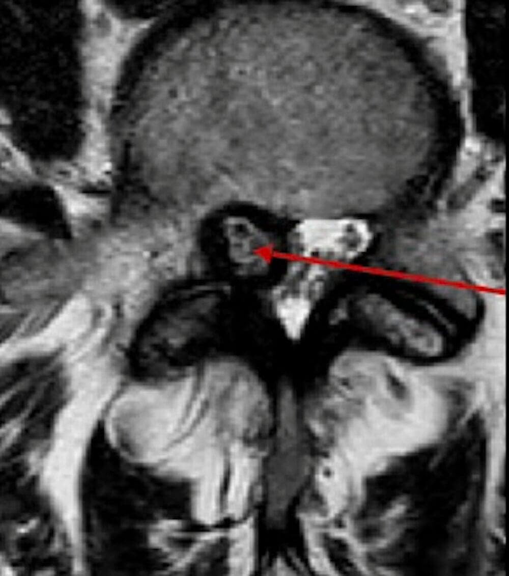
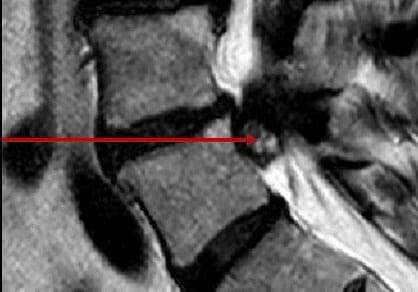
In addition to synovial cysts of the facet joints, patients in this group also presented with hemangiomas (Fig. 6), cystic lesions of internal organs (Figs. 7, 8), cystic formations in other joints (Fig. 9), as well as aneurysms.
Elevated IgG levels against CMV, HSV, and HHV-6 indicate prior infection, i.e., viral carriage. This is typical for the majority of adult patients and does not, in itself, establish a causal relationship with cyst formation or disc herniation.
Fig 6: Magnetic resonance imaging (MRI): (1) hemangioma of a thoracic vertebra
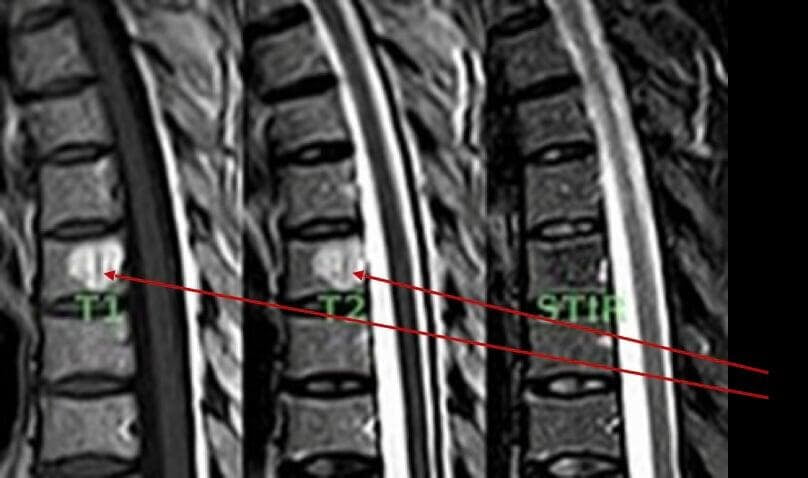
Fig 7: Ultrasound examination (US):(1) renal cyst
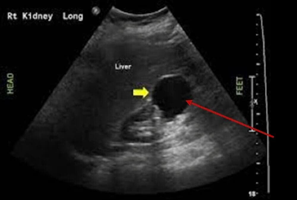
Fig 8: Ultrasound examination (US):(1) renal cyst
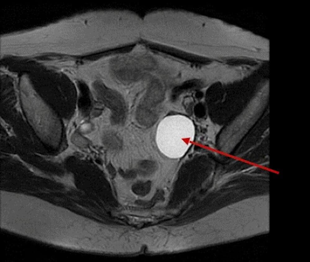
Fig 9: Magnetic resonance imaging (MRI): (1, 2) Baker’s cyst
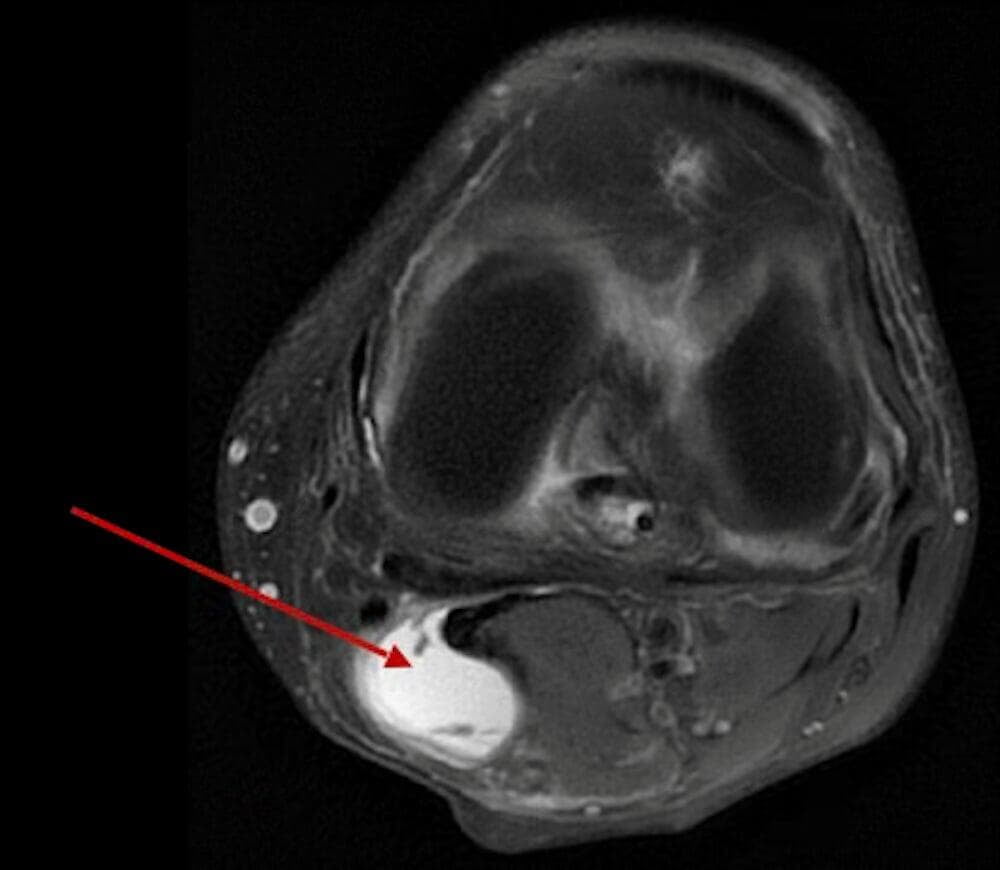
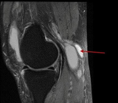
Demonstration of viral activity requires polymerase chain reaction (PCR) testing (in blood, cerebrospinal fluid, or affected tissue) in conjunction with clinical data; IgG avidity testing is used to determine the timing of CMV infection. (21 - 24) In our opinion, alterations of connective tissue manifested as volumetric, rounded formations, together with elevated IgG titers, may suggest the presence of such an association.
Following primary infection, herpesviruses (including CMV, HHV-6, and HSV) persist in the host for life. Under conditions of stress or immunosuppression, they may undergo reactivation. Even in the absence of detectable viral DNA in the analysed sample, herpesvirus persistence, particularly CMV, may be associated with prolonged immune activation. (25 - 29)
Table 1: Avidity changes in the course of infection
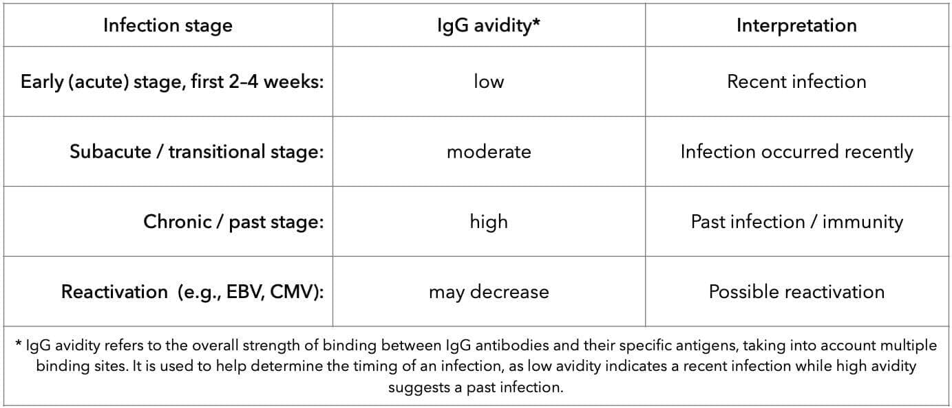
Macrophages normally perform a protective function (M1 phenotype); however, under conditions of chronic viral stimulation, they may shift toward an M2-like phenotype. Such a shift is associated with reduced antiviral immune responses and increased production of profibrotic cytokines. TGF-β, IL-13, IL-17, and IL-10). Resulting in fibroblast activation and collagen accumulation. Note: Cytokines are low-molecular-weight signalling proteins that mediate intercellular communication within the immune system and regulate inflammation, immune responses, and tissue remodelling processes.
A stiffer and less elastic connective tissue is formed, clinically manifested by pain, stiffness, and a tendency toward recurrence following physical нагрузки or surgical interventions; scar tissue may also develop, including in tendons, as an example of a localised fibrotic process (e.g., Dupuytren’s contracture). (30–40) The author observed softening of scar tissue following therapeutic correction with antihomotoxic agents.
Table 2: Key cytokines and their clinical significance
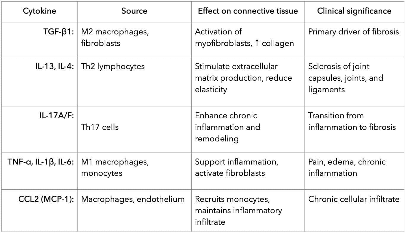
Clinical case 1
Patient B., 56 years old
Complaints of pain in the left thigh, aggravated by movement. The clinical presentation was consistent with left-sided L5 nerve root compression. Instrumental examination revealed a facet joint cyst (Figs. 10, 11). The patient also had a Tarlov cyst. Laboratory testing demonstrated elevated IgG antibodies to CMV and EBV (Fig. 12).
Surgical treatment was performed, consisting of removal of the facet joint cyst. In the postoperative period, resolution of pain in the left leg was observed.
Figs: 10, 11: Magnetic resonance imaging (MRI): (1)
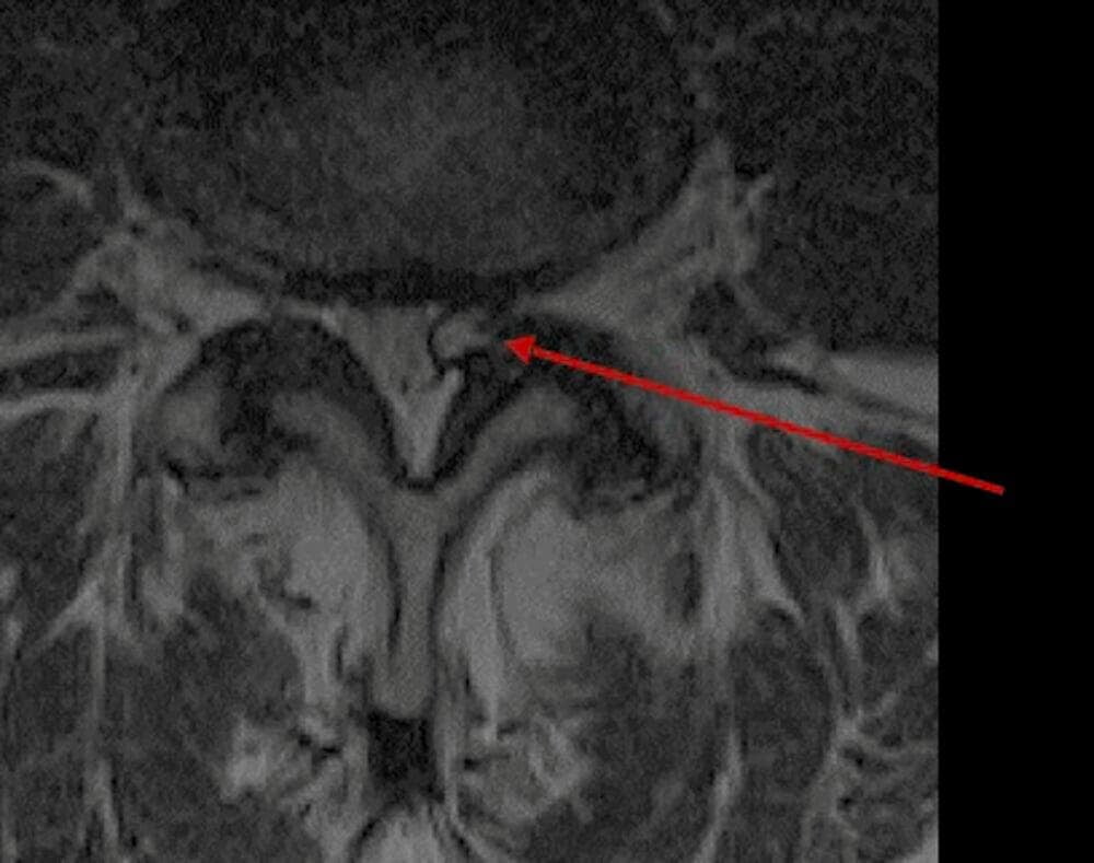
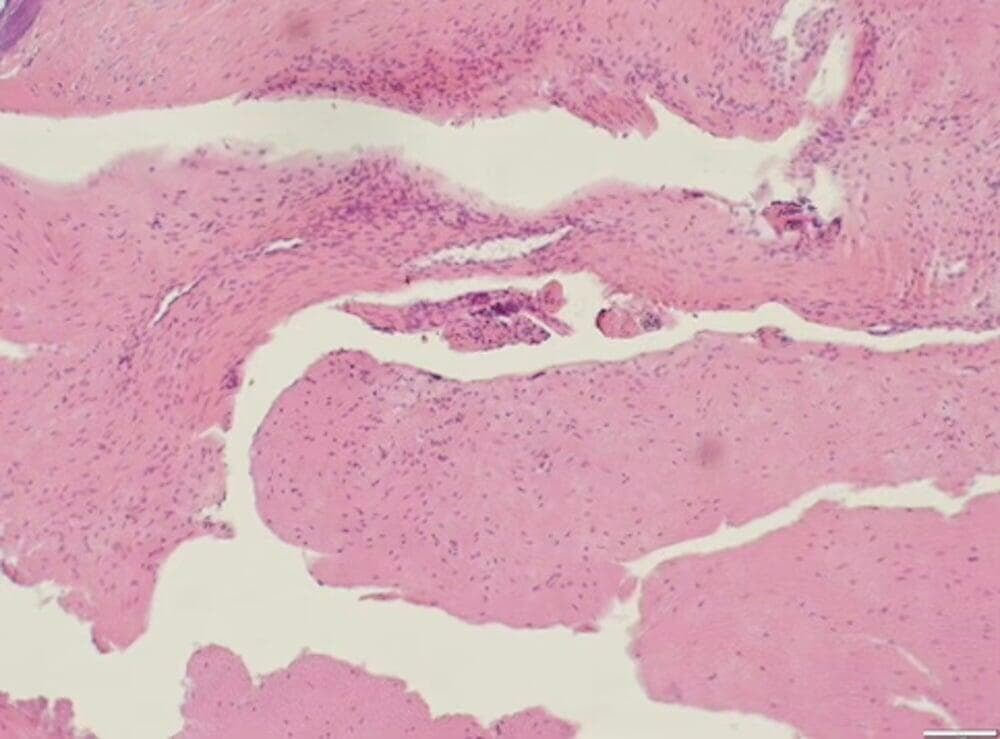
Fig.12: Laboratory findings of patient B.
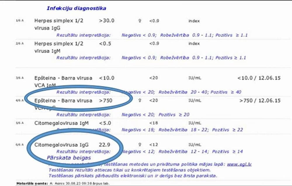
Clinical case 2
Patient T., 71 years old
Complaints of severe lower back pain radiating to the right lower extremity, with sensory disturbance in the right foot. Instrumental examination revealed a facet joint cyst (Fig. 13).
Patient T. underwent conservative treatment using high-dilution integrated phytotherapeutic preparations produced by Alfa-Omega. Drainage, symptomatic, detoxification, and preparations containing information related to EBV and CMV infection were selected. The selection was based on manual muscle testing (MMT). (48)
As a result of the treatment, a reduction in cyst size and resolution of nerve compression were observed (Fig. 14); subjectively, there was complete resolution of pain and restoration of sensation.
Fig. 13. Magnetic resonance imaging (MRI): (1) facet joint cyst before treatment 12/06/2024
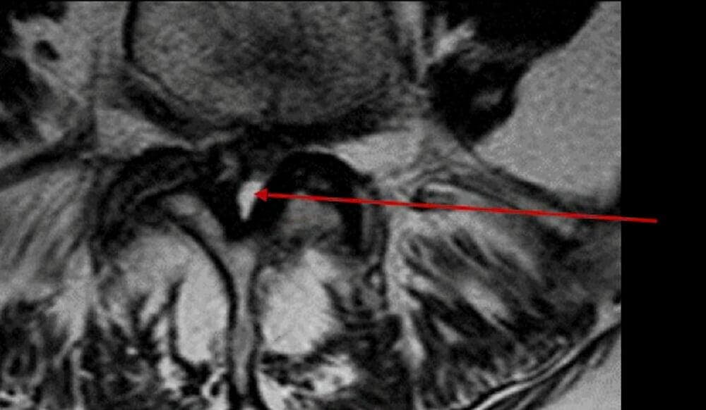
Fig. 14.Magnetic resonance imaging (MRI):(1) facet joint cyst after treatment, after 4 months, on 24/10/2024
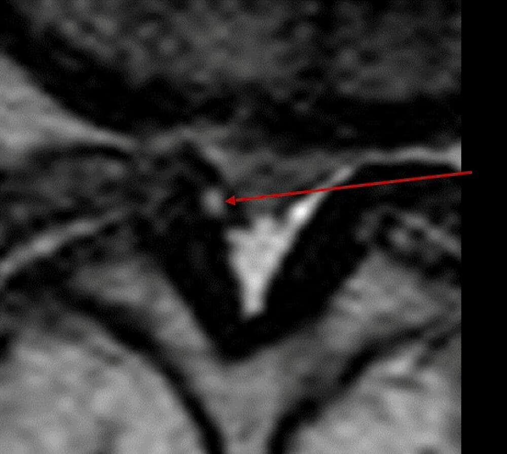
Empirical observations
The use of complex antihomotoxic therapy preparations aimed at suppressing herpesvirus infection leads to a reduction or even disappearance of cysts (Fig. 15) and resolution of synovitis. In several cases, lipomatosis, cystic-fibrotic formations in the breast, and ovarian cysts were resolved.
A significant reduction of fibrosis in Dupuytren’s contracture was also observed. Patients reported disappearance of tendon thickening in stenosing ligamentitis. Resolution of excess interstitial fluid and improvement of connective tissue weakness and hypermobility were also noted. Cases of arrest in the progression of bone cysts and dural cysts were documented.
Fig. 15. Magnetic resonance imaging (MRI):
(L) facet joint cyst before treatment; (R) the same facet joint cyst after treatment
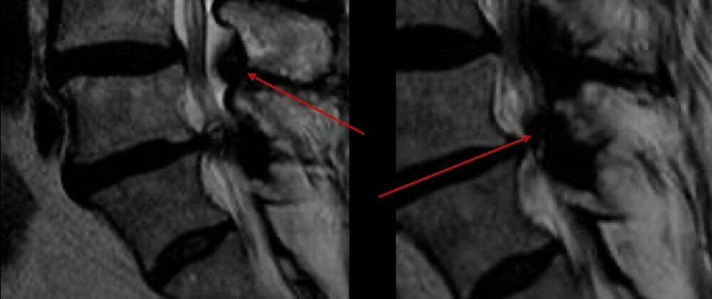
Figure 16 illustrates how fibroblasts, connective tissue cells, give rise to other specialised cells that form the structures of the body. If impairment of fibroblast function under the influence of herpesviruses is assumed to lead to alterations in multiple tissues, this provides a logical explanation for how CMV and EBV may initiate cystic formations in various anatomical locations.
The clinical diversity of manifestations associated with chronic consequences of exposure to these viruses suggests a profound level of viral impact at the level of fibroblasts and during the formation of different tissues.
Fig. 16. Differentiation of fibroblasts into other cell types of the body.
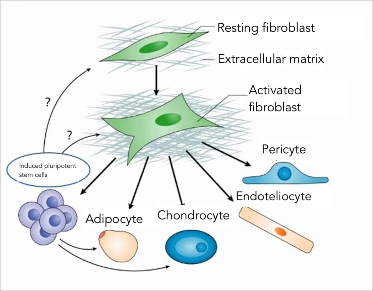
Cyst-like formations in various tissues of the body may reflect a common mechanism of their development: uniform pressure of the contents (blood, cerebrospinal fluid, synovial fluid, glandular secretions, etc.) under conditions of connective tissue weakness leads to the formation of spherical structures. The association between these viruses and connective tissue may serve as a unifying factor linking diverse clinical conditions and structural abnormalities.
Given the widespread distribution of connective tissue and its derivatives in the body, structural abnormalities potentially associated with herpesvirus infection may be expected to occur almost ubiquitously. The author has not identified references to this in the available literature. Further studies aimed at a more detailed investigation of this phenomenon may allow prediction of cyst development and assessment of connective tissue status, and in many cases may support a shift from surgical treatment toward more conservative and less invasive therapeutic approaches.
Conclusions
- Morphologically, cysts have a degenerative-fibrotic nature; no direct evidence of herpesvirus cytopathic effects has been identified within them.
- High seropositivity for herpesviruses (IgG) reflects prior infection and does not constitute evidence of an etiological relationship. This issue requires further investigation.
- Elevated IgG titers are observed in all patients with cystic formations, indicating the need for further study of the chronic consequences of CMV and EBV infection.
- Chronic viral burden maintains immune-mediated inflammation, promotes the shift of macrophages from the M1 to the M2 phenotype, and leads to excessive production of profibrotic cytokines. Consequences include chronic pain, stiffness, soft tissue fibrosis, risk of symptom recurrence after interventions, and cystic formations in various tissues.
- As a rule, patients present with cystic formations in multiple tissues rather than localised involvement.
- The presence of cystic formations indicates connective tissue weakness, which may be used as a prognostic marker in injury prevention in both athletes and other populations.
- The use of specific phytotherapeutic, homeopathic, and antihomotoxic preparations (containing information about the nature of the pathogen) may eliminate or, at least, reduce the manifestations of chronic viral impact, thereby suggesting CMV and EBV as factors contributing to significant changes in the body
- Herpesvirus carriage can only conditionally be considered asymptomatic. The presence of the virus in the body leads to continuous effects on connective tissue, with consequences affecting various structures of the organism.
- Patients with cysts of various localisations should undergo laboratory diagnostics aimed at detecting herpesvirus infection.
- The use of manual muscle testing (MMT) allows for diagnosis and individualised selection of antihomotoxic therapy regimens aimed at suppressing herpesvirus infection.
- The presented data are based on analysis of the literature and clinical observations. The mention of complementary therapeutic approaches does not imply a causal relationship from the perspective of evidence-based medicine and does not replace standard methods of diagnosis and treatment.
Bibliography
- Cohen JI. Epstein–Barr virus infection. N Engl J Med. 2000;343(7):481-92.
- Dunmire SK, Hogquist KA, Balfour HH Jr. Infectious mononucleosis. Curr Top Microbiol Immunol. 2015;390(Pt 1):211-40.
- Balfour HH Jr, et al. Age-specific prevalence of Epstein–Barr virus infection among individuals aged 6–19 years in the United States. Clin Infect Dis. 2013;56(10):1356-63.
- Chebib I, et al. Histopathology of synovial cysts of the spine. Histopathology. 2018;72(6):923-929. DOI 10.1111/his.13465.
- Seo DH, Park H-R, Oh J-S, Doh J-W. Ligamentum flavum cyst of lumbar spine: case report and review of literature. Korean J Spine. 2014;11(1):18-21.
- Taha H, Bareksei Y, Albanna W, Schirmer M. Ligamentum flavum cyst in the lumbar spine: case report and review of the literature. J Orthop Traumatol. 2010;11:117-22.
- Singh V, Rustagi T, Mahajan R, et al. Ligamentum flavum cyst: rare presentation and literature review. Neurol India. 2020;68(5):1207-10.
- El-Kader HEB. Juxtafacet spinal synovial cysts. Asian Spine J. 2016;10(1):46-51.
- Murphy KJ, Nussbaum DA, Schnupp S, et al. Кисты Тарлова: клинические проявления и патогенез. AJNR Am J Neuroradiol. 2016;37(2):373-9.
- Tarlov IM. Perineural cysts of the spinal nerve roots. Arch Neurol Psychiatry. 1938;40:1067-74.
- Patel MR, Louie W, Rachlin JR. Perineural cysts: diagnosis and management. Neurosurgery. 1997;40(1):168-74.
- 12. Fortuna A, La Torre E. Arachnoid diverticula: clinical and surgical aspects. Acta Neurochir (Wien). 1977;39:259-68.
- StatPearls Publishing. Tarlov Cyst. Treasure Island (FL); 2023.
- Young PM, Fenton DS, Czervionke LF. Postoperative annular pseudocyst: report of two cases with an unusual complication after microdiscectomy. Spine J. 2009;9(2):e5-e8.
- Kim HS, Ju CI, Kim SW. Hemorrhagic lumbar synovial cyst after microscopic discectomy. Neurospine (Korean J Spine). 2017;14(3):93-6.
- -Kusakabe T, Kasama F, Aizawa T, et al. Facet cyst in the lumbar spine: radiological and histopathological findings and possible pathogenesis. J Neurosurg Spine. 2006;5(5):398-403.
- Patgaonkar PR, et al. Postoperative discal pseudocyst: report of a case with an unusual complication after microlumbar discectomy and successful treatment by transforaminal endoscopic lumbar decompression. Surg Neurol Int. 2024.
- Obame FLO, et al. Postoperative discal pseudocyst: a case report and literature review. J Orthop Case Rep / J Clin Orthop Trauma (см. источник). 2023.
- Jadhav N, et al. Lumbar discal cyst and post-operative discal pseudocyst. J Surg Case Rep. 2022;2022(5):rjac239.
- Cao Z, et al. A review and summary of patients with symptomatic postoperative discal pseudocyst after discectomy. Orthop Surg. 2023.
- Centers for Disease Control and Prevention (CDC). Laboratory Testing for CMV and Congenital CMV. Updated 15 Apr 2024. (IgG avidity distinguishes primary vs past CMV infection; maturation over 2-4 months).
- ARUP Consult. Cytomegalovirus (CMV) | Choose the Right Test. Updated 10 Oct 2024. (Алгоритмы диагностики, роль серологии и IgG-авидности; когда использовать ПЦР).
- Prince HE, Lapé-Nixon M. Role of Cytomegalovirus (CMV) IgG Avidity Testing in Diagnosing Primary CMV Infection. Clin Vaccine Immunol. 2014. (Авидность эффективно отличает первичную от прошлой/реактивации).
- Fowler KB, et al. A systematic literature review of the global seroprevalence of cytomegalovirus. BMC Public Health. 2022. (Интерпретация CMV IgG без IgM как перенесённой, не острой инфекции; распространённость).
- Cohen JI. Herpesvirus latency. Cold Spring Harb Perspect Med. 2020. (обзор: пожизненная латентность и реактивация герпесвирусов)
- Forte E, Zhang Z, Thorp EB, Hummel M. Cytomegalovirus latency and reactivation: an intricate interplay. Front Cell Infect Microbiol. 2020.
- Griffiths P, Reeves M. Pathogenesis of human cytomegalovirus in the immunocompromised host. Nat Rev Microbiol. 2021. (латентность, реактивация, эндогенная реактивация)
- Semmes EC, et al. Cytomegalovirus as an immunomodulator across the lifespan. 2020. (длительное влияние CMV на иммунный фенотип)
- Jackson SE, et al. The immune response to human cytomegalovirus: impact of lifelong infection. Phil Trans R Soc B. 2025.
- Murray PJ et al. Macrophage activation and polarization. Nat Rev Immunol. 2014.
- Italiani P, Boraschi D. From monocytes to M1/M2 macrophages. Front Immunol. 2014.
- Wynn TA, Vannella KM. Macrophages in tissue repair, regeneration, and fibrosis. Immunity. 2016.
- Gieseck RL et al. Type 2 immunity in tissue repair and fibrosis. Nat Rev Immunol. 2018.
- Meng XM et al. IL-17 in fibrotic diseases. Cell Death Dis. 2012.
- Wynn TA. Cellular and molecular mechanisms of fibrosis. J Pathol. 2008.
- Rockey DC et al. Fibrosis - a common pathway. N Engl J Med. 2015.
- Schleip R et al. Fascial stiffness and fibrosis. J Bodyw Mov Ther. 2012.
- Stecco C et al. Fibrosis of deep fascia. Surg Radiol Anat. 2014.
- Shih B et al. Dupuytren’s disease - a fibroproliferative disorder. Lancet. 2012.
- Verjee LS et al. Unraveling Dupuytren’s disease. Proc Natl Acad Sci USA. 2013.
- Epstein NE. Lumbar synovial cysts: a review of diagnosis, surgical management, and outcome assessment. J Spinal Disord Tech. 2004;17(4):321-5.
- Boviatsis EJ, et al. Symptomatic lumbar synovial cysts: pathogenesis, diagnosis and surgical treatment. Neurosurg Focus. 2006;20(3):E6.
- Lyons MK, Atkinson JL, Wharen RE, et al. Surgical evaluation and management of lumbar synovial cysts. J Neurosurg. 2000;93(1 Suppl):53-7.
- Xu R, McGirt MJ, Parker SL, et al. Factors associated with recurrent back pain and cyst recurrence after resection of lumbar synovial cysts. Spine J. 2010;10(6):547-53.
- Kusakabe T, Kasama F, Aizawa T, et al. Facet cyst in the lumbar spine: radiological and histopathological findings and possible pathogenesis. J Neurosurg Spine. 2006;5(5):398-403.
- Trummer M, Flaschka G, Tillich M, et al. Diagnosis and surgical management of intraspinal synovial cysts: report of 19 cases. J Neurol Neurosurg Psychiatry. 2001;70(1):74-7.
- Jankowski R, Szymaś J, Nowak S, Dukiel R, Sokół B, Paprzy W. Synovial cysts of the lumbar spine. Neurol Neurochir Pol. 2012;46(5):456–461. DOI 10.5114/ninp.2012.31356.
- Applied Kinesiology Synopsis Paperback - January 1, 1988, by David S. Walther (Author) Triad of Health Publishing, ISBN-13978-0929721040.
- Kirdoglo G. Clinical Atlas of Manual Muscle Testing. United Association of Applied Kinesiology and Medical Rehabilitation (UAAK); 2025. ISBN 978-617-7762-07-1.
Bibliography
AU: Charles S Masarsky DC. Northern Virginia Community College, Private practice of Chiropractic, Vienna VA. e: viennachiropractic@verizon.net
Narrative: In a departure from the usual phoenix-like ability of olfaction to recover after trauma or infection, long COVID victims frequently suffer such disturbances as anosmia, hyposmia, and parosmia long after acute infection has abated. Recent research has implicated dysautonomia leading to a hyperinflammatory state of the nasal mucosa as a likely causative or aggravating factor.
Introduction
Like a physiological phoenix, the neurons of the olfactory nerve can be regenerated after they are destroyed by trauma or infection. This is made possible by stem cells, globose basal cells, closely embedded with the neurons of the olfactory epithelium.
The loss of these stem cells has been implicated as a major culprit in olfactory dysfunction during acute COVID-19 infection. That the repopulation of the olfactory mucosa with globose basal cells can remain incomplete long after the acute phase is suggested by the fact that as many as ten percent of long COVID victims are afflicted by anosmia, hyposmia, or parosmia according to recent estimates. (Kim et al, 2024)
The responses of patients in a 2023 case series suggest an important role for inflammation in general and rhinitis in particular in this delayed mucosal recovery. Stellate ganglion blocks have been shown to bring about restoration of olfactory function in some long COVID victims. (Kalava et al 2023) The investigators suggest that olfactory dysfunction in these patients was exacerbated by excessive sympathetic stimulation to the nasal mucosa, leading to a hyper-inflammatory state. Stellate ganglion block is hypothesised to reduce this sympathetic stimulation, allowing inflammatory responses to return to normal, with resulting restoration of basal stem cells.
Nasal inflammation due to dysautonomia takes on additional significance considering a 2026 paper. In a retrospective study of the medical records of 118 million patients a statistically significant increased risk of allergic rhinitis and chronic rhino-sinusitis was found in COVID-19 survivors with three months of follow up. (Olbrich et al, 2026) Long COVID rhinitis exacerbated by dysautonomia would create what amounts to a perfect storm, plausibly suppressing the replacement of globose basal cells.
Kalava et al mention several possible causes of this and similar forms of dysautonomia, including infection, diabetic neuropathy, obesity, and alcoholism. Absent from this discussion is the possibility of somato-autonomic disturbance due to aberrant vertebral mechanics, the vertebral subluxation complex. With this in mind, the following is offered:
Hypothesis: Long COVID olfactory dysfunction can be exacerbated by dysautonomia generated by vertebral subluxation.
Subluxation and Olfactory Dysautonomia
Within the Chiropractic profession the idea that vertebral subluxation can generate dysautonomia has usually enjoyed broad acceptance. In 1996, the Association of Chiropractic Colleges affirmed the long-standing chiropractic tenet that subluxation can ‘ … compromise neural integrity and may influence organ system function and general health’. (ACC, 1996) By implication, this statement clearly points to the possibility of dysautonomia generated by subluxation.
Of the multiple mechanisms for subluxation-generated dysautonomia several bear discussion here. Compression or distraction of spinal nerve roots within the intervertebral foramen could disturb the presynaptic fibres of sympathetic pathways. At some spinal levels in some individuals, the vulnerability of spinal nerve roots to this disturbance may be increased by the presence of transforaminal ligaments partially closing the foraminal ‘windows’. (Masarsky, 2025)
The aberrant motion and/or position of vertebra caused by subluxation would tend to stress the adjacent tissues, including the ligament connecting the anterior surfaces of lamina of adjacent vertebra, the ligamentum flavum. Resultant buckling or swelling of the ligamentum flavum has been shown to result in compression of the dorsal root ganglion. (Giles, 1994) The afferent neurons within this ganglion include those that serve the visceral sensorium. Therefore, disturbance of these neurons could distort autonomic efferent output.
The formation of osteophytes in response to the aberrant mechanics of a subluxated spinal motion segment could plausibly result in dysautonomia. Interference with spinal nerve roots when osteophytes grow into the intervertebral foramen is one mechanism by which osteophytes can disturb autonomic function. Furthermore, when osteophytes project in an anterolateral direction, they can penetrate the para-vertebral sympathetic chain. (Giles, 1992)
The existence of connective tissue bridges between cervical extra-spinal structures and the cervical dura mater has been noted since the 1990s. (Hack et al, 1995 and Mitchell et al, 1998) Cervical subluxation would be expected to disturb these myodural bridges with resultant disturbance to the dura mater. The connection between dural disturbance and dysautonomia is widely appreciated. (Henderson, 2015)
Autonomic innervation to the nasal mucosa is primarily via the sympathetic nervous system. Presynaptic sympathetic fibres emerging with the upper four or five thoracic spinal nerves enter the cervical sympathetic chain. They synapse primarily within the superior cervical sympathetic ganglion. Postsynaptic axons innervate the nasal mucosa. Therefore, subluxation-generated dysautonomia affecting the olfactory mucosa would most likely arise from cervical and upper thoracic levels.
Chiropractic adjustments and Long COVID Olfactory Dysfunction
Clinical vignette 1
In the early days of the COVID-19 pandemic (May, 2020) a middle-aged executive presented with a chief complaint of back pain. (Masarsky, 2021) The pain originated when she over-reached to flush the toilet. While Chiropractic intervention focused on lumbar subluxations, adjustments were also indicated and performed at C1 and T2.
Although her chief complaint was not olfactory in nature, she made parenthetical mention of having recently recovered from a COVID-19 infection, which also afflicted other members of her household. Her residual symptoms included loss of her sense of smell, anosmia.
When she was about to leave, she took a bit of hand sanitiser and was surprised that she could smell its fragrance. On her return visit two days later, she mentioned that she was able to smell garlic at her family’s dinner table the previous evening. This experience prompted a call for subjects to more systematically study the influence of Chiropractic care on long COVID signs and symptoms.
Clinical vignette 2
A 58-year-old real estate agent responded to our call for subjects. (Masarsky and Todres-Masarsky, 2022) Her long COVID manifestations included hyposmia. On a scale of olfactory sensitivity developed by Gupta et al, she rated her sense of smell at ‘1’ where ‘5’ is a completely normal sense of smell. (Gupta et al, 2013) She mentioned that aromas seemed to be less intense when she inspired through the right nostril than the left. On physical examination, Weber test was consistent with a mild conductive hearing loss in the right ear, suggesting the possibility of right sided ear-nose-throat congestion.
In addition to hyposmia, she also suffered from parosmia. This manifested itself as a transient ‘house on fire’ aroma, without any actual fire present. She was unable to identify a pattern or trigger for these parosmia episodes.
Intervention at her first visit included adjustments of C1 and the right first rib. On her second visit three days later, the patient reported the ability to definitively smell the aroma of bread at the dinner table.
After ten Chiropractic visits over a period of six weeks, she rated her sense of smell as 2 - 3 on the previously mentioned Gupta et al scale. Weber test was now negative. Her occasional parosmia episodes continued, but the ‘house on fire’ aroma had evolved into a less disturbing ‘burnt popcorn’ aroma. She reported restoration of her ability to smell several odours, including a litter box, mildew, garlic, tobacco, eucalyptus, and grass. The adjustments during this time included various cervical, thoracic, lumbar, and cranial levels according to the findings of the treating Chiropractor.
Clinical vignette 3
A 66-year-old retired military officer with a history of two COVID-19 infections presented with multiple long COVID manifestations. (Masarsky CS and Todres-Masarsky M, 2025) Regarding his sense of smell, he stated he had some loss after his first infection, which had not completely recovered. Currently he rated his sense of smell as ‘3’ on the previously mentioned Gupta et al scale. He also described one to two parosmia episodes per week, with each episode lasting several hours. During these episodes he perceived a ‘dumpster smell’ unrelated to the presence of garbage in his vicinity.
Chiropractic adjustments varied from visit to visit based on the treating Chiropractor’s findings. All adjustments included at least one cervical level. After twelve visits over a five-week period, the patient noted his sense of smell was now almost normal, almost ‘5’ out of ‘5’ on the Gupta et al scale. The frequency of his ‘dumpster’ parosmia episodes had been decreasing during this period of care.
Clinical discussion
Chiropractic care for all three cases summarised above included cervical adjustments and often involved upper thoracic adjustments, the vertebral levels most closely associated with sympathetic innervation of the nasal mucosa.
The asymmetry of olfaction reported by the real estate agent (aromas were less intense in the right nostril than the left) was consistent with the presence of a positive Weber test. The unilateral conductive hearing loss suggested by the positive Weber test could indicate a generalised ear-nose-throat congestion.
Such congestion could have been aggravated by rhinitis. That the resolution of the Weber test coincided with olfactory improvement further suggests the linkage between auditory and olfactory dysfunction in this patient. It also suggests the aggravation of long COVID auditory and olfactory dysfunction was aggravated by cervical and thoracic vertebral subluxation.
Testing the hypothesis.
While the findings of the cases published to date plausibly point to aggravation of long COVID olfactory dysfunction by the dysautonomia of vertebral subluxation, robust support for the hypothesis requires more evidence.
One approach would be to note the presence or absence of outcomes related to dysautonomia that exhibit covariance with olfactory dysfunction. Useful clinical signs would be those closely associated with sympathetic innervation from cervical and/or upper thoracic vertebral levels.
Such signs could include facial thermography, pupillometry, facial sweat gland activity, and heart rate variability. Symptoms related to these levels of sympathetic innervation could include salivary gland activity (dry mouth or excessive salivation), changes in light sensitivity (aversion to bright light or difficulty seeing in dim light), and changes in tear production (dry eye or excessive tearing). If improvement in these signs/symptoms and olfactory function covary with signs of vertebral subluxation and/or reduction of these signs and symptoms under chiropractic care, such evidence would be supportive of the hypothesis.
Practical implication of the hypothesis
Inter-professional cooperation on behalf of the long COVID patient could be encouraged by wide dissemination of easily applied examination measures that suggest the presence of cervical and/or thoracic subluxation. For example, a medical practitioner examining a patient with long COVID anosmia could notice restriction in cervical flexion/extension or significant asymmetry in cervical rotation/lateral flexion. This could prompt a referral for Chiropractic co-management of this patient. On the other side of the coin, a Chiropractic practitioner caring for a patient with long COVID olfactory dysfunction would be better equipped to explain the rationale for care to others in the patient’s health care team.
Adjuncts to Chiropractic care commonly include measures to counter hyperinflammatory states. Such adjunctive measures are within the common domain health care practice in general Therefore, these interventions can serve as a further arena of inter-professional cooperation.
Practices to counter hyperinflammatory states take on increased importance for patients with olfactory dysfunction related to vertebrogenic dysautonomia.
These include yogic and meditation techniques such as alternate nostril breathing, dietary advice to limit animal fat and emphasise fruits/vegetables/whole grains, regular exercise, and laughter. Such measures take on additional significance for a patient with olfactory loss related to a hyperinflammatory state.
Additional adjuncts to support the Chiropractic adjustment of cervical and thoracic subluxation are also common in chiropractic practice. These would include range of motion exercises for the relevant regions of the spine, postural advice, and consideration of the patient’s bed and pillow, among other factors. Again, these take on additional significance if nasal inflammation is related to vertebrogenic dysautonomia.
Acknowledgement
The author gratefully acknowledges the late Marion Todres-Masarsky, D.C. Her comments on an early draft of this paper significantly strengthened the final product. Her role in refining this manuscript was the last of her many contributions to the chiropractic professional literature.
About the author
Dr Charles Masarsky has been in the private practice of chiropractic with Dr. Marion Todres-Masarsky since 1983. Marion sadly passed August 2026.
Their office is located in Vienna, Virginia, USA in the suburbs of Washington, DC. He also offers continuing education programs for chiropractic colleges and associations and teaches at George Mason University (Fairfax, Virginia). For information about his practice or his CE programs, please e-mail viennachiropractic@verizon.net.
Citations for Dr Masarsky’s published papers are available at his website under ‘about the doctors’: www.viennachiropractic.com.
Dr Masarsky writes this frequent feature in the Journal called ‘The Wide Angle Lens’ in which he takes a broader than usual perspective on one issue or another, and has contributed much on clinical aspects of COVID.
Also by this author
- Masarsky CS. The Wide-Angle Lens. Hypothesis: Cranial Suture Complexity is influenced by Sensory Innervation. Asia-Pac Chiropr J. 2026;6.3. www.apcj.net/papers-issue-6-3/#MasarskySutureComplexity
- Masarsky CS. The Wide-Angle Lens: Transforaminal Ligaments, Unanswered questions. Asia-Pac Chiropr J. 2025;6.2. www.apcj.net/papers-issue-6-2/#MasarskyTransforminalLigaments
- Masarsky CS. Hypothesis: Chiropractic adjustments reduce the risk of stroke in Long COVID. Asia-Pac Chiropr J. 2025;6.1. www.apcj.net/papers-issue-6-1/#MasarskyLongCOVIDHypothesis
- Masarsky CS, Todres-Masarsky M. Long COVID with Obstructive Sleep Apnea: A case report. Asia-Pac Chiropr J. 2025;6.1. www.apcj.net/papers-issue-6-1/#MasarskySleepApnea
- Masarsky CS. Respiratory RCTs: What Are We Missing? [The wide-angle lens]. Asia-Pac Chiropr J. 2025;5.3. www.apcj.net/papers-issue-5-3/#MasarskyRespiratoryRCTs
- Masarsky CS, Todres-Masarsky M. Long COVID with Parosmia and Exertional Malaise: A case report. Asia-Pac Chiropr J. 2025;5.4. www.apcj.net/papers-issue-5-4/#MasarskyLongCOVID
- Masarsky CS, Todres-Masarsky M. Long Haul COVID-19 and Subluxation: A case report. Asia-Pac Chiropr J. 2022;2.6. apcj.net/papers-issue-2-6/#MasarskyLongCovid
- Masarsky CS, Todres-Masarsky M. Long COVID Hyposmia/Parosmia and subluxation: A case report. Asia-Pac Chiropr J. 2022;3.2. apcj.net/Papers-Issue-3-2/#MasarskyHyposmia.
- Masarsky CS, Todres-Masarsky M. Long COVID: A Risk Factor for Scoliosis Incidence and Exacerbation? [Hypothesis] Asia-Pac Chiropr J. 2023;3.3. apcj.net/Papers-Issue-3-3/#MasarskyLongCovidScoliosis.
- Masarsky CS, Todres-Masarsky M. Long COVID Pharyngitis, Possible Glossopharyngeal Neuralgia, and Subluxation: A case report URL Asia-Pac Chiropr J. 2023;4.1. apcj.net/Papers-Issue-4-1#MasarskyTodreaMasarskyLongCovid
- For others, search ‘Masarsky’ atapcj.net or go to www.viennachiropractic.com.
Bibliography
ACC. Issues in Chiropractic. Position Paper #1: The ACC Chiropractic Paradigm: Association of Chiropractic Colleges, July, 1996.
Giles LGF. Paraspinal Autonomic Ganglion Distortion Due to Vertebral Osteophytosis: A Cause of Vertebrogenic Autonomic Syndromes? J Manipulative Physiol Ther, 1992; 15: 551-555.
Giles LFG. A Histological Investigation of the Human Lower Lumbar Intervertebral Canal (Foramen) Dimensions. J Manipulative Physiol Ther, 1994; 17:4.
Gupta N, Singh PP, Goyal A, Bhatia D. Assessment of Olfaction Using the “I-Smell” Test in an Indian Population: A Pilot Study. Indian J Otolaryngol Head Neck Surg, 2013; 65(1): 6-11. Full text: https://www.ncbi.nlm.nih.gov/pmc/articles/PMC3585564/
Hack GD, Koritzer RT, Robinson WL, Hallgren RC, Greenman PE. Anatomic Relation between the Rectus Capitis Posterior Minor Muscle and the Dura Mater. Spine, 1995; 20(23): 2484-2485. https://journals.lww.com/spinejournal/abstract/1995/12000/anatomic_relation_between_the_rectus_capitis.3.aspx
Henderson FC. Dysautonomia – A co-morbidity or consequence of crani ocervical instability and ventral brainstorm compression? In: Batzdorf U,
Henderson FC. 2015 CSF Colloquium Proceedings: Mechanisms and Pathophysiology of Headache in the Population of Patients with Chiari Malformation and Hypermobility Connective Tissue Disorders. Chiari and Syringomyelia Foundation, New York, 2019. https://sa1s3.patientpop.com/assets/docs/243375.pdf
Kalava A, Benyahia SA, Tico Calzada R, Staat CM. Efficacy of Stellate Ganglion Block in Treating Long-Term COVID-19-Related Olfactory and Gustatory Dysfunction: A Case Series. Cureus. 2023 Jun 25;15(6):e40929. doi: 10.7759/cureus.40929.
Kim S, Finlay JB, Ko T, Goldstein BJ. Long-term olfactory loss post-COVID-19: Pathobiology and potential therapeutic strategies. World J Otorhinolaryngol Head Neck Surg. 2024 Mar 21;10(2):148-155. DOI 10.1002/wjo2.165.
Masarsky CS. The wide-angle lens: The post-pandemic research era. Asia-Pac Chiropr J. 2021;1.4. www.apcj.net/papers-issue-2-1/#MasasrskyLongHaulers
Masarsky CS. The Wide-Angle Lens: Transforaminal Ligaments, Unanswered questions. Asia-Pac Chiropr J; 2025;6.2. https://www.apcj.net/site_files/4725/upload_files/Masarsky%20Trannsforaminal%20Ligaments.pdf?dl=1
Masarsky CS, Todres-Masarsky M. Long COVID Hyposmia/Parosmia and Subluxation: A Case Report. Asia-Pac Chiropr J, 2022; 3:2. Full text: https://www.apcj.net/Papers-Issue-3-2/#MasarskyHyposmia
Masarsky CS, Todres-Masarsky M. Long COVID with Parosmia and Exertional Malaise: A case report. Asia-Pac Chiropr J. 2025;5.4. www.apcj.net/papers-issue-5-4/#MasarskyLongCOV
Mitchell BS, Humphreys BK, O’Sullivan E. Attachments of the Ligamentum Nuchae to Cervical Posterior Dura and the Lateral Part of the Occipital Bone. J Manipulative Physiol Ther, 1998; 21(3): 145-148. Abstract: http://www.ncbi.nlm.nih.gov/pubmed?term=((nucchal%20ligament)%20AND%20dura%20mater)%20AND%20J%20Manipulative%20Physiol%20Ther
Olbrich H, Preuß SL, Kridin K, Hernandez G, Thaçi D, Ludwig RJ, Curman P. COVID-19 infection raises respiratory type 2 inflammatory disease risk, whereas vaccination is protective. J Allergy Clin Immunol. 2026 Feb;157(2):517-524. DOI 10.1016/j.jaci.2025.07.030.
Clinical evidence as case reports

About evidence
Case reports are observational in design and thus limited by lacking controls; the effect of potential confounding factors, including comorbidities, cannot be excluded. The findings could support a clinically relevant hypothesis that the identification and correction of spinal subluxation and associated functional dysfunction are modifiable contributors to the effective management and resolution of the specific topic of the case report. In other words, they have meaning.
However case reports are eligible for inclusion as ‘expertise’ bringing clinical insights into the JBI FAME evidential ring (JBI Manual for Evidence Synthesis; 2024) to inform evidence-based healthcare in general and the science of Chiropractic in particular. This Journal specifically recognises that subluxation identification and correction is the art of the individual Chiropractor.

Alexandra Zoda, Anthony Pellegrino, Ruth Postlethwaite and Clare McIvor
This case report followed an 8-year-old boy with ADHD, dyslexia, emotional dysregulation, sensory-seeking behaviour, poor coordination, and retained primitive reflexes. Across eleven months, he received low-force, neurologically focused Chiropractic care incorporating Torque Release Technique, paediatric Sacro Occipital Technique, and craniopathy. Surface electromyography and fine-motor control improved, while heart-rate variability and thermography fluctuated; retained Moro, ATNR, STNR, and Spinal Galant reflexes persisted. Parents reported better sleep, mood, emotional regulation, social engagement, coordination, task completion, and executive function. These changes were concurrent with care, but causation is unsupported because this uncontrolled case used proprietary measures, environmental modifications, and no formal behavioural testing. (Manus)
AU1: Alexandra Zoda MSc (Nutrition), DC, CSSPP, CSCPP. Private practice of Chiropractic, Sea Girt, NJ, US e: drallie@getabsolutehealth.com
AU2: Anthony Pellegrino DC, DACCP, CSSPP, CSCPP, CBS. Private practice of Chiropractic, Sea Girt, NJ, USA.
AU3: Ruth Postlethwaite BBiomedSc. Writer, ASRF.
Background: An 8-year-old male with a diagnosis of ADHD, dyslexia and suspected oppositional defiant disorder presented for Chiropractic care. His presenting concerns included severe resistance to academic tasks and daily transitions, defiance and emotional outbursts, sensory-seeking behaviour with chewing and mouthing, drooling and mouth breathing, poor motor tone with clumsiness and limited spatial awareness, separation anxiety, and difficulty with rules and social connection. His family had initiated, but not yet completed, a diagnostic assessment for Autism Spectrum Disorder.
Intervention: The patient received chiropractic care over approximately eleven months. Analysis and adjusting drew on the Paediatric Experience (PX) framework, Torque Release Technique (TRT) with the Integrator instrument, paediatric Sacro Occipital Technique (SOT), and craniopathy protocols.
Outcomes: A broad range of objective findings improved significantly over the course of chiropractic, though the retained Moro, ATNR, STNR and Spinal Gallant reflexes remained detectable at every evaluation, with variable laterality across time points. Notably, parents reported consistent and significant gains across sleep, mood, emotional regulation, outburst frequency, social engagement, dietary variety, multi-step instruction following, and executive function. This contributed to a significant increase in quality of life concomitant with decreases in behavioural and developmental challenges associated with his comorbidities.
Conclusion: Chiropractic care for paediatric neurodevelopment has yet to benefit from large-scale clinical studies or randomised controlled trials, despite case report data increasingly indicating improvements in individual cases. Further research into this issue is therefore warranted.
Indexing Terms: Chiropractic; Subluxation; paediatrics; ADHD; dyslexia; emotional regulation; socio-emotional functioning.
Cite: Zoda A, Pellegrino A, Postlethwaite R, McIvor C. Improvements in emotional regulation, motor coordination and executive function in an 8-year-old male with ADHD and dyslexia concomitant with Chiropractic care: A case report. Asia-Pac Chiropr J. 2026;7.2. https://www.apcj.site/ZodaADHD.pdf
Take-away: 'Over the course of care, the parents described a child who became calmer, more resilient and more engaged. They reported substantial improvement in sleep quality, mood and emotional regulation, with markedly fewer outbursts and less rigidity in daily routines ...'
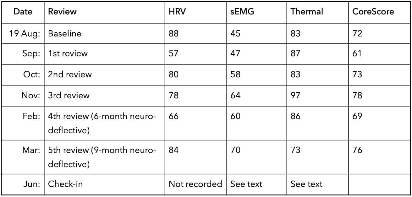
At the six-month evaluation (February 2025), Moro, ATNR, STNR and Spinal Gallant reflexes were all retained, with Spinal Gallant now positive bilaterally where it had been positive on the left only at baseline. The heel-to-toe walk elicited a Moro response. Tracking deficits persisted. The alternating hand slap remained difficult with eyes closed.At the nine-month evaluation (March), Moro was retained and left-dominant, ATNR and STNR were retained, and Spinal Gallant was positive on the right. The heel-to-toe walk continued to elicit a Moro response together with balance difficulty. The left eye fatigued during convergence and divergence testing. The alternating hand slap with eyes closed had improved.No primitive reflex tested was integrated during the reported period. However, significant improvements were noted. The MORO, ATNR, and STNR reflexes improved significantly when subject to repeated tests. Interestingly, the Spinal Gallant moved from left, to bilateral, to right. However, this shift in laterality makes some degree of sense in that the patient has Dyslexia, and is not fully lateralised to one side of their body. This, plus the improved, but still positive ATNR reflex indicates that the patient may have been attending more to one side of his body than the other depending on other factors during the day.It goes without saying that the Chiropractic assessment is a picture of a distinct moment in time, and higher demands would cause reflexes to express in different ways on different days. Primitive reflexes do not just disappear either, but rather integrate. The clear positive finding on this measure is the improvement in the alternating hand slap performed with eyes closed, which is consistent with better proprioceptive fine motor control.Parent-reported outcomesAcross the care period, the parents reported consistent and cumulative gains, recorded through the Brain Blossom framework.At the first review (September) they reported improvements in sleep and mood, decreased outbursts, emerging social interest, and improved comprehension and communication.At the second review (October) they reported increased emotional regulation, more actively sought social contact, improved fine motor skill and concept understanding, and early Flower stage gains in academic and social domains.At the third review (November) they reported reduced anger and rigidity, improved mood, better processing of emotions, better task follow-through, and improvements they attributed to respiratory, digestive, immune and muscular function.At the six-month evaluation (February) they reported better sleep, mood, self-regulation, abstract thought and resilience.At the nine-month evaluation (March) they reported improved balance, fine motor skill and multi-step processing, reduced stress around routine changes, and executive function gains.Over the course of care, the parents described a child who became calmer, more resilient and more engaged. They reported substantial improvement in sleep quality, mood and emotional regulation, with markedly fewer outbursts and less rigidity in daily routines. The child showed a greater desire to connect with family and peers, easier recall and processing of past events, and growing curiosity about new activities and environments. He also showed increased dietary variety, improved coordination, and an ability to follow multi-step instructions that reduced frustration at home and at school.In the later stages of care, the parents described their child as more confident and more capable, with emerging skills in planning, organisation and emotional resilience that made social and academic situations considerably less stressful.Functional gains were reported as maintained at the June check-in, despite the upper cervical and thoracolumbar stress indicators recorded at that visit.Notably, the family avoided ADHD medication throughout the reported period, meeting the secondary goal stated at intake.DiscussionWhile on face value, this case report contains contrary findings of notable functional improvements without full integration of retained primitive reflexes or resolution of subluxation findings. Despite this, the case report may offer a possibility that sustained care and input into the nervous system has the potential to deliver positive functional outcomes over time. During the initial stages of care, Chiropractors reported observing significant improvements, but these were not consistent in CLA readings. A feasible explanation for this may lie in the timing of dropping care frequency (from three visits per week to two) and adding additional therapies back in before the nervous system was ready.This factor alone means the clash in objective findings and parent-reported improvements are not necessarily cut and dried.Over approximately eleven months the parents reported large, consistent and cumulative functional gains across sleep, mood, emotional regulation, social engagement, diet, coordination and executive function. Over the same period, one objective index improved and held, one declined and recovered without exceeding baseline, one rose and then fell below baseline, and no primitive reflex test was integrated. When looking at the core score findings, it becomes clear that overall nervous system coherence was attained.Several explanations for the dissociation are feasible, though none are proven and only further research could explain the phenomena.
- The first is that the parent-reported gains are real and that the objective measures used are not sensitive to those changes. Sleep, emotional regulation and executive function are not indicated by paraspinal surface electromyography or by a composite thermal score, and a child might improve substantially in the former while the latter moved for unrelated reasons.
- The second is that the objective indices are measuring something real that did not improve, and that the parent reports reflect, in part, the recognised tendency of engaged caregivers to observe improvement in a child they are actively and hopefully monitoring. The Brain Blossom framework was explicitly described to the parents as a means of reinforcing progress awareness and engagement. This should be considered as a potential mechanism of improvement, especially when juxtaposed against Hock and Spoelstra (2022) who undertook a retrospective analysis of 37 children undergoing Chiropractic care. The brain blossom framework was utilised in this population, and there was a positive correlation between chiropractic care and the areas measured.
This case occurred within a subluxation-based, neurologically focused paradigm, in which care outcomes were conceptualised not as resolution of a discrete symptom but as improved expression of neurological function of multiple developmental domains. The Brain Blossom assessment serves as a multi-domain outcome measures, and a structured communication tool, translating changes in neurological function into observable, parent-relevant developmental markers.
- A third possibility emerges due to the first 12 weeks of the protocol, in which the parents withdrew the patient from all other programs and supports. This was intentional, and the treating chiropractors regard it as a key contributor to the early functional gains (which must be tested through further research). It is equally possible that the removal of demand (i.e. the withdrawal of therapy sessions pitched above the child's functional level, in a child whose presenting complaints centred on resistance to academic tasks and transitions) may have produced substantial behavioural improvement independent of any adjustment delivered. While this may seem separate from Chiropractic ‘treatment’ per se, it is respectful of a nervous system approach in which we give the nervous system what it needs, remove what it does not need, and allow adaptability to emerge accordingly.
- First, does the withdrawal of concurrent therapy load, on its own, produce measurable behavioural improvement in children presenting this way?
- Second, does the addition of cranial adjusting alter outcomes relative to spinal adjusting alone?
- Third, do parent-reported functional gains in this population track objective autonomic and reflex measures, or do they diverge as they did here? i.e, is this case an anomaly, or par for the course? Larger scale studies would be beneficial.

ASRF Case Report project
Three Case Reports in this issue are part of the ASRF Case Report Project, a project designed to gather client studies from chiropractors and transform them into much-needed case reports, focused on the effects of chiropractic care on clinical presentations highly relevant to chiropractic, such as stress, immunity and adaptability.
This valuable project is made possible by the generous fundraising and contributions of ASRF supporters.
The three latest case reports are:
- Zoda A, Pellegrino A, Postlethwaite R, McIvor C. Improvements in emotional regulation, motor coordination and executive function in an 8-year-old male with ADHD and dyslexia concomitant with Chiropractic care: A case report. Asia-Pac Chiropr J. 2026;7.2. https://www.apcj.site/ZodaADHD.pdf
- Pryjma J, Postlethwaite R, McIvor C. Improvements in mood, latching, range of motion and gastrointestinal function in 3-month-old twins: A Chiropractic case report. Asia-Pac Chiropr J. 2026;7.2. https://www.apcj.site/PrjymaTwins.pdf
- Rai S, Postlethwaite R, McIvor C. Chiropractic management of a 49-year-old female with Arnold-Chiari Malformation and associated neurological sequelae: A case report. Asia-Pac Chiropr J. 2026;7.2 https://www.apcj.site/RaiACMalformation.pdf
Jacey Pryjma, Ruth Postlethwaite and Clare McIvor
Three-month-old male twins, delivered via caesarean section, presented with birth-related strain patterns affecting the craniocervical junction, thoracic spine, and shoulder girdle. Twin A exhibited severe gastro-oesophageal reflux, latching difficulty, and constipation; Twin B demonstrated neuromuscular hypertonicity, an exaggerated Moro reflex, and feeding dysfunction secondary to residual posterior tongue-tie. Vertebral subluxations were identified at multiple spinal levels. An eight-week multimodal, low-force care plan, comprising Activator adjustments, drop-piece technique, sustained-pressure contacts, craniosacral therapy, myofascial release, and oral motor stretches, yielded resolution of hypertonicity, reflex integration, improved range of motion, reduced reflux, and attainment of age-appropriate motor milestones, supporting chiropractic's role in infant neurofunctional regulation. (Manus)

Shaan Rai, Ruth Postlethwaite and Clare McIvor
A 49-year-old female with Arnold-Chiari Malformation Type 1, status post posterior fossa decompression fifteen years prior, presented with chronic migraines, facial palsy, cervicogenic pain, positional dizziness, and impaired activities of daily living. Analysis incorporating paraspinal thermography, leg length assessment, postural evaluation, and radiography identified subluxations at the craniocervical junction, primarily involving the atlas and axis. Subluxation-based care utilising the Knee Chest Upper Cervical technique was delivered over thirty-six visits. Objective improvements included thermographic symmetry, cervical range of motion, and leg length equality, while the Bournemouth Questionnaire score reduced from 43 to 7. Migraine frequency, dizziness, sleep, and functional capacity improved markedly, supporting further prospective research into chiropractic management of ACM-1. (Manus)

Max P Bento
Bento reports a 28-year-old man with a two-year history of right otalgia and aural fullness, cervicalgia, exertional cephalgia, dysphagia, hypoacusis, and left upper limb paresis. Examination identified impaired left arm motor control, balance deficits, right craniofacial hyperesthesia, mastoid-provoked coughing, upper cervical restriction, right temporal bone dysfunction, and a posterior-right-inferior C2 listing. Chiropractic management incorporated Sacro-Occipital Technique blocking, sacral procedures, C2 adjustment, and cranial corrections. By week four, otologic, pharyngeal, cephalgic, and upper limb symptoms had resolved; intermittent cervicalgia remained improved at week five. This case suggests potential clinical benefit, although its single-case design precludes causal conclusions and warrants further investigation. (Manus)

JD Dudum and Bryan Gatterman
An 18-year-old female presented with a two-year history of progressive low back pain, left lower-extremity numbness, and sciatica secondary to an L5–S1 disc protrusion. Following unsuccessful epidural steroid injection and impending surgical consultation, she completed 13 sessions of accelerated nonsurgical spinal decompression with adjunctive cold laser, electrical muscle stimulation, and cryotherapy over 18 days. Follow-up MRI demonstrated a 26.5% reduction in Herniation Index, accompanied by complete pain resolution, restored ambulation, and resolution of radicular symptoms. Although clinically compelling, this single uncontrolled case cannot establish causation or generalisability; controlled trials with serial imaging are required to evaluate protocol efficacy and durability. (Manus)
AU1: JD Dudum DC. Private practice of Chiropractic. Walnut Creek, CA. e: jrdudum2@gmail.com w: https://www.drjddudum.com
AU2: Bryan Gatterman DC, DACBR. Life Chiropractic College West, Hayward. Private practice of Chiropractic Radiology, Castro Valley, CA.
Objective: Lumbar disc herniation (LDH) is a major contributor to chronic low back pain and lower extremity radiculopathy, posing significant disability burden globally. Nonsurgical spinal decompression (NSD) therapy has demonstrated efficacy in reducing disc herniation size in prior research; however, the minimum time required to achieve measurable MRI-confirmed disc reduction following an intensive NSD protocol has not been previously reported. This case report presents MRI-confirmed disc herniation reduction at the L5-S1 level following only 18 days of accelerated NSD therapy in a young female patient with a two-year history of failed conservative and interventional treatment, highlighting a potentially accelerated pathway to structural disc improvement.
Clinical Features: An 18-year-old female presented with a greater than two-year history of low back pain, left leg numbness, and sciatica radiating to the left calf, rated 8/10 on the Visual Analog Scale (VAS). She had failed epidural steroid injection and was scheduled for spinal surgery. Pre-treatment MRI taken 4 months prior to her appointment confirmed a disc protrusion at L5-S1. The patient’s condition progressively worsened including functional limitations such as the inability to walk more than five minutes.
Intervention and Outcome: The patient underwent an accelerated NSD protocol of decompression sessions combined with cold laser therapy, electrical muscle stimulation (EMS), and cryotherapy over an 18-day period. Post-treatment MRI, obtained 18 days after the initiation of treatment, demonstrated a 26.5% reduction in the Herniation Index at L5-S1. The patient reported complete resolution of pain (0/10 VAS) and was walking greater than 20,000 steps per day at discharge.
Conclusion: This case documents structural disc reduction and alleviation of symptoms after 18 days of an accelerated NSD protocol. The findings suggest that high-frequency NSD therapy may substantially accelerate disc size reduction, even in cases of prolonged chronicity and failed prior intervention. Further controlled investigation is warranted to evaluate the disc resorption timeline in NSD management of low back pain.
Indexing Terms: Chiropractic; Low Back Pain; MRI; Lumbar Disc Herniation; Spinal Decompression; Radiculopathy; Herniation Index; Sciatica.
Cite: Dudum JD, Gatterman B. Rapid MRI confirmed reduction of lumbar disc herniation following an accelerated nonsurgical spinal decompression protocol in an 18y female: A case report. Asia-Pac Chiropr J. 2026;7.2. https://www.apcj.site/DudumLxDisc.pdf
Introduction
Low back pain (LBP) remains the leading cause of disability worldwide, affecting more than half a billion people globally and projected to exceed 800 million by 2050. (1) The economic and societal burden is substantial, with direct spine-related healthcare costs in the United States estimated at US$315 billion annually, with surgical procedures accounting for most expenditures. (2) Lumbar disc herniation (LDH) is among the most common structural aetiologies of LBP and lower extremity radiculopathy, and it disproportionately affects younger populations whose functional demands and long-term disability risk are greatest.
Standard management of LDH includes pharmacological therapy, physical rehabilitation, epidural steroid injections, and surgical discectomy. While surgery provides short-term relief in severe cases, 5 to 40% of patients develop failed back surgery syndrome in the years following lumbar surgery, with estimated annual treatment costs ranging from US$18,195 to US$21,402 per patient. (3, 4) Given this risk, identifying effective nonsurgical alternatives capable of producing measurable structural disc improvement is a high clinical priority.
Nonsurgical spinal decompression (NSD) therapy is a computer-controlled motorised traction system designed to apply segmental distraction forces to the lumbar spine, creating negative intradiscal pressure (-100 to -600 mmHg) within the nucleus pulposus. (9) This negative pressure gradient is theorised to facilitate retraction of herniated disc material, reduce nerve root compression, and promote disc rehydration through improved nutrient diffusion. (9, 10)
Several randomised controlled trials and case series have demonstrated the effectiveness of NSD therapy in reducing pain, improving function, and producing MRI-confirmed reductions in disc herniation size. (11 - 14)
Our prior published case series documented MRI-confirmed herniation index reductions ranging from 9.4% to 78% following eight weeks of NSD therapy combined with Chiropractic care in four patients with LBP and radiculopathy. (14) A critical unanswered question is the minimum treatment duration required to achieve measurable structural disc reduction, and whether an accelerated, high-frequency NSD protocol can compress the resorption timeline beyond what is observed with standard conservative management.
This case report describes an 18-year-old female with a greater than two-year history of chronic LBP, left leg numbness, and sciatica who had failed epidural steroid injection and was scheduled for surgical intervention.
The patient
An 18-year-old female presented with a chief complaint of low back pain, left leg numbness, and sciatica radiating to the left calf. The patient had experienced these symptoms for greater than two years with progressive functional decline and no sustained relief from prior interventions. The patient’s clinical condition progressively worsened. Pain intensity increased, and ambulation tolerance declined further. She rated her pain intensity at 8/10 on the Visual Analog Scale (VAS) at initial presentation. Her quality of life was severely compromised, with functional limitations including the inability to walk more than five minutes continuously. The patient was unable to participate in normal daily activities commensurate with her age.
Prior treatment history included an epidural steroid injection at the L5-S1 level, which provided no sustained improvement. Given the failure of conservative and interventional management, the patient had been recommended for lumbar spinal surgery.
Pre-treatment MRI of the lumbar spine confirmed a disc protrusion at the L5-S1 intervertebral level. The pre-treatment AP dimension measured 11.3 mm, yielding a pre-treatment Herniation Index of 471.5. The patient consented to participation in this case report; all identifying information has been de-identified in accordance with patient privacy standards.
The patient underwent the accelerated NSD protocol, consisting of twice-daily decompression sessions combined with cold laser therapy, EMS, and cryotherapy over 18 consecutive days. This technique is discussed in the section on ‘NSD Treatment Protocol’. Compliance with the protocol was complete for the full treatment course.
Follow-up MRI of the lumbar spine was obtained 18 days following the initiation of treatment. Radiological review confirmed a measurable reduction in disc herniation size at L5-S1. The post-treatment AP dimension measured of 10.97 mm, indicating a post-treatment Herniation Index of 346.44, representing a 26.5% reduction from baseline.
Figure 1: Pre-treatment axial MRI image at the L5-S1 intervertebral disc level demonstrating the disc protrusion. Posterior disc protrusion with encroachment on the spinal canal is noted, consistent with the patient’s clinical presentation of low back pain, left leg numbness, and sciatica radiating to the left calf.
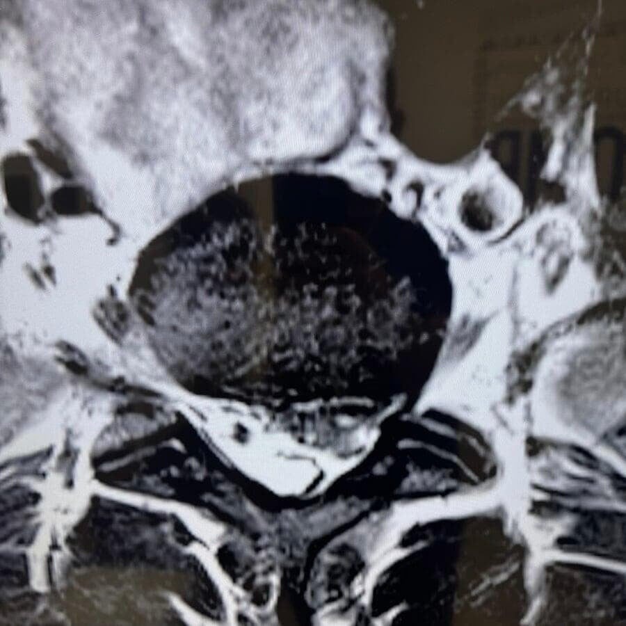
Clinically, the patient reported complete resolution of pain at discharge, rating pain intensity 0/10 on the VAS, a reduction of 8 points from baseline.
Functional recovery was dramatic: the patient progressed from an inability to walk more than five minutes to walking greater than 20,000 steps per day. Resolution of left leg numbness and sciatic symptoms was reported. The patient avoided spinal surgery.
Figure 2: Post-treatment axial MRI image at the L5-S1 intervertebral disc level demonstrating reduction in disc protrusion size following 18 days of accelerated nonsurgical spinal decompression therapy. Reduced posterior disc protrusion and improved visualisation of the spinal canal are noted compared to Figure 1.
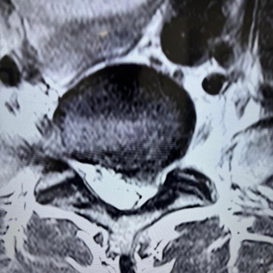
Imaging protocol
To evaluate the change in the herniated disc following NSD therapy, magnetic resonance imaging (MRI) was performed 4 months prior to the initiation of treatment and repeated 18 days following the commencement of therapy. MRI of the lumbar spine was performed utilising a 1.5 T magnet. Standard imaging with sagittal T1, T2, and STIR sequences along with an axial T2 sequence were obtained. The type of herniation was categorised as a protrusion.
Disc herniation size was quantified using the herniation index methodology (HI) described in our previously published research. (14) The HI was calculated utilising the axial image demonstrating the greatest degree of distortion of the posterior intervertebral disc margin. The calculation involved multiplying the maximal anteroposterior (AP) dimension of the intervertebral disc herniation by the width of the herniated disc material and then dividing this value by the product of the sagittal diameter of the spinal canal and the width of the spinal canal at the same axial level, multiplied by 1,000.
HI Formula: (AP distance × Width distance)/ (AP sagittal canal distance× Width sagittal canal distance) × 1000 = Herniation Index (HI)
All MRI measurements and HI calculations were performed by a spinal radiologist. The measurements were obtained 3 times and averaged to obtain a final value. See Table 1 for the values obtained in this patient.
Table 1: The values used to calculate the HI.
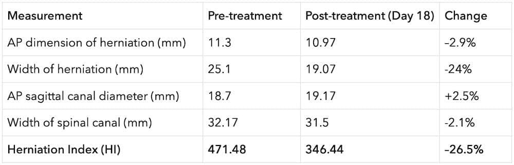
NSD treatment protocol
The accelerated NSD treatment protocol in this case consisted of twice-daily decompression sessions administered in 13 sessions delivered over an 18-day period. Two (2) to 3 sessions were clustered on treatment days, spaced 30 minutes to 3 hours apart, based on patient scheduling. Each session included 20 to 30 minutes of active spinal decompression on a computerised motorised traction table. At the initiation of each session, the patient was positioned supine on the decompression table and fitted with adjustable body harnesses. The computerised algorithm gently applied distraction force to the lumbar spine; as paraspinal musculature resisted the applied tension, sensors detected resistance and modulated tension until relaxation occurred before reapplying the decompression force.
The ratio of hold to rest time was 1:1, with oscillation ranging from 10 - 50% decrease between hold and rest phases. Each session was supplemented with adjunctive therapies including cold laser therapy, electrical muscle stimulation (EMS), and cryotherapy (ice application). No Chiropractic spinal manipulation was performed during the acute treatment phase. The treating clinician supervised all sessions.
Discussion
The primary finding of this case report is MRI-confirmed structural disc herniation reduction at L5-S1 after 18 days of an accelerated NSD protocol. The patient had experienced disc herniation related symptoms for greater than two years with progressive functional decline and no sustained relief from prior interventions. This change in the disc herniation and patient symptoms has important implications for the understanding of how NSD therapy may mechanistically influence the disc resorption process, and for the clinical management of young patients with chronic LDH who have failed conventional and interventional treatment.
Several biomechanical mechanisms may explain why an intensive, twice-daily NSD protocol could accelerate disc size reduction beyond what is achievable with standard conservative care. NSD therapy generates significant negative intradiscal pressure (-100 to -600 mmHg) within the nucleus pulposus, a magnitude of decompressive force that exceeds conventional traction and that cannot be replicated by rest, physical therapy, or injection alone. (9)
This negative pressure gradient is theorised to facilitate mechanical retraction of herniated disc material, restoration of disc height, and enhanced diffusion of oxygen and nutrients into avascular disc tissue, thereby promoting biological remodelling. (9, 10) The twice-daily administration of these decompressive forces in this case doubled the mechanical stimulus compared to standard once-daily protocols, potentially amplifying both mechanical retraction and biological rehydration effects.
The biological mechanisms underlying disc resorption, including macrophage-mediated phagocytosis, neovascularisation, inflammatory cascade activation, and matrix protease activity, are relevant to the accelerated response observed in this case. (5, 6) It is well established in the resorption literature that protrusion-type herniations, in which the annulus fibrosus remains intact, demonstrate lower spontaneous resorption rates compared to extrusion and sequestration types, as intact annular containment limits contact between disc material and the epidural venous plexus necessary to trigger immune-mediated degradation. (8)
The change in the disc protrusion in this patient suggests that the mechanical decompressive forces generated by the twice-daily NSD protocol, rather than immune-mediated biological resorption alone, may be the primary driver of the observed structural change, potentially through direct mechanical retraction of contained disc material and restoration of negative intradiscal pressure gradients. This proposed mechanism warrants direct investigation in future controlled studies.
The chronicity of this patient’s presentation adds additional significance to the rapidity of the observed response. The patient had been symptomatic for greater than two years. a duration associated with structural disc changes, neurological sensitisation, and reduced likelihood of spontaneous resorption in the epidemiological literature. (5) The patients symptoms were progressively becoming worse prior to NSD intervention. Despite this unfavourable chronicity profile, and despite prior failure of epidural steroid injection, the patient achieved complete pain resolution and dramatic functional restoration within 18 days of the commencing treatment.
This outcome challenges the clinical assumption that prolonged symptom duration predicts resistance to conservative disc intervention and suggests that the intensity of the NSD protocol may be a more important determinant of outcome than chronicity alone.
The results are consistent with and extend the findings of our previously published case series, in which four patients with LBP and radiculopathy achieved herniation index reductions ranging from 9.4% to 78% following eight weeks of standard-frequency NSD therapy combined with chiropractic care, with all four patients achieving complete pain resolution. (14) The present case demonstrates that comparable or superior structural and clinical outcomes may be achievable in a fraction of the treatment duration when a high-frequency protocol is employed, at least in select patients. The 26.5% reduction in HI at 18 days compares favourably with the eight-week outcomes reported in our prior series and with findings from other published NSD studies demonstrating disc reduction following standard protocols. (11 - 14)
Additional published NSD research is consistent with the clinical improvements observed here. Amjad et al. demonstrated that NSD combined with routine physical therapy produced superior reductions in pain intensity and functional disability compared to physical therapy alone in patients with lumbar radiculopathy. (11) Gaowgzeh et al. found NSD combined with core stabilisation significantly outperformed core stabilisation alone in patients with lumbar disc prolapse. (12) The present case extends this literature by documenting the potential of an accelerated NSD protocol to produce rapid structural disc change in a clinically complex, previously treatment-resistant patient.
Several limitations of this case report merit acknowledgment. As a single case, the findings cannot be generalised, and the absence of a control condition precludes attribution of disc reduction solely to the NSD protocol. The possibility that some degree of spontaneous regression contributed to the observed structural change is unlikely, since the patient presented with greater than two years of chronicity. The MRI measurements are subject to the inherent limitations of HI calculation methodology, including inter-reader variability, and the use of different magnets and the imaging protocols of different facilities.
Prospective controlled trials comparing accelerated versus standard-frequency NSD protocols with serial MRI monitoring are needed to confirm these preliminary findings.
Conclusion
This case report documents MRI-confirmed 26.5% reduction in lumbar disc herniation at L5-S1 following 18 days of accelerated nonsurgical spinal decompression therapy in an 18-year-old female with greater than two years of chronic LBP, sciatica, and radiculopathy who had failed epidural steroid injection and was facing spinal surgery.
As of the initiation of the NSD treatment, the patient’s condition was progressively becoming worse. The 18-day treatment period for measurable disc reduction is significant when considering the timeline reported in the spontaneous resorption literature.
This study extends our prior published case series by suggesting that high-frequency NSD protocols may substantially accelerate the disc reduction process. The patient achieved complete pain resolution and returned to full functional activity, avoiding surgery.
These findings support the continued investigation of accelerated NSD protocols as a time-efficient, nonsurgical intervention for LDH, particularly in younger patients with chronic, treatment-resistant disc pathology. Prospective controlled trials with serial MRI assessment are warranted to confirm whether intensive NSD frequency is a determinant of the rate of structural disc improvement.
References
- Global Burden of Disease Collaborator Network. Global, regional, and national burden of low back pain, 1990–2020, its attributable risk factors, and projections to 2050: a systematic analysis of the Global Burden of Disease Study 2021. Lancet Rheumatol. 2023;5(6):e316–e329. https://doi.org/10.1016/s2665-9913(23)00098-x
- Yelin E, Weinstein S, King T. The burden of musculoskeletal diseases in the United States. Semin Arthritis Rheum. 2016;46(3):259–260. https://doi.org/10.1016/j.semarthrit.2016.07.013
- Stanton EW, Chang KE, Formanek B, Buser Z, Wang J. The incidence of failed back surgery syndrome varies between clinical setting and procedure type. J Clin Neurosci. 2022;103:56–61. https://doi.org/10.1016/j.jocn.2022.06.027
- Hollingworth W, Turner JA, Welton NJ, Comstock BA, Deyo RA. Costs and cost-effectiveness of spinal cord stimulation (SCS) for failed back surgery syndrome. Spine. 2011;36(24):2076–2083. https://doi.org/10.1097/BRS.0b013e31822a867c
- Chiu CC, Chuang TY, Chang KH, Wu CH, Lin PW, Hsu WY. The probability of spontaneous regression of lumbar herniated disc: a systematic review. Clin Rehabil. 2015;29(2):184–195.
- Zhong M, Liu JT, Jiang H, et al. Incidence of spontaneous resorption of lumbar disc herniation: a meta-analysis. Pain Physician. 2017;20(1):E45–E52.
- Farrag A, Koura HA, Darwish H. Spontaneous resorption of herniated lumbar disk: observational retrospective study in 9 patients. World Neurosurg. 2019;122:e1252–e1257. https://doi.org/10.1016/j.wneu.2018.11.104
- Han Z, Yin H, Liu J, et al. Prevalence, clinical predictors, and mechanisms of resorption in lumbar disc herniation: a systematic review. Orthop Rev. 2023;15:96437.
- Ramos G, Martin W. Effects of vertebral axial decompression on intradiscal pressure. J Neurosurg. 1994;81(3):350–353. https://doi.org/10.3171/jns.1994.81.3.0350
- Apfel CC, Cakmakkaya OS, Martin W, et al. Restoration of disk height through non-surgical spinal decompression is associated with decreased discogenic low back pain: a retrospective cohort study. BMC Musculoskelet Disord. 2010;11:155. https://doi.org/10.1186/1471-2474-11-155
- Amjad F, Mohseni-Bandpei MA, Gilani SA, Ahmad A, Hanif A. Effects of non-surgical decompression therapy in addition to routine physical therapy on pain, range of motion, endurance, functional disability and quality of life versus routine physical therapy alone in patients with lumbar radiculopathy; a randomized controlled trial. BMC Musculoskelet Disord. 2022;23(1):255. https://doi.org/10.1186/s12891-022-05196-x
- Gaowgzeh RAM, Chevidikunnan MF, BinMulayh EA, Khan F. Effect of spinal decompression therapy and core stabilization exercises in management of lumbar disc prolapse: A single blind randomized controlled trial. J Back Musculoskelet Rehabil. 2020;33(2):225–231. https://doi.org/10.3233/bmr-171099
- Choi E, Gil HY, Ju J, Han WK, Nahm FS, Lee PB. Effect of nonsurgical spinal decompression on intensity of pain and herniated disc volume in subacute lumbar herniated disc. Int J Clin Pract. 2022;2022:6343837. https://doi.org/10.1155/2022/6343837
- Dudum J, Gatterman B. Reduction of the size of a lumbar disc herniation using non-surgical spinal decompression combined with chiropractic care. J Contemp Chiropr. 2024;7(1):146–155.
- Hornung AL, Barajas JN, Rudisill SS, et al. Prediction of lumbar disc herniation resorption in symptomatic patients: a prospective, multi-imaging and clinical phenotype study. Spine J. 2023;23(2):247–260.
Book review
Quick-take

Hidden plastic hazard in cups
From The Australian: Single-use paper cups used for takeaway coffee and other beverages release millions of microplastic particles when exposed to hot liquid, a new Australian study has found.