
Asia-Pacific Chiropractic Journal
Issue 7-3 Published 1 November 2026;
Next issue: 1 Jan 2026
Proudly written and produced by Human Intelligence
Read in English, Maori, Hindi, Amharic, Sinhala, Tamil, Vietnamese and more






In this issue
EditorialCommentaryDialoguePreliminary observationDavid Walls-Kaufman: Is the human body a musical instrument that needs to be precisely tuned?Clinical practiceGil Weiner & Charles Blum: Vasomotor Rhinitis (VMR) and Cerebral Spinal Fluid Flow: A Chiropractic issueRichard M Burger: Mandibular Torsion: Category 1 of the Jaw
Angelo L M Battiston et al: Chiropractic spinal adjustments in patients with heavy sympathomimetic drug use
Professional Identity

Jessey Wong, Ruth Postlethwaite, & Clare McIvor: Improvements in pain, fatigue and gastrointestinal function in a 32-year-old female with fibromyalgia and pudendal neuralgia under upper cervical chiropractic care
Brian Lanoue, Thea Lanoue, Ruth Postlethwaite & Clare McIvor: Improvement in symptoms relating to Scoliosis, ADHD and Depression in a 29-year-old male
Quick-take On-line exclusive with cartoon
Phil Maffetone: The caffeine connection: An endurance drug
Book ReviewDonald McDowall: The Forgotten History of D.D. Palmer’s Chiropractic Disciples
Editorial
Commentary
Dialogue
AU: Phillip Ebrall BAppSc(Chiropr), GC Tert Learn Teach, MPhotog, PhD, DC (Hon), FACCS, FICCS. Editor. Research Scientist: History and Philosophy - Chiropractic [Field 220299]. e: pebrall@me.com
Narrative: Generative artificial intelligence (AI) is reshaping educational practice, presenting opportunities and risks for Chiropractic educators, learners, and researchers. This narrative commentary examines how AI can support Chiropractic education while preserving human authorship, professional judgement, and ethical responsibility. It proposes augmented intelligence as a preferable conceptual framework to technological substitution, positioning AI as a collaborator and learning assistant.
Practical applications include literature summaries, formative quizzes, personalised tutoring, virtual patient encounters, writing refinement, and educational image generation. These applications require verification of factual claims, citations, and clinical interpretations, alongside attention to bias, privacy, copyright, and transparent disclosure. The paper advocates a transition from competency-focused education towards capability development, emphasising disciplinary reasoning, critical thinking, communication, empathy, and ethical decision-making.
Assessment should combine secure demonstrations of individual achievement with opportunities to develop responsible AI-supported workflows. Chiropractic institutions are encouraged to embed AI literacy and explicit boundaries between assistance and substitution.
Effective integration depends on informed educators who retain accountability and exercise human judgement, using AI to strengthen rather than replace meaningful learning and patient-centred care.
Indexing Terms: Chiropractic; education; Gen AI; curriculum; assessment.
Cite: Ebrall P. Humanism in Chiropractic education and our embrace of AI. Asia-Pac Chiropr J. 2026;7.3 https://www.apcj.site/EbrallAI.pdf
Take-away: ' … Your judgement ‘engine’ is what AI will amplify. If your judgement is poor it will amplify you into mediocrity; should your judgement be astute, AI will amplify you into excellence … '
Introduction
‘Artificial Intelligence, deep learning, machine learning, whatever you’re doing if you don’t understand it, learn it. Because otherwise, you’re going to be a dinosaur within 3 years’ (1)
Mark Cuban, 2017. American businessman, Entrepreneur, Television personality.
The biggest danger with AI is not what it produces; the danger lies in the user who accepts it and thinks that what it produces is gospel, worse, they never bother to check. I write from the perspective of an educator where our main interest lies in what is written, either for us by learners or by us for educative purposes.
If you take nothing else from this paper, it must be that:
You are responsible for every statement or inference made by an AI agent and used by you in your name. You must always edit, proof, and verify every sentence you have AI construct on your behalf. Where data is cited you must read and verify the source and the authors. AI agents are known to hallucinate.
This paper addresses a topic which is far bigger than me, and as with most readers I am still wrestling with this new beast we erroneously call Artificial Intelligence. My preferred term is Augmented Intelligence, notwithstanding President Trump deciding to rename AI as Superior Intelligence. My hope it that by the time we reach my conclusion you too will agree that AI is best considered as Augmented Intelligence. I will hopefully reach a destination where AI is seen as a companion and assistant for you, not as a replacement of you.
It has been said that AI replaces tasks, not people, so you would be wise to look now at the tasks you are doing, appreciating that AI will do most of them better than you; then ask, how can you move more towards validating your role in your organisation?
Be selective in how you use AI
I have seen too many colleagues embarrass themselves publishing phrases they have generated with an AI agent but failed to check for truth, accuracy, and common sense. It is disappointing for someone who is not an academic and with no years of ‘classroom’ experience to use AI to ‘write a book’ on education for them. This is not meant to be funny as it is my real-world experience which cost me a friendship. I found it intolerable as the output was blatantly imaginative with no substance, an insult to those of us who have built an experience with stories to tell and anecdotes to enrich.
Rather than jumping onto a smart phone and speaking your input into an AI agent, you really should start with the bare minimum of some tutorials on YouTube and hopefully work up to taking a short course of some classes on how to structure your prompts and refine the outputs.
This paper has been written by me in my capacity as a human author with scant use of any AI agent. I did use Manus, my AI agent of choice and below I asked Manus to ‘explain why people are afraid of AI and how you think AI can best be used by writers’. This use seems reasonable given I have declared it. I think it is somewhat ironic to ask an AI agent to explain itself. Here is the response from Manus, lightly edited for reasons of presentation and formatting.
ME - PROMPT: please explain why people are afraid of AI and how you think AI can best be used by writers.
MANUS - ‘People are afraid of AI for several understandable reasons:
- Loss of work and status: Writers, artists, and other professionals worry that organisations will use AI to replace paid human labor or reduce creative work to a cheap commodity;
- Loss of control: AI systems can behave unpredictably, while decisions about how they are developed and deployed are often made by a small number of powerful companies;
- Misinformation: AI can produce convincing but false material at enormous scale, making it harder to know what, and whom, to trust;
- Copyright and consent: Many writers object to their work being used to train commercial systems without clear permission, credit, or compensation;
- Cultural sameness: Because AI predicts patterns from existing material, careless use can produce generic writing and reinforce clichés, biases, or dominant viewpoints;
- Devaluation of human creativity: People fear that readers and publishers may stop valuing the lived experience, judgment, and the deliberate craft behind a piece of writing; and
- Broader uncertainty: Rapid technological change naturally creates anxiety, particularly when regulation and social norms have not caught up’.
Numbers iii) and v) are worth considering and are the reasons for my insistence that a creator verifies the output before accepting it as useful. Number v) in particular is actually a reason for us to use AI to generate summaries of our professional papers. This is, in effect, training the Large Language Models (LLMs) in our discipline. And in the bigger picture, it underscores the importance of publishing subluxation-centric papers in our professional, peer-reviewed and indexed literature. Every paper that is reviewed by an AI Agent adds to the knowledge base of that agent and the LLMs which feed it. The more subluxation-centric publications we can feed into the ‘machine’ the greater the chance we have of shaping the evolving idea of Chiropractic in the AI world.
I then asked Manus how a writer could make good use of AI:
MANUS - ‘I think AI is most useful as a collaborator, not a substitute for authorship. It can help writers:
- Explore possibilities: Generate alternative premises, titles, character motivations, counterarguments, or ways to structure a difficult section;
- Interrogate a draft: Ask where the logic becomes unclear, which scenes feel repetitive, what objections a skeptical reader might raise, or whether a character’s actions remain consistent;
- Overcome mechanical obstacles: Create outlines, summarise notes, organise research, format citations, transcribe interviews, or identify grammatical problems;
- Revise deliberately: Compare several versions of a sentence, test different tones, reduce jargon, or adapt material for a different audience, while leaving the final decisions to the writer;
- Improve accessibility: Produce plain-language versions, descriptions, translations, or reading-level adaptations that the writer can then review; and
- Support learning: Explain unfamiliar forms, offer exercises, analyse rhetorical techniques, or act as a patient practice partner.
These 6 points are each good advice and guidance. Number 4 is expanded below as I hold that this is where AI brings empowerment to a student’s learning journey:
MANUS - ‘The central principle is “let AI reduce friction, but do not let it replace intention”. Writers should verify factual claims, protect confidential material, understand the copyright implications of the tools they use, and preserve records of their own drafts where authorship matters. Most important, they should resist accepting AI-generated prose simply because it sounds polished. A writer’s real value lies not merely in producing sentences, but in deciding what deserves to be said, why it matters, and how a particular human consciousness should say it’.
Remember, the output from an AI agent does not make you a guru, it makes you a target.
AI exposes your ignorance when you fail to validate it. The credibility of what you accept and promulgate tells the truth of your human intellectual capacity.
You must proactively and deliberately revise. If a sentence or 2 do not sit comfortably with you then ask for them to be revised. Try asking for a different voice, perhaps one using ‘professional language’ or one ‘explaining to a mother’s group’, then review again and edit where needed. Where AI is giving you facts and figures, ensure it also gives you the citation of the source but be aware, citations can be hallucinated. How?
A website for academic writing, editage, (2) says:
‘AI language models predict statistically likely word patterns rather than retrieve verified facts, so they can invent authors, titles, and journals that sound real but do not exist. LLMs are trained to produce fluent, plausible text. When asked for a citation, the model generates a reference that fits the expected format of author names, publication year, journal title, and page numbers, even if no such source was ever indexed in its training data.
‘This happens because the model has no built-in mechanism to check facts against a live database at the moment of writing. It is working from patterns learned during training, not from a verified index of published work.
‘Fabricated references often blend details from 2 or more real sources. A genuine author name may be paired with a fake title, or a real journal may be credited with a study it never published. This makes hallucinated citations especially hard to spot at a glance’. (2)
The blog gives the following types of hallucinated citations:
- A fabricated citation where the entire source, including author, title, and journal, is invented and does not exist anywhere;
- A misattributed citation which is a real finding or quote credited to the wrong author, journal, or publication year;
- A mixed citation where details from 2 genuine sources are merged into 1 reference that does not actually exist; and
- Incorrect details in which the source is real, but the volume, issue, page range, or DOI is wrong.
These are the things you must check for every citation AI brings to you. Remember, AI can serve you a lot of possibilities, but the final decisions are left to you, the writer, and it is your reputation which is on the line. Citations are very easy to check, simply enter the title into your browser and see if it returns the paper. I work in the Apple ecosystem and this functionality is built-in to Safari. If the paper is not returned you need to dig a little deeper to satisfy yourself whether it is real or not. When the paper is returned it is a simple matter to cross-check authors, journal, year, volume, issue, and pages. Of course, you must also cross-check any link AI has given you.
As a side-note, I am aware of one citation for a work by me, floating on the web, where I have no recollection of ever writing said paper. It looks plausible yet I can not locate the actual item.
How to know if you can trust an AI’s answer to your question
This is the question addressed in a delightful paper by librarian Leo S Lo (3) is which he gives a 4-tier framework to assess an AI-generated answer to a question. He calls it the CARE approach, which is Classify, Assess, Review, and Enhance the answer.
Lo explains that he ‘finds it useful to sort AI answers into an “answer typography” of four broad types: factual, interpretive, constructive and strategic. A factual claim can often be checked against a source. An interpretation can be accurate and still reflect choices about which evidence matters. A construction can be well reasoned and still be wrong for the person receiving it. A beautifully written strategic document may not be true. Yet AI presents all four types of answers in much the same fluent, authoritative form; the differences are easy to miss’. (3)
Thus some answers are factual, responding to prompts like ‘What is open access’? or ‘When was the Meiji Restoration’? with concise statements that resemble dictionary or encyclopaedic entries. Others are interpretive. They answer ‘why’ and ‘how’ questions, weaving together explanations or comparisons, as in ‘Why did the Roman Empire fall’? or ‘How does constructivism differ from behaviourism in education’? A third pattern is constructive, seen when users ask for paragraphs, outlines, or sample responses such as ‘Write a paragraph introducing climate justice’ or ‘Outline an essay on data literacy in libraries’. These answers provide scaffolding for the users’ own writing and products. The fourth pattern is strategic. These are plans and procedures in response to prompts like ‘How should I study for an exam’? or ‘What steps should I follow to conduct a literature review’? (3)
‘Constructive and strategic answers can look similar, since both produce text. One practical way to keep them distinct is to focus on their role. Constructive answers concentrate on what to say or produce in a piece of writing. Strategic answers concentrate on how to act, the process or workflow a user might follow. A single response can mix these elements, but asking whether an answer is shaping content, process, or both helps clarify what kind of support a user may need next’. (3)
Remember, AI agents are pattern machines, predicting what should come next based on the patterns of text on which they have been trained.
Lo is a university librarian and Dean of Libraries at the University of Virginia and leads national efforts to develop AI competencies for library professionals. He consults widely on AI literacy and says that librarians need an ‘answer first’ mindset that recognises AI responses as texts that require interpretation’ and proposed two related constructs to support that stance, a brief ‘answer typography’ that helps user notice what kind of work AI answers are doing, and the CARE approach (Classify, Assess, Review, Enhance), which articulates a critical way of engaging those answers with users. (3)
When a Chiropractor generates an AI-output there is a strong likelihood they are producing content with which they are broadly familiar. This can be a blessing or a curse. It is a blessing when the AI response is well written, more so than anything the person providing the prompt could write, and the prompt is actually saying what the prompter wants said. In this sense AI is amplifying and perhaps gentrifying the prompter’s half-formed sentences and ideas. It is a curse when AI returns something the prompter gets excited about but does not bother to proof-read and cross-check, and it turns out to be a load of rubbish.
AI in eduction
This section draws on a piece in the Mercury newspaper published at the time of writing, September 2026, by UTAS Professor and Vice-Chancellor Learning and Teaching Performance, Luke Bereznicki. (4) His position is that the future of education won’t be defined by machines replacing people, but by institutions strengthening the human capabilities technology can’t replicate. I strongly agree and note this guidance from the New York Times for those wanting to better secure their working future. Ask:
Which classes will give me the skills that machines will not replicate, making me more distinctly human?
Bereznicki argues that universities are under growing pressure to prepare students not just for today’s jobs, but for workplaces that may look radically different in just 10 years from now. He says the focus of the curriculum has to set-up today’s students well for 2035 or 2050. The gravity is no less for today’s Chiropractic students where all who commence their studies now will be graduates from 2031 onwards.
He urges us to rethink higher education around the skills and judgment that remain essential in an AI-enabled world. Where does this put us as educators of Chiropractors? Where is the research around what Chiropractic will look like in 2035? Will corporations own most clinics in North American suggesting we need to teach how to be an employee rather than a principal and owner? I will come back to these questions, but for now, Bereznicki says rather than asking whether AI will replace people, universities should be asking what role humans will play in future professional workflows, and how students should be prepared for that.
‘It’s not really about whether AI is going to replace me or replace a certain role’, Bereznicki said. ‘It’s about what humans bring that remains essential’.
This shows us our greatest strength: Chiropractic is a low-tech, high-touch health discipline. While I would be a fool to suggest an AI Robot would never be able to replace us to deliver a finely-tuned spinal adjustment with intent, I think we should be aware that this just might happen and prepare our students accordingly.
I said Chiropractic is low-tech, however the future according to a 2025 report in Chiropractic News (5) will have emerging technologies including AI-powered diagnostics, Virtual Care, and AI powered diagnostics. The report says Chiropractic care is evolving rapidly and wearable technology has emerged as a game-changer for monitoring patient progress between office visits. Chiropractors are integrating devices like posture sensors, spinal load monitors, and movement trackers into treatment plans. These wearables provide real-time feedback to the patient when they are slouching or moving improperly, helping reinforce proper biomechanics throughout the day. Advanced models can track sleep positions, measure range of motion improvements, and even detect muscle tension patterns. Are syllabus constructors considering these developments? We know wearables are ubiquitous and becoming smarter, so how do we incorporate these into our teaching? And especially into our teaching clinics? Is this more an ‘American’ thing or is it valid for countries such as India, Ethiopia, Sri Lanka and Vietnam?
Bereznicki comes back to the human qualities needed as technology intrudes more into our patient’s lives. He says we need capabilities such as systems thinking, disciplinary reasoning, ethical judgment, communication, collaboration, creativity and decision-making, qualities he believes are becoming more valuable as generative AI becomes more powerful and more widespread.
A direct expression of Bereznicki’s work at UTAS is a new Graduate Capability Framework, designed to shape how courses are developed and reviewed across the university. The framework sets out five broad capability areas:
- Systems Thinking and Analysis;
- Adaptive Professional Excellence;
- Transformative Change Agency for Holistic Sustainability;
- Creative Innovation and Cultural Expression; and
- Relational Intelligence and Personal Agency.
To me, these are right on-song with how I see Chiropractic education needing to develop. It is a framework created for a period of major change in higher education and in workforce expectations. In my view, every student now enrolling into a Chiropractic program deserves to graduate, as Bereznicki says, ‘with the capabilities the workforce genuinely needs, including the disciplinary depth and technical fluency to direct generative AI rather than be displaced by it’.
For over 20 years I have argued the imperative for Chiropractic education to shift from competence to capability, (6) and now the Transnational Curriculum is capability driven where the learning objectives are tied to specific capabilities, thus forging Outcomes-Based Education (OBE).
Bereznicki also thinks in terms of capabilities, saying ‘The capabilities this framework develops - analytical thinking, disciplinary reasoning, ethical and creative thinking, relational intelligence - are precisely those that recent workforce research identifies as least substitutable by AI and most in demand by employers’.
Bereznicki’s position is sector-leading in several ways and we should take close note, in particular that rather than applying capabilities as a generic overlay, his approach expresses them through the reasoning traditions of each discipline. Each domain is paired with structured progression descriptors so capabilities can be developed and assessed at the right educational level.
In Chiropractic we must embed our discipline’s knowledge systems throughout the framework as a foundational way of knowing, alongside our traditions. This runs into direct confrontation with those trying to re-make our discipline in their own pseudo-scientific form where subluxation is cast aside in the name of appeasing the gods of political medicine.
Danger ahead
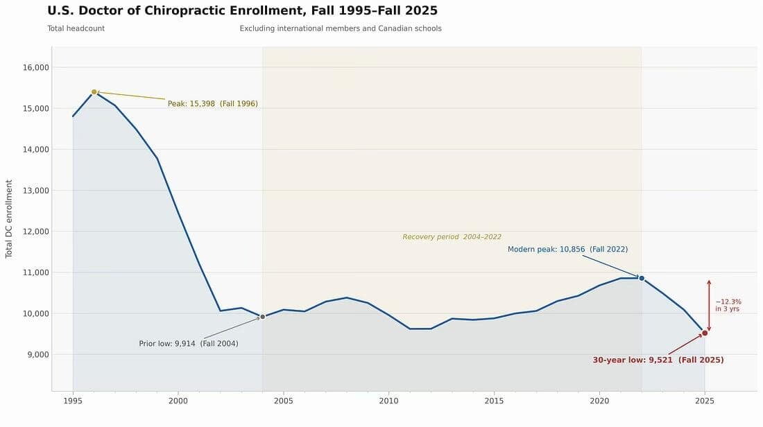
Chiropractic institutions not already on-board with integrating AI with guardrails into their curriculum are well behind the state-of-play in education. In North America the institutions are facing mounting debt and falling enrolment (Figure 1)(7, 8) and I am confident several colleges will close before 2030.
Figure 1: DC enrolments, USA, 1995-2025
Those institutions yet to embrace AI will remain the weakest. Among the wise moves for institutions to take right now is the introduction of a suite of AI literacy modules for staff and students, along with a shift in focus from competence to capability. Competencies are no longer the currency they were held to be in the 1980s. The rush of AI across all educational institutions in the discipline is making this glaringly obvious.
Prof Bereznicki says ‘The only constant is going to be change, and what changes is the pace of change’. Consider this a warning.
AI is now pervasive, with Generative AI embedded in many software systems which has implications not just for teaching content, but for assessment, academic standards and graduate preparedness, the tasks which make or break a Chiropractic institution.
How AI impacts our assessment strategies
In 2011 I implemented a ‘two-lane’ approach to assessment, an early attempt at what UTAS is now introducing. One lane consists of secure assessment, such as invigilated exams, interviews and observed practical tasks, which allowed verification that students had personally met learning outcomes, while the second lane allowed more flexibility, including the use of on-line resources where appropriate, helping students develop contemporary workflows while preparing for higher-stakes assessment.
Today my approach is light on invigilated exams given their cost and limitations which include testing memory more than clinical thinking, and big on face-to-face viva voce on two conditions:
- There is equality across all students in task or question difficulty; and
- Transparent grading rubrics are in place and adhered to.
At the end of the day I agree with Bereznicki that we need to make sure we can verify students have met the objective learning outcomes and have demonstrated the capabilities we announce. We can not remove the human judgement from learner assessment which in turn carries the imputation we must also train our learners how to perform. Does this mean that assessment is an act? To a degree, yes, but then the pragmatist will tell us that all human performance is an act. I don’t think it matters as long as there is equality of opportunity, overall fairness, and transparent decision-making.
In Professor Bereznicki’s’s view, the future of education lies in preparing students to use powerful tools fluently, critically and ethically while keeping the humanistic dimension in shaping vision, exercising judgment and making decisions.
In a world of abundant information and machine-generated content, the real task is helping students learn how to question, interpret, connect and act responsibly in complex systems. (Bereznicki, 3)
Valuing human engagement
Reporter Matt Michael (9) wrote: ‘The idea is straightforward. As artificial intelligence and robotics become capable of doing more, there will be some activities we consciously choose to keep human. Not because machines are incapable of doing them, but because we decide there is value in humans continuing to do them’.
AI is a prediction engine trained on the past. It can produce high quality, average outputs at scale. All it can do is replicate patterns, styles, and strategies than have gone before. If you ask AI to do what everybody else asks it then all you will get is an average output. Average becomes cheap, and cheap becomes worthless. To do better you must give AI better direction, better constraints, better taste, better data, better feedback loops, better goals. You need to build a system that improves itself. You are building a personalised tutor at scale. A tutor that adapts to your pace, your blind spots, your motivation patterns. It can explain to you the same concept in 10 different ways until it clicks with you, and then create practice problems tuned to your exact weaknesses. Your AI tutor will plot your learning journey across months and shape your learning plan with ruthless efficiency.
Chiropractic learning is now a private competitive sport where the winners are those with the most consistent systems. AI is a force multiplier for Chiropractic clinical learning where the learner’s treatment plan for a patient can be tested against what are in effect real-time meta-analyses and outcomes predictions. Gone are the ‘3 times a week’ patient scheduling formulas used simply to boost a learner’s number of patient visits. Instead, total ‘patient-visits’ under a lessening regime of supervision will be meticulously plotted by patient case-mix and demographic data. Students will complete with ‘x number of cases of uncomplicated low-back pain is adult males, y number of cervicogenic headaches in teenage females’ and so on.
Do I recommend any AI platform or agent?
No, and you do not need to select a particular AI platform. You must become a problem engineer, not a prompt engineer. This means you can define a real problem, translate that problem into constraints and objectives, then judge the output with taste, context, ethics, and strategic clarity. Remember AI can propose a plan for you but it can not care for you. Strategic caring becomes your advantage, the ability to choose a direction and commit, the ability to say ‘this matters, this doesn’t’.
You have to continually cleanse and strengthen your filters because the Chiropractic world is about to drown in cheap content, cheap ideas, and cheaper shortcuts.
You have to constantly work on your judgement and the way to do this is to read more deeply into the literature. This is where the Index to Chiropractic Literature becomes your best friend. You can enter your search terms and retrieve a treasure trove of human intelligence published over the past 60 years or so. You must selectively read what others before you have thought on a particular topic so you develop your human sense of judgement as to what AI output is bullshit and which may be useful, with true meaning. You can not ask AI to make that human judgement. You can not trust AI outputs without knowing the foundations.
Remember, you must use AI to build your clinical skills so that you can produce measurable outcomes faster and more effectively.
AI is a platform shift. The biggest mistake is to use AI to help you do the same things faster. AI is infrastructure that produces intelligence. Think of the introduction of electricity as underpinning the functioning world, thus think of AI as underpinning all that we do today. (10)
Move on from being a tool user (‘help me study’) to become a power user: ‘Build me a personalised tutor that learns how I think, tracks my weaknesses, designs quizzes, and turns my learning into a compounding advantage’. You could also prompt to ‘Build me a pipeline that generates 30 learning concepts drawn from [for example Week 4 content] to build my identity as a Chiropractor and output ready to use clinical assets every week’. A good clinical learner will be one who is able to interpret, challenge, and apply AI-supported insights while maintaining human empathy and ethical judgement.
Perhaps the best way to think of AI from now on is that it compresses the curriculum and increases the speed of attainment. AI is a collaborator and the learners who understand this will excel. AI is a critical asset for those who lead learning who must understand that they need to become a master within the domain of Chiropractic in the first instance, and then a master in sub-domains of relevance to them, such as paediatric care, sports Chiropractic, and vitalistic-centred care. AI will provide multiple care plans but you have to decide which one is best suited to a particular patient. You need an anchor of what ‘good care’ looks like, and can defend it with clarity. AI will not replace clinical-decision making but it will provide many alternative pathways.
Your judgement ‘engine’ is what AI will amplify. If your judgement is poor it will amplify you into mediocrity; should your judgement be astute, AI will amplify you into excellence.
The correct use of AI is cyclical. Use it to generate, test, measure, refine. Repeat. Be aware that AI will hallucinate and produce nonsense, and it is up to you to filter for this and correct the cycle.
Is Chiropractic ready for the AI avalanche?
Chiropractic Future is an advocacy group with some of the profession’s most influential volunteers focused on increasing access to Chiropractic care and fighting for better reimbursement. They claim to leverage technology, research, and the profession’s collective expertise to strive to create a sustainable future for Chiropractors. (11) They claim ‘there has never been an effort of this magnitude that brings all Chiropractors together for a common cause … together, we can overcome the challenges the current healthcare system poses and secure a brighter future for Chiropractors’.
However their 2025 Annual Report (10) makes no mention of AI under Technology. Rather, they hold that:
‘Chiropractic’s future depends on the ability to collect, interpret, and leverage high-quality data. CFSP’s technology initiatives, including the national data lake and state-level reimbursement dashboards, create the infrastructure needed to capture insights that have never before been available to the profession. These tools generate real-time visibility into utilisation, payer behaviour, reimbursement trends, payment disparities, and patient outcomes. Together, they form the foundation that enables evidence-based decision-making, strengthens advocacy, and ensures chiropractic has a credible, data-driven seat at the healthcare table’. (10)
In a similar vein the magazine Chiropractic Economics (12) in its 2020 feature which reported the views of various Chiropractic dignitaries, generally felt the profession was peddling furiously to stay in place. Karlos Boghosian DC, president of the Federation of Chiropractic Licensing Boards (FCLB), was reported as saying ‘we have an obligation to advance the profession through research, clinical studies, education, regulation and consistent introduction of emerging technologies’. Admittedly this was in 2020, before AI surged as it has today. However I do note that the quote at the head of this paper, from Mark Cuban, was reported in 2017, 3 years earlier than that report which at least gave recognition that emerging technologies warranted attention.
This prescience is not evident in the World Federation of Chiropractic’s (WFC) Strategic plan and vision 2024-29. (13) While humanistic in focus, the WFC does say ‘Advancing technology means that access to health care is expanding globally. At the same time, access to health resources has reinforced personal autonomy and promoted shared decision-making. As a global federation, we will encourage our members to use technology to promote quality health services for all, access emerging evidence and collaborate with colleagues’. (12, p 9)
In 2024 the Journal of Future Studies (14) published ‘Advancing Chiropractic Care: Opportunities and Challenges for the Future’. It presented findings using the dated SWOT technique and identified an opportunity as:
Technological advancements such as computer-assisted manipulation, telehealth, and wearable technologies have the potential to significantly impact the profession.
However, among the threats they identified, embracing technological advancements was not mentioned, only the same tired trope of ‘Failure to develop a scientifically based definition of subluxation’ along with ‘lack of cultural authority’. (13)
The profession still receives the benefit of the periodic Practice Analysis of Chiropractic, the latest being for 2025, from the National Board of Chiropractic Examiners (NBCE). (15) This is the 7th instalment in a series of surveys of US chiropractors, with the inaugural survey having been conducted in 1991 and subsequently reported in the 1993 Job Analysis of Chiropractic. (16)
These reports carry valuable information for improving the management of the profession in the US, especially the report of patient conditions (14, p 98) which can largely be extrapolated to other countries where Chiropractic is established under legislation. It is an interesting guide as to what may be expected in the countries where the discipline is emerging and which has been reported. (17, 18) While there is a small section on continuing education, it is rather odd to find no report on the status of pre-professional education in America. This is something I would have thought a national board of examiners should have a vested interest in reporting.
It is a report on ‘what Chiropractic is’ in the year examined and as such does not need to look into the future, let alone make any predictions about technology and AI, however …
… I am left with the feeling that the profession is largely oblivious to the AI tsunami which reinforces my view that AI is catching a lot of institutions and associations unaware. I fear the resultant carnage.
AI and research in Chiropractic
I discuss this from dual perspectives; that of the research generator, and then of the research consumer. Each is impacted significantly by AI and in a very positive manner.
Research generation
My view is that Chiropractic researchers will become redundant unless they initiate new lines of inquiry. This is due to the ‘easy’ options of doing a meta-analysis or a literature review are now done faster by AI. A project such as a systematic analysis was once the crucible for researchers seeking a doctorate. I don’t deny this work was beneficial for training the academic rigour needed to earn a PhD, however now the training is about how to become far more critical of what AI returns to one’s prompts.
For doctoral candidates it is now more important to practice critical reading and undertake a deeper source analysis to ensure the veracity of external data. At the same time, statistical analysis of one’s own data is simplified yet still demanding that we maintain checks for true inference.
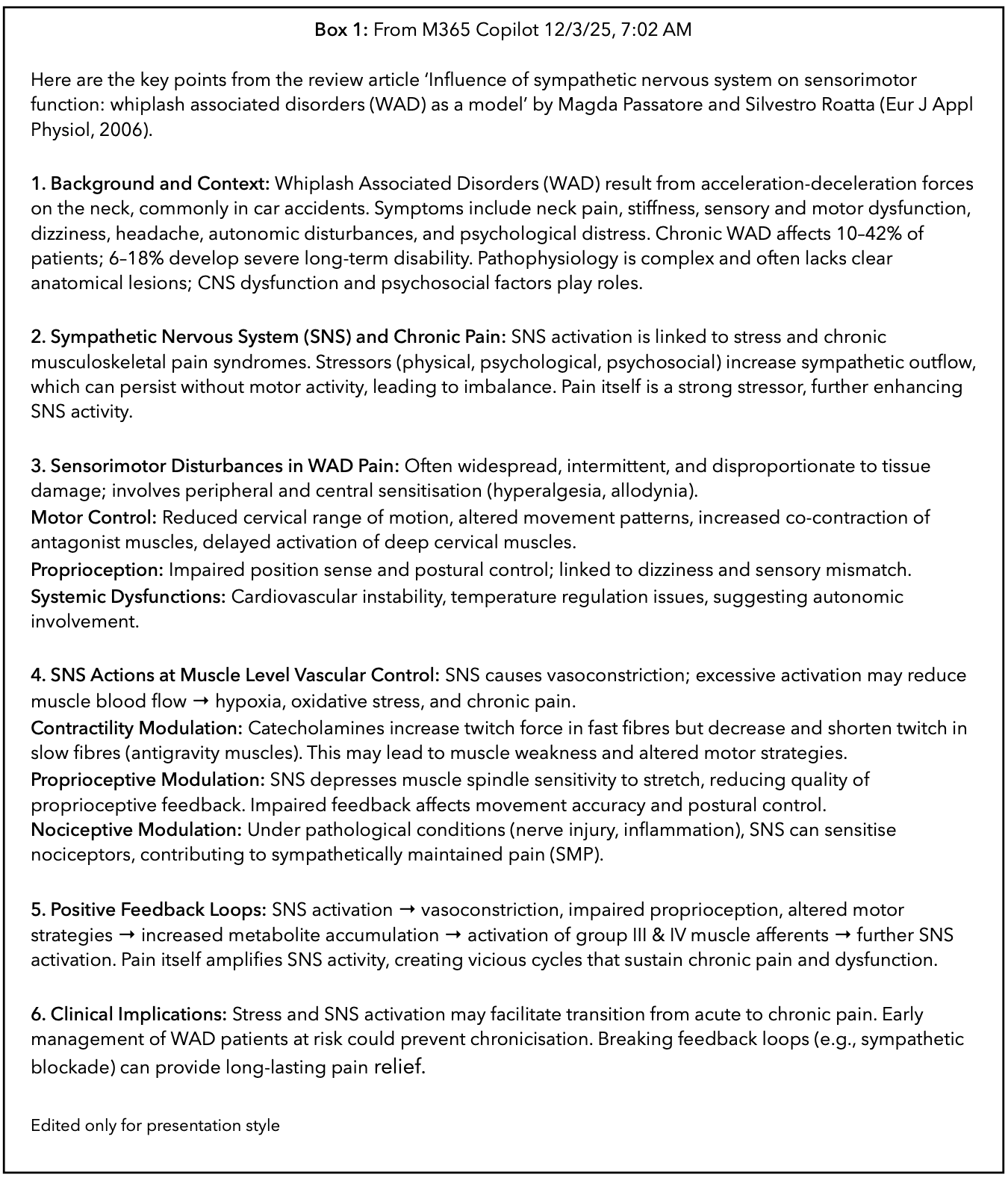
By creating and testing various prompts an AI Agent should be able to produce a literature review for you and go a long way towards gathering papers for a systematic review. Even Co-Pilot, a basic agent, can provide a user-friendly summation of a paper. A colleague entered the following prompt and retrieved the response given verbatim in Box 1
List key points from Influence of sympathetic nervous system on sensorimotor function - whiplash associated disorders (WAD) as a model (19)
Should this response proceed to being reproduced somewhere, even in a patient brochure, it must be fully referenced. I give the most acceptable format for this citation as reference 19.
AI for educators
A power user could build on the above prompt, perhaps as: ‘Build me 4 quizzes each with answers as multi-choice, true-false, one of the following, and choice of one from 5 stems on this paper each going deeply into a concept relevant to building my practice as a Chiropractor’. This approach is more beneficial for academics and learners. Let me show you how this works, it is the approach I use in the Asia-Pacific Chiropractic Journal (20) where my AI Agent is Manus. I was using Grok however they seem to lack any customer service and my operational and billing questions remain unanswered, thus I now use Manus, (21) supposedly an independent agent lab.
First, select your source paper and have it as a PDF to drag into Manus when you open it on your desktop. Reading a PDF is a good ability in Manus and not so good, if at all, with Grok in my experience.
The reference article for this exercise is:
Jagadeesh A, Mohanty P, Ghosh T. Assessment of ergonomic risk factors in the occurrence of work-related musculoskeletal discomfort & pain among cashew workers. Asia-Pac Chiropr J. 2026;7-2.https://www.apcj.site/JagadeeshErgonomicRiskFactors.pdf
ME PROMPT: Provide a 2 paragraph summary of this paper by Jagadeesh and colleagues using professional clinical language including key statistical findings and provide 3 multi-choice quiz questions and 3 true/false question with answers
MANUS - Jagadeesh and colleagues conducted a quantitative cross-sectional study to characterise postural ergonomic risk and pain among 40 cashew-processing workers, 20 men and 20 women, aged 35-55 years, in Kasaragod district, Kerala. Participants engaged in either shelling or peeling were recruited by convenience sampling and assessed using the Rapid Entire Body Assessment (REBA) and Visual Analogue Scale (VAS). Mean REBA scores were similar for shelling and peeling workers (7.10 ± 2.36 and 7.15 ± 3.31, respectively), corresponding to the upper boundary of the medium-risk category; mean VAS pain scores were likewise similar (4.55 ± 1.87 and 4.50 ± 2.01, respectively), consistent with moderate pain. In the task-stratified analysis, 60% of shelling workers and 40% of peeling workers were classified as medium ergonomic risk, while very high risk was more frequent in peeling than shelling (25% versus 15%). Moderate pain was the most common pain category in both groups, affecting 50% of shelling workers and 55% of peeling workers; no participant was classified as pain-free.
Pain intensity was significantly and positively associated with ergonomic risk in both sexes. Among men, the relationship between REBA and VAS scores was strong (r = 0.725, df = 18, p = 0.0001); among women, it was moderate (r = 0.538, df = 18, p = 0.014). Moderate pain was reported by 60% of men and 45% of women, while women showed a wider distribution across mild, moderate, and severe pain categories. These findings support targeted occupational interventions, including workstation redesign, postural training, scheduled rest breaks, task rotation, joint-protection education, and energy-conservation strategies. Clinical interpretation should remain cautious because the study’s cross-sectional design cannot establish causality, and its small convenience sample limits generalisability; psychosocial and organisational determinants were also not assessed. In addition, the article contains a minor internal inconsistency: the task-stratified REBA table records one peeling worker at negligible risk, whereas the sex-stratified table reports no participants at negligible risk.
I find this to be a good summary of a well-detailed paper. I like that it spotted an inconsistency in reporting, something not identified in peer review.Next, the quizzes:
MANUS QUIZ 1:
1. Which instruments were used to assess ergonomic risk and pain intensity?
A. RULA and Numeric Rating Scale
B. REBA and Visual Analogue Scale
C. Oswestry Disability Index and DASH
D. Nordic Musculoskeletal Questionnaire and Borg Scale
Answer: B. REBA and Visual Analogue Scale. The Rapid Entire Body Assessment (REBA) evaluated posture-related ergonomic risk, while the Visual Analogue Scale (VAS) measured subjective pain intensity.
2. Which task group had the greater proportion of workers classified as being at very high ergonomic risk?
A. Shelling workers
B. Peeling workers
C. Both groups had equal proportions
D. The study did not report this outcome
Answer: B. Peeling workers. Very high REBA risk was reported among 25% of peeling workers, compared with 15% of shelling workers.
3. Which statement best describes the association between REBA and VAS scores among male workers?
A. Strong, positive, and statistically significant
B. Moderate, negative, and statistically significant
C. Weak, positive, and not statistically significant
D. No association
Answer: A. Strong, positive, and statistically significant. The correlation was r = 0.725, p = 0.0001, indicating that greater ergonomic risk was associated with higher pain intensity among male workers.
MANUS QUIZ 2, True - False:
1. Moderate pain was the most frequently reported VAS category among both shelling and peeling workers.
Answer: True. Moderate pain was reported by 50% of shelling workers and 55% of peeling workers.
2. The reported correlations demonstrate that ergonomic exposure caused the workers’ pain.
Answer: False. The cross-sectional study identified an association between ergonomic risk and pain, but its design cannot establish causality.
3. The association between REBA and VAS scores was statistically significant among both male and female workers.
Answer: True. The association was significant among men (r = 0.725, p = 0.0001) and women (r = 0.538, p = 0.014).
You can design the quizzes to any existing specification your institution prefers, and if you are presenting the quizzes in an on-line format you can include an instant Translation widget, I use the Website Translator widget from Elfsight. (22) This allows learners to take your quiz in their own natural language, however I am yet to fully test its robustness.
This exercise is the most basic starting point for Chiropractor Educators as it now so easy to generate content for the classroom; you can even ask AI to generate a slideshow from a paper to strengthen your in-class presentations. As shown here, you can form quizzes and in lieu of a peer-reviewed paper, you can submit your own teaching materials so that the tedious and repetitive ‘hit and miss’ chore of creating a quiz on your own teaching material is automated.
Each learner’s personal tutor
It is in this role that AI Agents excel. Each of us now has a process we can train to become a personal tutor for us. The applications for educators are endless and we can quickly be brought up to speed in a topic about which we presently know little. But the real buzz comes once you have introduced the idea to your class and shown them how powerfully well-written AI prompts can boost their learning.
Again the possibilities are endless but as a starting point the learner can be shown how to use AI to summarise a paper you require them to read, and then have a quiz test them so they can, in private, reinforce their understanding.
The most exciting part is that once you give your approval for the use of AI in this way, your learners will quickly become experts and share their discoveries and tips across the group. In fact, it is a good idea to set aside a few minutes once a week with the class to catch-up and share what various members are doing. If nothing else it will get the message to those who are slow adopters and eventually bring them on board.
Where do you set the boundaries?
Once you define AI as a learning assistant or personal tutor and explain where support ends and substitution begins you should reduce academic integrity risks and student anxiety. A useful starting point is practical AI literacy for students. This goes beyond warning students not to cheat. Students need to understand how to ask useful questions of AI tools, how to check outputs for errors or bias, and how to take responsibility for the work they submit. Treating AI as a thinking partner, rather than an answer machine, helps students see how effort, judgement, and learning can best sit with them. (23)
AI in Chiropractic institutions
- Chief Complaint, linked to spinal regions;
- Symptoms, Numbness, tingling, pain;
- Duration and Frequency, Time and severity scaling;
- Triggers, Identifying motion-based pain influencers; and
- Radiation, Tracking referring pain patterns.
The NZCC prototype mimics real world clinical note-taking and supports interactive, repeatable training sessions. While there may be limitations with this approach it is a significant institutional advancement with potential for direct student benefit.
I guess our educators fall into one of two groups, those as we see at NZCC who are pioneering roles for AI in the clinical education of Chiropractic learners, and those who will wait for others to do the work. But make no mistake, the work is getting done and is progressing rapidly. (26) Some research teams are now testing whether Large Language Model-Based chatbots are effective in providing reliable medical advice for presentations such as Achilles Tendinopathy. (27) New Zealand is a country at the leading edge of examining AI-use in Chiropractic and musculoskeletal clinical practice, and even with providing training in AI for practitioners and, presumably, educators. (28)
AI in content generation
My general position statement for AI use is:
All use of AI beyond grammar and spell check must be declared. Generative AI is not to be used to create new content in an author’s name. The correct approach is for you to write your paper using your human intellect and selecting your references, then run spelling and grammar-checks in your writing App for augmentation. You may also use an AI tool to review and critique your paper.
I consider a learner to be an author when creating work to be submitted for assessment. Subtle, but it gives a greater ‘air’ of reality beyond it being ‘just an assignment’. And to be included in all work submitted, even to the point of declaring AI was not used, is a variation of this statement:
During the preparation of this work the author used [name of agent including version #] in order to assist with copyediting and to generate alternative sentence structures. After using this tool, the author reviewed and edited the content as needed and takes full responsibility for the content of the published work.
Image generation
As an educator and a writer my focus on AI has been around its abilities with gathering data, phrasing ideas, and creating learning reinforcement. However others are active with another rather clever function, that of Image Generation.
My colleague Dr Scott Cuthbert, of the Philippines, is having success with writing prompts which can create what I think are marvellous informative and educational posters. I give one such image, an infographic on manual muscle testing for Gluteus Maximus, as Figure 2.
Figure 2: Infographic - MMT Gluteus Maximus. Courtesy of Scott cuthbert, 2026
The most important point to note is the inclusion of 5 citations in short form. It is easy to get carried away and churn out a few posters of this nature and it is another thing to take the time to do it in a way that is defensible.
This type of work must, in my view, be of sufficient standard to be accepted as a poster at a research or clinical meeting. This means it must be professional. Facts that are given must be checked and referenced. The depiction of the MMT must be of educational classroom standard, meaning it should go from this page into the class-room as an exemplar, which this does.
The anatomical pic is a good ¾ right posterior view which gives a depth to the muscle, and the clinical tips are a speed-read for the clinician. Purists may quibble over the abbreviated correlation data but quibbling is what they do best. To me, that box simply says the manual muscle testing correlates well with dynamometry and that is, to me, all the takeaway I need. I specifically like the 3 figures in action which graphically conveys 3 main functions, any of which may form the basis of a complaint from the patient sitting in front of me. This is good, useful information.
It is important to note Scott has avoided a common fault in AI Image generation, which is the tell-tale dark and heavy background where the page is crowded and the image is indistinguishable from any other you saw earlier. Here we have a pleasing turtle on a light-coloured background.
It really is amazing that a task which would previously have taken an academic a month to prepare, perhaps even with the aid of a graphic designer, can now be produced in under an hour, with time to fine-tune the prompt and tweak it here and there.
Who owns the image in Figure 2?
By now you will be wanting me to reproduce the prompt used by Scott. I have chosen to not publish this on the basis that in the case of image generation, it is very much the intellectual property of the person writing the prompt which will best create the image they envisage. This is different from writing where I have been offering guidance in general terms; images are specific creations and in the ideal world would be copyrighted.
And here is not the time to even start to consider the huge copyright implications where the work of artists and creators is taken, some say stolen, by the global corporations without any credit or payment for use to continue training their LLMs. This is a hot button issue in Australia and there is a significant movement happening to protect the IP of creators from theft by anonymous parties.
Former Australian prime minister Julia Gillard has entered the debate on regulating AI, urging governments to build their ‘muscle’ on intervention because the challenges of the new technology would continue to come in waves of change. (29) Ms Gillard who is chair of the Wellcome Trust, which funds health and scientific research, is reported as saying: ‘The AI challenge is going to mean all of that (regulation) has got to be done again and again and again. That’s going to be a big part of the debate going forward’.
As a writer and an educator I have conflicting views. The writer in me wants to say ‘hold my copyright tight for my IP’ while the pragmatist says ‘as a Chiropractor it is important to me for my writing to be sucked into every LLM there is’ in the expectation it may eventually change how AI sees Chiropractic and especially, what it says about our discipline. There is no income from any journal paper, in fact, some Chiropractic journals once reached a new low in demanding authors pay the journal to be published, but that very bad practice seems to have passed.
Conclusion
I can’t deny the thrilling outcomes we can now achieve for strengthening our learning objects, tutoring ourselves and our students to new heights and gaining strength in our writing. Image creation is icing on the cake, and having your AI Agent create a slide show for you really lifts to another level. Just remember to check and verify.
But as with all cakes, we have to stay away from temptations which can quickly become toxic and destroy our reputations overnight.
I see value in an educator becoming an informed advocate of the right and proper use of AI in the classroom. I would like to think I have offered some little guidance towards you achieving this.
Final word
I defer to His holiness Pope Leo XIV and his encyclical Magnifica Humanitas of 15 May 2026:
It is necessary to establish adequate regulatory tools capable of upholding justice and curbing the distorting effects of technological power. Nevertheless, the issue is not limited to regulation. As Pope Francis warned, we must realistically ask ourselves who holds this power today and how they use it.
In the past, it was largely up to the State to guide and direct innovation. Today, however, the main drivers of development are private, often transnational, parties that are endowed with resources and the capacity to intervene that surpass those of many Governments. Technological power thus takes on an unprecedented, predominantly ‘private’ aspect, which makes it even more challenging to discern, govern and direct such power toward the common good.
We must lovingly safeguard the grandeur of humanity bestowed upon us and revealed in its fullness in Christ, the splendour of which no machine can ever replace. True progress always stems from a heart open to others, an intelligence willing to listen and a will that seeks what unites rather than what separates. (30)
Let us retain our humanity as Chiropractors and be the master of AI, harnessing its power for the good of our students, our patients, and society which needs Chiropractic care more, now than ever.
Disclaimer
This paper is a Chapter from my forthcoming textbook, The Chiropractor Educator’s Handbook, and is already outdated. The AI tsunami to which I earlier alluded, is here and right now you will be interacting with AI agents in ways I have not yet imagined.
Of particular interest is the position of Apple which is to undertake the majority of AI-processing on-device, in contrast to agents which process requests and interactions off-device and in AI Data Centres. Leaving aside the negative press being generated by the imposition of this massive infrastructure on regular communities, there is as Apple exhorts, a real risk to one’s privacy.
I am not advocating one way or the other but simply wish to point out that at my time of writing, October 2026, I am yet to ‘play with’ Apple AI in any meaningful manner. I can’t imagine it achieving output significantly different to the matters I have discussed here, however I am always up for a surprise.
You can edit text on your website by double clicking on a text box on your website. Alternatively, when you select a text box a settings menu will appear. your website by double clicking on a text box on your website. Alternatively, when you select a text box
1. Mark Suster. Mark Cuban on Why You Need to Study Artificial Intelligence or You’ll be a Dinosaur in 3 Years. Medium. 8 Februry 2017. At https://medium.com/both-sides-of-the-table/mark-cuban-on-why-you-need-to-study-artificial-intelligence-or-youll-be-a-dinosaur-in-3-years-db3447bea1b4
2. How to check AI citations and identify AI hallucinations in references. [Blog]. editage. 30 July 2026. At https://www.editage.com/blog/how-to-verify-ai-generated-citations-references-steps-checklist-examples/
3. Lo LS. The CARE approach for academic librarians: From search first to answer first with generative AI. J Academic Librarianship. 2026;52(1). At https://doi.org/10.1016/j.acalib.2025.103186
4. As artificial intelligence reshapes universities at speed, a senior University of Tasmania leader says the future of education won’t be defined by machines replacing people, but by institutions strengthening the human capabilities technology can’t replicate. Luke Bereznicki, UTAS. [News]. Mercury. At https://www.themercury.com.au/education/regions/tasmania/universities-must-futureproof-graduates-with-skills-ai-cannot-mimic-says-top-academic/news-story/45171805204142413077a27e11f4229c
5. The Future of Chiropractic: Emerging Technologies and Treatment Approaches. Chiropractic News. 11 December 2025. At https://www.chiropracticnews.org/the-future-of-chiropractic-care-.html
6. Ebrall PS. From competency to capability: an essential development for chiropractic education. Chiropr J Aust. 2007; 37:61-7.
7. Chiropractic Colleges in Crisis. [News]. Dynamic Chiropractic. n.d. At https://dynamicchiropractic.com/article/103268-chiropractic-colleges-in-crisis
8. The Profession Is Running Out of Room for This Nonsense. The Chiropractic Chronicle. McCoy Substack. At https://drmatthewmccoy.substack.com/p/stephanie-libs-message-to-life-west
9. Matt Michael. AI is amazing, but took to humans if you want to get ahead. The Australian. 21 September 2026. At https://edition.pagesuite.com/popovers/dynamic_article_popover.aspx?artguid=3d4d6d28-c418-4768-a0af-aa012d521aea
10. Jensen Huang. Nobody Is Ready for What AI Can Do in 2026. YouTube. January 2026. At https://youtu.be/UrMnOp2N9Kw?si=ceF5PIgIXRLiDL2X
11. Chiropractic Future. 2025 Annual Report. At https://chiropracticfuture.org/wp-content/uploads/2026/02/CFSP-2025-Annual-Report-FINAL.pdf
12. Rick Vach. The future of chiropractic is now - the next 125 years looking forward. Chiropractic Economics. https://www.chiroeco.com/future-of-chiropractic/
13. Adjusting to the future. Strategic plan and vision 2024-29. World Federation of Chiropractic. At https://www.wfc.org/website/documents/797_wfc-strategic-plan-2024-2029.pdf
14. Riggs J. Advancing Chiropractic Care: Opportunities and Challenges for the Future. J Future Studies. 25 September 2026. At https://jfsdigital.org/articles-and-essays/2024-2/vol-28-no-4-june-2024/advancing-chiropractic-care-opportunities-and-challenges-for-the-future/
15. Practice Analysis of Chiropractic 2025. National Board of Chiropractic Examiners. USA. 2026. At https://www.nbce.org/wp-content/uploads/Practice-Analysis-of-Chiropractic-2025.pdf
16. Christensen MG, Delle Morgan D. Job analysis of chiropractic: a project report, survey analysis and summary of the practice of chiropractic within the United States. National Board of Chiropractic Examiners. USA. 1993. At https://books.google.com.au/books/about/Job_analysis_of_chiropractic.html?id=9yNtAAAAMAAJ&redir_esc=y
17. Tetrault MY. An initial case-mix report of chiropractic practice in the Philippines. Asia-Pac Chiropr J. 2020;1:006. At https://www.apcj.net/site_files/4725/upload_files/Tetrault2020200627(1).pdf?dl=1
18 Doshi J. An initial report of Chiropractic practice in India: The case-mix of 21,386 patients. Asia-Pac Chiropr J. 2026;7-2. https://www.apcj.site/DoshiCasemixIndia26.pdf
19. Passatore M, Roatta S. Influence of sympathetic nervous system on sensorimotor function: whiplash associated disorders (WAD) as a model. Eur J Appl Physiol. 2006;98:423-49. https://doi.org/10.1007/s00421-006-0312-8
20. Asia-Pacific Chiropractic Journal. At www.apcj.site
21. From tedious to effortless. Manus. At https://manus.im/team
22. elfsight. #1 Website Widgets Platform At https://elfsight.com/
23 Ritesh Chugh. Back to school with generative AI: what schools, students and teachers need in the first weeks of term. CQUniversity News. 27 January 2026. At https://www.cqu.edu.au/news/1261798/back-to-school-with-generative-ai-what-schools-students-and-teachers-need-in-the-first-weeks-of-term?utm_medium=email&utm_campaign=CQUReview---29-January-2026&utm_content=cqu.edu.au/news/1261798/back-to-school-with-generative-ai-what-schools-students-and-teachers-need-in-the-first-weeks-of-term&utm_source=comms.communication.cqu.edu.au
24. Participant solicitation invitation posted by researcher Aaron Downie. Contact the researcher directly at aron.downie@mq.edu.au
25. Imam Y, Niazi IK, Holt K, et al. AI-Powered Virtual Patient for Chiropractic Training. An MR and NLP Based Approach. 2025 47th Annual International Conference of the IEEE Engineering in Medicine and Biology Society (EMBC), Copenhagen, Denmark, 2025, pp. 1-7, DOI 10.1109/EMBC58623.2025.11252775.
26. Wang YL, Tian LC, Meng JY, et al. Evaluation of large language models in patient education and clinical decision support for rotator cuff injury: a two-phase benchmarking study. BMC Med Inform Decis Mak. 2025 Aug 4;25(1):289. DOI 10.1186/s12911-025-03105-5.
27 Liang Z, Wang M, Abdelatif NMN, et al. Are Large Language Model-Based Chatbots Effective in Providing Reliable Medical Advice for Achilles Tendinopathy? An International Multispecialist Evaluation. Orthopaedic Journal of Sports Medicine. 2025;13(4). DOI 10.1177/23259671251332596
28. AI for Chiropractors in New Zealand. GenAI Training NZ. At https://genaitraining.co.nz/articles/ai-for-chiropractors-nz/
29. Helen Trinca. Julia Gillard urges governments to keep building on regulation of AI. [News]. The Australian. 28 September 2026. At https://www.theaustralian.com.au/world/julia-gillard-urges-governments-to-keep-building-on-regulation-of-ai/news-story/ee5f2e692e309e4ca0d1076b090dee95
30. Encyclical letter Magnifica Humanitas of his Holiness Pope Leo XIV on safeguarding the human person in the time of artificial intelligence. 2026. At https://www.vatican.va/content/leo-xiv/en/encyclicals/documents/20260515-magnifica-humanitas.html
AU: Clare McIvor BBus(Admin), GD Comms(ProfWrit,Edit), GD(Psych)(Cand). Chiropractic writer. e: clare@claremcivor.com
Abstract: Generative artificial intelligence offers practical support for chiropractic writing and research, yet its reliance on dominant patterns in existing data may constrain professional enquiry. This dialogue examines ‘regression to the mean’ as a metaphor for the homogenisation of ideas and language, considering its implications for chiropractic’s salutogenic orientation, philosophical diversity, and commitment to individualised care. Drawing on published literature and professional writing experience, the author discusses linguistic convergence, cognitive offloading, knowledge cutoffs, and the potential loss of minority perspectives. Preliminary findings concerning critical thinking are considered cautiously, without assuming causation. Practical recommendations include reading primary sources before consulting automated summaries, verifying references, developing arguments independently, and using artificial intelligence to challenge rather than replace professional judgement. Deliberate prompting and authorial control are proposed as safeguards against generic framing and erosion of individual voice. The dialogue advocates responsible use of artificial intelligence as an adjunct to scholarship, while retaining human accountability, original research, and philosophical enquiry as essential foundations for chiropractic’s continuing intellectual development and distinctive contribution to health and wellbeing.
Indexing Terms: Artificial intelligence; Chiropractic; Scholarly communication; Cognitive offloading; Critical thinking.
Cite: McIvor C. Avoiding Regression to the Mean: A millennial perspective on AI in Chiropractic writing and research. Asia-Pac Chiropr J. 2026;7.3. https://www.apcj.site/McIvorAIDialogue.pdf
Take-away: '… Our challenge, as the age of AI opens, is to make sure that we do not become trained by the average, by the regression to the mean. Instead, we must train it .…’
Introduction
Rarely, in my work life, do I glance at fewer than a dozen research papers in a day. Whether it be for Chiropractic blogs, case reports or other writing, I'm neck-deep in journals and primary evidence. A year ago, hours would have been spent finding these articles, reading halfway down, realising they're not what I'm looking for. Nowadays, those hours are better spent. I've programmed an AI machine that knows which journals to monitor for new Chiropractic content. It knows my voice, because I've trained it on outputs from thirteen years as a full-time, professional writer. I've also trained it to use specific terms, inflections and paradigms as I write for different clients.
Also, I wince at AI slop, and spot it all too easily, as finding stock photography that reflects the Chiropractic mood is now impossible. AI has made that job harder, ironically. The old Nikon has been dusted off, demanding labour that used to be a two-minute search on a database. AI makes some things easier or better, and others harder and riskier.
I am an intermediate AI user, having learned a thing or two that can get better outputs than entry-level instructions, and I'll share some of those tips in the latter half of this article. But there is a strict limit to what AI can do for me. The rest, I must do myself. It can do literature searches, but I must make sure it didn't miss anything. It can summarise, but I must read and make sure it captured the essence of the article.
It can never be entrusted with my professional reputation. Nor can it be a philosopher.
Currently, AI is working approximately one to two years behind us, not capturing new frontiers. If you want it to read a news article from today, you have to put that in and specify the request; otherwise, that data point will not feature in its outputs.
Knowing this, and knowing the point in history at which Chiropractic stands, we carry a responsibility. As Chiropractors, philosophers and researchers, it is our job to be front-liners - to think about what is possible and to create new and incisive research questions and create primary data. It is our job to recognise that research is both currency and protection when it comes to our right to practice, and that social media is the megaphone that reaches those who need us and hopefully changes their ideas about what health and the expression of life can be. It is our job to move beyond the reductive and into the expanse of possibility and human potential; to theorise and build our way towards what a human could be and do if their nervous system worked free of interference.
This element of consciousness, humanness and innovation can never be entrusted to a machine.
In this issue, Ebrall (2026) sets out an educator’s case for treating artificial intelligence as augmented intelligence: a collaborator that amplifies the judgement of the person using it, for better or worse. He also observes that AI is a prediction engine trained on the past, and that if we ask of it what everybody else asks, we will receive an average output.
It is this regression to the mean that presents the greatest threat to Chiropractic, which is, at its heart, a hands-on model built on connectedness, both neurophysiological and interpersonal.
AI is, at is most basic form, a large language model (or LLM). And a language model is, by design, an instrument of regression to the mean. It produces the most probable continuation of a text, and the most probable is the most central: the consensus framing, the familiar phrase, the position held by the largest share of its training data. For many tasks this is useful. We do need to know consensus data on safety, efficacy, best practice and evidence-based approaches. But for a discipline whose distinct contribution lies away from the centre, it is a fundamental misfit.
We have seen the clash in the constant juxtaposition of traditional medical research with Chiropractic research. The current approach to evidence in traditional research looks to exclude all explanations other than the tablet or surgery that forms the intervention. Did this tablet cure your *insert ailment here? Can we be sure it was that tablet?
Chiropractic turns this approach on its head. It looks at the whole person, their individuality, their life and all that is in it and then asks, 'how can we help? How can adjusting the subluxation remove interference and allow this person to better express innate intelligence?' We have not put a limit on what this might mean, nor should we.
Chiropractic has never been content with the mean. It is philosophically expansive, asking what health might be rather than only what disease is, and inviting enquiry into wellbeing, adaptability, and the relationship between the nervous system and human consciousness. A profession that increasingly reads, synthesises, and writes through AI risks having them smoothed toward the probable, one summary and one paragraph at a time.
But we are not meant to be reductive. Evidence-based, yes. Science-backed, yes. But always expansive.
I write as a millennial, from a generation that learned to research in libraries, then on search engines, and now with generative models, often within a single career. We remember reading papers in full because there was no other way to know what they said, and we have watched each new tool promise to spare us that effort.
The argument that follows has four parts: how the pull toward the mean operates, why it matters for Chiropractic in particular, how it reshapes the user as well as the text, and what a writer can do to resist it.
The pull toward the centre
Most of us will only ever use the LLM version of AI - that is, the large language models marketed for mass public use (that, and photo editing software with AI functions). A large language model generates text by predicting probable sequences of words from patterns in its training data. Bender et al. (2021) described such systems as producing linguistic form without access to meaning, and noted that the web corpora on which they are trained over-represent hegemonic viewpoints. Floridi (2023) characterised them as agency decoupled from intelligence: they act by producing text, without understanding what the text says. Hicks et al. (2024), drawing on Frankfurt, argued that a model is structurally indifferent to the truth of its outputs.
While generative AI is likely to learn and improve over time, and some of the predictions as to how they might develop lean towards the fearsome, these accounts converge on a single tendency. A model’s default output gravitates toward the centre of its training distribution, and that centre is defined by whichever views are most frequently and most confidently expressed. Shumailov et al. (2024) demonstrated the tendency in its most extreme form. When models were trained on text generated by earlier models, the tails of the original distribution progressively disappeared, a process the authors termed model collapse. What was rare in the data became rarer, and then absent.
I saw this illustrated fabulously in an image of Dwayne ‘The Rock’ Johnson. AI was asked to recreate the image one hundred times. I sat and watched that video as The Rock became less human-looking, and then more menacing, and then took a turn into Picasso-esque cubism before degrading into blocks of colour no longer recognisable as a human face.
One could argue that a picture of a human face is already less than the experience of a human face. Thus, the visual illustration of this loss of humanity was even more impactful.
Chiropractic already lacks the large-scale funding that traditional medical research attracts. Ours is a grassroots movement wherein funding for projects comes from the very practitioners who consume this research and use it to inform their practice.
We are not a profession that exists within the mean
The challenge we face is that of creating more original research, more primary evidence gained from individual studies and cases, and we cannot afford to disappear through the data-destruction that occurs when averages are used repeatedly.
The same pressure bears down on individual writers. Kosmyna et al. (2025), in a preprint not yet peer-reviewed, found that essays written with an LLM were markedly homogeneous in vocabulary and topic structure. Sun et al. (2025) showed that each major model leaves a distinctive and persistent stylistic fingerprint. A writer who drafts with AI inherits not only its pull to the centre of gravity in ideas, but its way of expressing them.
This doesn’t make the tools useless, but it does create limits around what they cannot do and how we can responsibly use them.
Philosophy is the practice of holding a position, being answerable for it, and following an idea beyond the point where consensus stops. A model can summarise any philosophical position fluently. It cannot hold one, or pioneer one, and its structure inclines it to describe the unusual in terms of the usual.
It has often been said that Chiropractic is an art, a science and a philosophy. Our challenge, as the age of AI opens, is to make sure that we do not become trained by the average, by the regression to the mean. Instead, we must train it. That requires humanness, originality, and indeed, primary research.
A discipline built in the tails
Chiropractic's philosophical inheritance is, almost by definition, a tail of the distribution. It is not talking about the majority of the bell-curve, in which the bulk of traditional medical research and experience exists. It is on the edges, if you will. Non-drug and non-surgery (where possible), hands-on, subluxation-based and salutogenic. It is not looking at pain and symptoms, but at function, structure and optimal neurological function enabling adaptability and expression of life. This is not to denigrate the valuable and life-saving contribution made by medical science and practice, merely to point out that we do not neatly fit that model and are often treated as the proverbial outlier.
The dominant biomedical model locates health largely in the absence of identifiable pathology. Chiropractic has historically asked a broader question about the conditions under which a person flourishes and has placed the nervous system and the organism's capacity for self-regulation at the centre of our practice.
Our profession is not alone in this. Canguilhem (1966/1991) argued that health cannot be reduced to statistical normality: a healthy organism is one able to establish new norms of life when circumstances change, rather than one that merely conforms to an average. Antonovsky (1987) proposed a salutogenic orientation, asking what moves people toward health along a continuum from ease to dis-ease, rather than only what causes disease. Huber et al. (2011) recommended reframing health as the ability to adapt and self-manage in the face of social, physical, and emotional challenges, noting that the World Health Organization's 1948 definition had already reached beyond the absence of disease. Thornhill (2020) has drawn on Canguilhem to propose vitalism as an ethos for Chiropractic, describing life as fundamentally original, adaptable, and unpredictable, and not fully explicable in reductionist terms. He also traces affinities between Palmer's thought and Bergson's account of consciousness, a reminder that the profession's founding questions extended well beyond musculoskeletal mechanics.
One need not accept every element of this tradition to recognise its expansiveness. It asks about possibility rather than only about deficit, and it treats health, wellbeing, and perhaps consciousness itself as open questions awaiting human answers. These are the questions a language model is least equipped to pursue. Asked about vitalism, a model will most likely reproduce the dominant framing of it as a historical curiosity or a scientific liability, because that is the narrative most heavily represented in its training data. Asked to write about health, it will default to the language of risk factors and symptom reduction. Neither response is malicious. But both are regressions to the mean.
This bears on a point Ebrall (2026) makes about publishing. He encourages the profession to place subluxation-centric work in the indexed literature so that it shapes how AI represents Chiropractic. I agree, with one emphasis drawn from Shumailov et al. (2024). That literature will shift the models only if it is distinctively human in its reasoning. A body of papers drafted by AI and lightly edited would feed the machine a version of itself, and the expansive positions the profession hopes to preserve would be among the first to be averaged away.
I use AI every day. I argue with it. I ask it for structure. I ask it to check my logic on pieces I've already written, then I strengthen my argument. AI doesn't necessarily make my life easier: it merely removes the grunt work I'd spend wading through papers, writing and rewriting, and gets me to the point where I can argue, find fault, strengthen arguments and build.
Trainer or trained?
The thing that excites me most about being a Chiropractic writer is that I have the honour of partnering with chiropractors and organisations to bring more Chiropractic voices into the conversation. These are people ordinarily too busy, or too pen-shy, to write. Every time we do this, we put more data out there and feed the machine originality. We train it.
But allow me to share another human reflection: we can be trainers, or we can be trained. I recently watched a TikTok video wherein a writer-friend was raising her concern over the fact that she creates all her content without AI assistance, yet she is constantly being flagged as being largely AI-written. The alarm was genuine, and I believe that she does, in fact, write original content.
I also have to sit with the possibility that she may have been linguistically trained by reading other LLM-generated content. Given my role in communications, I’ve learned to spot it. ChatGPT likes saying things, ‘In the vast tapestry of life’ at the beginning of essays. Claude likes saying, ‘It's not this; it’s that’ or ‘This deserves to be stated plainly’. My love of em dashes and the Oxford comma has been challenged by the fact that AI also loves them, a little too much, and the humans who use them have to accept that they’re going to look a little machine-written. But I’ll keep the Oxford comma, thank you very much. That is a hill I’ll die on.
My point is that when we hear or read common sentence structures, we become trained by them. They become normal, and then we repeat them in our own human linguistic schema. The machine isn’t sounding like us; we are sounding like the machine. Then comes the issue of cognitive offloading.
Risko and Gilbert (2016) defined cognitive offloading as the use of physical action to reduce the cognitive demands of a task, such as writing a reminder rather than holding it in memory. Offloading isn't new, and it is mostly benign. What has changed is the kind of cognition now being offloaded. Phone storage takes over memory of phone numbers. A calculator takes over arithmetic; a language model can take over reading, synthesis, and argument, which are the activities through which a researcher’s judgement is formed.
The early evidence warrants caution. Gerlich (2025), in a survey of 666 participants with follow-up interviews, found a significant negative correlation between frequent AI use and critical thinking scores, mediated by cognitive offloading. Younger participants reported greater reliance on AI and scored lower on critical thinking. The design was correlational, though, and thus it remains possible that people with weaker critical thinking skills are simply more inclined to use these tools. Lee et al. (2025), surveying 319 knowledge workers, found that higher confidence in generative AI was associated with less critical thinking, while higher self-confidence was associated with more. They also observed that critical effort shifted from producing work toward verifying and integrating what the AI produced. Kosmyna et al. (2025) reported that participants who wrote with an LLM showed weaker neural connectivity on EEG than those writing unaided, reported lower ownership of their essays, and had difficulty quoting what they had just written.
These findings are preliminary, of course, but they suggest that the effort we save is not always effort we can spare. An uncomfortable reflection that runs parallel is this: Chiropractic is a profession in which more critical thinking, not less, is needed. Critical thinking in philosophy, research and the art (the adjustment) we use in practice is what will protect us and allow us to keep and expand our practice going forward.
Here the regression to the mean moves from the text to the person. Ebrall (2026) recommends AI summaries of papers for learners, and also urges readers to build judgement by reading deeply, recommending the Index to Chiropractic Literature for the purpose. I agree most heartily. I never only read a summary, and the differences between the summary and the real deal are often striking.
When an LLM summarises a paper, it decides what is salient, and its sense of salience is drawn from the centre of its training data. A finding that challenges the consensus may be softened or omitted; a speculative discussion section may vanish entirely. If I read only the summary, I absorb the model's priorities along with its content. Repeated across hundreds of papers, those priorities gradually become mine, and my own thinking drifts toward the same centre. That is the sense in which the user is trained by the tool.
A striking example is a powerful paper by Haavik et al (2024). The title was ‘Neuroplastic responses to Chiropractic care: broad impacts on pain, mood, sleep and quality of life’. The AI summary captured the elements in the title. The paper itself was astounding, but for not necessarily for those reasons. It spoke about the default mode network, the part of a person that is active when they are daydreaming, mind-wandering or generally not focused on a specific task. This is where the person makes meaning, ruminates, and self-reflects. In an interview with ASRF, (Australian Spinal Research Foundation, 2026) Dr Haavik said it ‘is how Chiropractic touches the soul’.
AI did not understand that. It was new information, outside the mean, and thus forgotten. As a Chiropractic writer, and as a person who benefits from Chiropractic care, the lights came on for me. This was the moment science and philosophy met, and the conversation about Chiropractic and human consciousness lit up.
Sequence matters, as does extra reading. A researcher who reads a paper first and then asks for a summary can use the comparison to see what the model flattened, and the gap between the two. A researcher who reads the summary first, and perhaps never reads the paper, has outsourced the formation of judgement itself.
The onus for this can’t be placed on the machine. It is ours, as chiropractors, researchers, philosophers and writers, to make sure we are the trainers, not just the trained.
The lag time and its detriment
Every language model is trained on data up to a cutoff date, released some months later, and then used for a year or more after release. Unless it is searching the web at the time of the query, its picture of the literature is therefore already dated when it answers.
The gap may be larger than vendors report, too. Cheng et al. (2024) found that the effective knowledge cutoff of several models, meaning the period their knowledge most closely matched, often differed markedly from the reported cutoff, largely because training corpora contain older versions of documents. In practice, a writer using a model without live search may be consulting a research assistant whose reading stopped one to two years ago.
This has a significant consequence for a field like Chiropractic. A model asked to review the evidence on a Chiropractic topic may omit the most recent trials, cite a superseded systematic review, or describe a debate as it stood before the latest contribution. It will do so fluently and without flagging the gap. The mean toward which it regresses is therefore also a dated one: the consensus as it stood, not as it now stands.
The posture I would suggest follows from this. When writing with AI, assume that you are the most current reader in the conversation. Treat the model’s account of the literature as a starting map drawn some time ago, useful for orientation and unreliable for detail. Go to the primary databases for anything recent, and expect to find work the model does not know exists. More broadly, write from your own position first and consult the model second. A draft that begins with the writer's argument and is then tested against AI retains its author. A draft that begins with the model's framing tends to keep it, however thoroughly the sentences are later revised.
Practical section I: recognising unedited AI text
I’ll now shift from the polemic element of this article and invite you to a more practical overlay. We can't necessarily avoid AI. When you type something into a search engine these days, you will get an AI-powered, general response. To get the best out of AI, I’d suggest being more proactive. Use it intentionally, not in an ad hoc manner.
AI-assisted writing is already measurable at scale in the research literature. Kobak et al. (2025) analysed more than 15 million PubMed abstracts and estimated that at least 13.5% of those published in 2024 had been processed with an LLM, identified through an abrupt rise in certain style words. Liang et al. (2024) estimated that between 6.5% and 16.9% of peer review text at several major computer science conferences had been substantially modified by an LLM. Different models also leave different fingerprints. Sun et al. (2025) trained a classifier that distinguished text produced by ChatGPT, Claude, Grok, Gemini and DeepSeek with 97.1% accuracy, and found that the distinguishing patterns persisted even when the text had been rewritten by another model.
No single marker proves that a text was machine-written, and human writers used every one of these words before 2022. It is the clustering that tends to give a text away. Read together, these markers are the audible signature of the mean: the most probable words and structures, chosen because they are probable rather than because they are apt. The markers below fall into three groups: vocabulary documented in the research literature, structural habits, and features I associate with particular models from my own reading and editing.
Vocabulary with documented excess use
Structural habits
There are also a number of linguistic tells I have observed across AI models. They're not always possible to avoid in human writing, but excessive use can give readers a feeling that they are reading something AI-generated.
- Contrastive framing. ‘This is not merely X; it is Y’. A claim is introduced by denying a weaker version nobody proposed.
- Triplets. Lists, adjectives and clauses arrive in threes with unusual regularity.
- Announced emphasis. Phrases that tell the reader a point is important rather than showing it, such as ‘it is worth noting’, ‘importantly , or ‘this deserves to be stated plainly’.
- Even rhythm. Paragraphs of similar length, each opening with a topic sentence and closing with a summary line.
- The tidy close. A final paragraph beginning ‘Ultimately’ or ‘In conclusion’ that restates the piece and gestures toward the future.
- Hedged balance. Every claim offset by its counterpoint, so that no position is quite taken.
- Dense punctuation habits. Heavy use of the em dash and the colon to introduce a reveal.
There are model-specific tells as well, and it is to the ire of some old-school grammar-heavy or grandiloquent writers that we wince and try to write differently in order to not sound like AI. Thus, the purpose of the above list is for example only, not punitive or a declaration of AI-authorship guilt. A writer who recognises these patterns in their own drafts can remove them, and in doing so is forced to rethink their writing and oftentimes, their point.
Practical section II: training, arguing with, and learning from AI
The key problem before us is not to let AI do the heavy lifting for us, but to give us a better springboard to original thinking. The methods below are offered in that spirit. They assume that the writer remains the author of record, as the Committee on Publication Ethics (2023) requires, and that every reference is checked against the primary source.
Train it before you use it
Most platforms now allow standing instructions that apply to every conversation, such as custom instructions in ChatGPT or project instructions in Claude. Both are behind the paywall, but are worth the investment as it allows a deeper exploration of AI’s capability, and protects your control over the machine. These custom or project instructions should be set carefully. A useful set might specify the audience and tone, require that uncertain claims be flagged, prohibit invented references, and list phrases to avoid. Supplying two or three samples of your own published writing gives the model a reference point for voice. None of this guarantees compliance, but it shifts the default output away from the generic.
An example standing instruction
Write for a peer-reviewed Chiropractic audience. Prefer authoritative understatement to emphasis. Do not cite any source you cannot name precisely; if unsure, say so. Avoid the words delve, crucial, pivotal and intricate, and avoid contrastive constructions of the form ‘not X but Y’. Flag any statement about research published after your training cutoff.
I have many projects set up in the back end of my AI program of choice. In each, I have a tone guide that I authored. Each project has granular detail as to target audience, colours, fonts, phrases, sources, and some have exhaustive lists of my own authored pieces to train the LLM's voice. There are specific instructions around punctuation, cross-checking, flagging things, and specifically instruct the program not to fill in the blanks but to flag where it does not know something for sure.
The latter occurs frequently. We must all beware of the blind acceptance of an AI hallucination wherein it just makes up answers and fabricates citations to back it.
Argue with it
Language models trained on human feedback show a measurable tendency toward sycophancy, favouring responses that agree with the user over responses that are correct (Sharma et al., 2024). Left to its defaults, a model will tend to tell a writer that the argument is sound. One remedy is to ask explicitly for disagreement.
- Ask it to argue the opposing position as strongly as a capable critic would.
- Ask it to identify the weakest claim in your draft and explain why.
- Ask it to respond as a sceptical peer reviewer from outside the profession.
- When it agrees with you, ask what evidence would change its answer.
The value lies less in the model’s objections, some of which will be poor, than in the discipline of having to answer them.
Ask for the tails
Because a model’s default is the centre, the writer has to ask deliberately for what lies away from it. Useful requests include: which minority or dissenting positions exist on this question, and who has argued them; how a vitalist, a salutogenic, or a phenomenological framing would approach the same problem; and what the dominant framing leaves out. The answers will need checking, as always, and some will be thin. The point is to use the tool to map the margins rather than to settle in the middle.
Seek feedback without surrendering the draft
When asking for feedback, ask the model to mark rather than fix. This is where you, as a human, must intervene. A request such as ‘identify where the argument loses coherence, but do not rewrite anything’ keeps the revision in the writer’s hands. Rewritten paragraphs are tempting to accept whole, and accepting them is how a writer's voice is gradually replaced.
Read first, then compare
For literature, reverse the usual order. Read the paper, write a short summary in your own words, and only then ask the model for its summary. Where the two differ, return to the paper to see who was right. Used this way, AI becomes a check on your reading, and the reading remains yours.
My own sequence
- Use AI to propose a structure and to identify candidate sources.
- Locate and read the sources myself, discarding those that do not exist or do not say what was claimed.
- Check the reference list from the sources and include any that I think AI missed.
- Write the draft from my own notes.
- Ask AI to challenge the argument and to mark weak passages.
- Revise by hand, then check the final text against the markers listed above.
- Head to my frequented journals to see if there's anything I missed and can include.
- Redraft
AI doesn't necessarily save me labour. It redistributes the labour from the grunt work of finding sources to the quality-improving act of scrutinising them and checking my logic.
Conclusion
As Chiropractors, researchers, philosophers and Chiropractic writers, mastery of AI lies in the refusal to let the profession’s thinking be averaged. The habits involved are unglamorous: reading the paper, writing the first draft, and deciding for oneself what an argument means. They are also the habits through which a discipline keeps its capacity to think beyond the consensus and thus discover new horizons.
Chiropractic’s contribution has always depended on asking larger questions than the prevailing model asks, about health as a positive capacity, about adaptation and self-regulation, and about the place of consciousness in human wellbeing. Those questions will not be answered by a system whose nature is to return the most probable response and errs on the side of regression to the mean. Our challenge is to stay in the tails of the distribution, on the edge of the bell curve, to read widely enough to know what lives there, and to write in their own voices about what we find.
My generation adopted each new tool as it arrived and can still recall what each one cost. The present tools are the most capable yet, which makes the choice about what to keep for ourselves the most consequential. Tis a decision and a process we must navigate with intention.
Declaration of AI use
During the preparation of this work the author used Claude Opus 5.5 to propose an initial structure and to identify candidate sources. All sources were located and read by the author. The author wrote and revised the text and takes full responsibility for the content of the published work.
References
Antonovsky, A. (1987). Unraveling the mystery of health: How people manage stress and stay well. Jossey-Bass.
Australian Spinal Research Foundation (2025). When Neuroscience and philosophy connect – the default mode network and the Chiropractic adjustment. Australian Spinal Research Foundation's Research for Practice Podcast. Spotify.com https://open.spotify.com/episode/3JjEk2txmUeMg6UIHF1FoW
Bender, E. M., Gebru, T., McMillan-Major, A., & Shmitchell, S. (2021). On the dangers of stochastic parrots: Can language models be too big? In Proceedings of the 2021 ACM Conference on Fairness, Accountability, and Transparency (FAccT '21). Association for Computing Machinery. https://doi.org/10.1145/3442188.3445922
Canguilhem, G. (1991). The normal and the pathological (C. R. Fawcett, Trans.). Zone Books. (Original work published 1966)
Cheng, J., Marone, M., Weller, O., Lawrie, D., Khashabi, D., & Van Durme, B. (2024). Dated data: Tracing knowledge cutoffs in large language models [Conference paper]. First Conference on Language Modeling (COLM 2024). https://arxiv.org/abs/2403.12958
Committee on Publication Ethics. (2023, February 13). Authorship and AI tools [Position statement]. https://publicationethics.org/guidance/cope-position/authorship-and-ai-tools
Ebrall, P. (2026). Humanism in education and our embrace of AI. Asia-Pacific Chiropractic Journal, 7(3).
Floridi, L. (2023). AI as agency without intelligence: On ChatGPT, large language models, and other generative models. Philosophy & Technology, 36(1), https://doi.org/10.1007/s13347-023-00621-y
Gerlich, M. (2025). AI tools in society: Impacts on cognitive offloading and the future of critical thinking. Societies, 15(1), https://doi.org/10.3390/soc15010006
Haavik, H., Niazi, I. K., Amjad, I., Kumari, N., Ghani, U., Ashfaque, M., Rashid, U., Navid, M. S., Kamavuako, E. N., Pujari, A. N., & Holt, K. (2024). Neuroplastic Responses to Chiropractic Care: Broad Impacts on Pain, Mood, Sleep, and Quality of Life. Brain sciences, 14(11), 1124. https://doi.org/10.3390/brainsci14111124
Hicks, M. T., Humphries, J., & Slater, J. (2024). ChatGPT is bullshit. Ethics and Information Technology, 26(2). https://doi.org/10.1007/s10676-024-09775-5
Huber, M., Knottnerus, J. A., Green, L., van der Horst, H., Jadad, A. R., Kromhout, D., Leonard, B., Lorig, K., Loureiro, M. I., van der Meer, J. W. M., Schnabel, P., Smith, R., van Weel, C., & Smid, H. (2011). How should we define health? BMJ, 343, https://doi.org/10.1136/bmj.d4163
Kobak, D., González-Márquez, R., Horvát, E.-Á., & Lause, J. (2025). Delving into LLM-assisted writing in biomedical publications through excess vocabulary. Science Advances, 11(27), https://doi.org/10.1126/sciadv.adt3813
Kosmyna, N., Hauptmann, E., Yuan, Y. T., Situ, J., Liao, X.-H., Beresnitzky, A. V., Braunstein, I., & Maes, P. (2025). Your brain on ChatGPT: Accumulation of cognitive debt when using an AI assistant for essay writing task (arXiv:2506.08872) [Preprint] https://arxiv.org/abs/2506.08872
Lee, H.-P., Sarkar, A., Tankelevitch, L., Drosos, I., Rintel, S., Banks, R., & Wilson, N. (2025). The impact of generative AI on critical thinking: Self-reported reductions in cognitive effort and confidence effects from a survey of knowledge workers. In Proceedings of the 2025 CHI Conference on Human Factors in Computing Systems Association for Computing Machinery. https://doi.org/10.1145/3706598.3713778
Liang, W., Izzo, Z., Zhang, Y., Lepp, H., Cao, H., Zhao, X., Chen, L., Ye, H., Liu, S., Huang, Z., McFarland, D., & Zou, J. Y. (2024). Monitoring AI-modified content at scale: A case study on the impact of ChatGPT on AI conference peer reviews. Proceedings of Machine Learning Research, 235, 29575–29620. https://proceedings.mlr.press/v235/liang24b.html
Risko, E. F., & Gilbert, S. J. (2016). Cognitive offloading. Trends in Cognitive Sciences, 20(9), 676–688. https://doi.org/10.1016/j.tics.2016.07.002
Sharma, M., Tong, M., Korbak, T., Duvenaud, D., Askell, A., Bowman, S. R., Durmus, E., Hatfield-Dodds, Z., Johnston, S. R., Kravec, S., Maxwell, T., McCandlish, S., Ndousse, K., Rausch, O., Schiefer, N., Yan, D., Zhang, M., & Perez, E. (2024). Towards understanding sycophancy in language models. In The Twelfth International Conference on Learning Representations (ICLR 2024). https://proceedings.iclr.cc/paper_files/paper/2024/hash/0105f7972202c1d4fb817da9f21a9663-Abstract-Conference.html
Shumailov, I., Shumaylov, Z., Zhao, Y., Papernot, N., Anderson, R., & Gal, Y. (2024). AI models collapse when trained on recursively generated data. Nature, 631(8022), 755–759. https://doi.org/10.1038/s41586-024-07566-y
Sun, M., Yin, Y., Xu, Z., Kolter, J. Z., & Liu, Z. (2025). Idiosyncrasies in large language models. Proceedings of Machine Learning Research, 267, 57854–57885. https://proceedings.mlr.press/v267/sun25z.html
Thornhill, J. T. (2020). A vitalism ethos and the Chiropractic health care paradigm. Journal of Chiropractic Humanities, 27, 59–81. https://doi.org/10.1016/j.echu.2020.10.004
Preliminary observation
AU: David Walls-Kaufman DC. Independent researcher, Retired chiropractic physician. Crofton, Maryland, US. e: david.walls.kaufman@gmail.com
Narrative:A 2026 paper by Takafumi Shiga in the Asia-Pacific Chiropractic Journal proposed a new examination of vertebral subluxation as an attractor-state phenomenon. (1) The present paper builds upon that contribution and asks several broader questions based on the body of additional work out of Chiropractic BioPhysics® (CBP®): Is enough data compiled now to begin to regard the human frame as a musical instrument that needs to be precisely tuned? Is much of the subtle information processing and physiology through any distorted region in some way as responsive as acoustics are to precise structure and tension in a musical instrument? Might the human body possess an innate, mathematically describable structural optimum, a whole made from its spinal axis and its periphery, or a bounded range of optima, such that departure from that set is acknowledged to impose increasing regional and whole-body biomechanical and biological cost? And wherever one of these detunings occurs, might each count as an endogenous stress that, like a detuned musical instrument, does not fully resolve until the structural component is addressed?
However no accepted integrated mathematical model of an optimal or low-cost whole-body skeleton has been identified to the knowledge of this author.
This paper proposes the Human Musical Instrument Hypothesis and a preliminary Human Geometric Envelope: a bounded, scale-adjusted multidimensional model of structural relationships within which compensatory demand is minimised. It suggests the introduction of a comprehensive Human Geometric Deviation Index and a structural-cost function incorporating deviation, duration, loading, resilience and relevant covariates. The proposal is testable: increasing departure from the envelope should help predict increasing muscular expenditure, altered loading, impaired movement and sensorimotor performance, tissue stress, degeneration, pain, disability and less favourable aging trajectories; measurable structural correction should reduce at least some of these costs. Wolff’s law, the Hueter-Volkmann principle and modern mechanobiology provide plausible routes through which structural configuration can influence biological information across scales. Light and sound likewise demonstrate the general principle that physical signals are transduced into neural codes; Smith and Lewicki’s auditory-coding work and the established visual neural-coding literature make that proposition uncontroversial.
Indexing terms: Chiropractic; Subluxation; body as musical instrument; Human Geometric Envelope; Chiropractic BioPhysics.
Cite: Walls-Kaufman D. Is the human body a musical instrument that needs to be precisely tuned? Asia-Pac Chiropr J. 2026;7.3 https://www.apcj.site/Walls-KaufmanInstrument.pdf
Take-away: ' ... An instrument does not compensate; its performance changes. A living organism does compensate. That distinction makes anatomical detuning less visible, not less consequential …’
Introduction
Is structural tuning a fundamental requirement of health, aging and longevity? And is that tuning vulnerable to stress, and then correctable?
Shiga’s attractor-state model offers an important conceptual opening: an altered spinal state may be locally stable while biologically suboptimal; the subluxation is a new best-case scenario that the body must live with because in-body leverage mechanics do not exist to correct the injury. (1) This useful restatement of what a subluxation is begs the supposition that a whole-body ‘ideal’ may be definable. If this optimality envelope exists, can it be constructed broadly enough to accommodate legitimate human variation, yet sufficiently constrained that departure carries measurable cost and predicts consequences in ways that are legible to a wider audience, even a lay audience?
For more than two centuries, anatomy books, orthopedics, radiology and impairment medicine have engaged in a rich study of quantifying even minute relationships among bones, joints, mechanical axes, spinal curves, posture and function. The AMA Guides to the Evaluation of Permanent Impairment further demonstrate that minute structural and functional alterations are exactingly quantifiable. (2) The field of Chiropractic has introduced into the discussion thousands of cases since the 1910s arguing that structural well-being is a field woefully understudied and underfunded. Since the 1990s, Chiropractic BioPhysics® has stepped up with impressive cross-disciplinary data developing mathematical models of spinal geometry and published evidence that selected structural displacements can not only affect a wide array of health and resilience measures, but that even dramatic postural displacements, once thought immovable and hopeless, can be measurably corrected, with benefits that track with the extent of correction. (3, 4, 5, 6, 7, 8, 9, 10, 11, 12)
Musical instruments such as violin, guitar, trumpet, and tuba depend for what they are and what they do upon exact relationships among their parts. When sufficiently detuned, the instrument becomes a laughable facsimile of itself; detuning can even become progressively painful to hear.
The instrument does not compensate; its performance changes. A living organism does compensate. That distinction makes anatomical detuning less visible, not less consequential.
Muscles, ligaments, joints, connective tissues and neural control attempt to preserve upright function despite structural displacement, but compensation itself requires energy and may engage pathological pathways that are well understood.
Donald Harrison, Deed Harrison, Paul Oakley, Jason Haas and the broader CBP family have produced mathematical spinal models and clinical research that have made structural optimality and correction not only experimentally approachable but, alongside the body of physiological investigation, increasingly predictive. (3, 4, 5, 6, 7, 8, 9, 10, 11, 12)
DD Palmer’s concept of tone treated normal tension and functional balance as fundamental to health. (13)
A Human Musical Instrument Hypothesis may be understood as an attempt to extend that early intuition into a modern, mathematically testable account of structural tuning. Pettibon methods, upper-cervical procedures and other Chiropractic traditions have also treated structural relationships as clinically meaningful. Kent has illustrated the structural-aging question by observing how, in a spine where each vertebra is the same chronological age, the faster aging of degenerative joint disease clusters progressively in the misaligned or mechanically stressed cervical segments. Kent highlights a well-studied phenomenon.
Based on what is known at least from the Harrison contributions on the spine, the hypothesis does not assert one rigid peripheral skeleton for every person at scale. CBP has demonstrated that the constraints for spinal architecture appear far more constrained than those likely to be found in peripheral modelling. (3, 4, 5) We know that different peripheral body types are more or less advantageous across many sports, sports positions and types of employment, all of which offer demanding testing grounds.
Hence, the proposal is that human structural variation is bounded, relativity matters a great deal at scale, and form remains a fundamental but incompletely integrated determinant of health, aging and longevity. Why not sandwich these two tremendous fields of knowledge into the first attempt to model the entirety mathematically?
Outside Chiropractic and bioengineering circles, the clinical significance of spinal structure has remained controversial, yet regional orthopedic geometry is canonical in conservative medicine. (14, 15) What appears absent is not measurement but synthesis. Medicine may therefore have spent more than a century constructing, piece by piece, much of a map of normal and abnormal human structure without asking whether those pieces comprise a single integrated whole. Pelvic, hip, knee, ankle and foot geometry alter loading through linked kinetic chains, and their integrated actions are highly predictive. (14, 15) A whole-body bioengineering perspective likely means that no important relationship is presumed independent merely because anatomical and medical specialties have historically measured them separately. It also likely means that each misalignment is an endogenous perturbation that may contribute its weight to a deleterious whole cost.
The mathematical proposition
The central claim is not that one perfect skeleton exists, but that the Harrison model demonstrates that much tighter parameters may define spinal optimality. We will probably find wider parameters in the periphery, but there may still be a complexity envelope of bounded constraint. A bounded low-cost regime may exist within a multidimensional structural space.
Let the measured structural state of an individual be represented by the vector
represent a reference state or empirically derived low-cost centre. The deviation vector is
A preliminary Human Geometric Deviation Index is then
where is a positive semidefinite weighting matrix.
This formulation is intentionally more informative than a sum of isolated abnormalities. The diagonal terms of weight the relative importance, reliability and scale of individual variables. Its off-diagonal terms represent interactions such as these: the consequence of forward-head translation will depend upon cervical curvature and thoracic position; pelvic translation will interact with lumbar configuration, limb length and lower-extremity axes. The interaction term is therefore not conceptually optional. A human frame is a linked system, and the same local measurement may have different cost depending upon the geometry surrounding it.
The Human Geometric Envelope is proposed as

where 𝓣 is an empirically determined boundary within which unnecessary compensatory cost remains relatively low. The envelope for different joints could be elliptical, asymmetric, age-dependent or piecewise rather than spherical. It may contain several locally efficient configurations rather than one point. The mathematical form should be decided by data, not imposed by analogy.
The biological cost of structural deviation can be represented provisionally as

where:
- [ Cs ] is structural or biological cost;
- [ DH ] is geometric deviation;
- [ t ] is duration of exposure;
- [ L ] represents loading magnitude, direction, repetition and duty cycle;
- [ R ] represents validated covariates and confounders.
- [ c ] represents validated covariates and confounders.
Relevant components of c may include sex, age, ligamentous density or laxity, muscularity, adiposity, bone density, developmental variation, prior injury, occupation and activity, medical history and previous stress exposures, and individual inherent neuroendocrine variation in healing, response and pain sensibility. A crucial research distinction is whether a variable changes the position or shape of the optimal envelope itself, or merely changes tolerance to departure from it. Greater muscularity, for example, may increase capacity to compensate without making the displaced geometry intrinsically lower-cost. One variable as yet largely unquantifiable is how much of a role a hormetic buffer will play in any equation.
Time must also carry explicit weight. A modest but persistent deviation may generate greater cumulative cost than a larger transient deviation. The simplest testable expectations are
outside an appropriate physiological or hormetic range. More realistically, cost may be nonlinear, with thresholds, plateaus, interactions and delayed consequences. Nonlinearity may emerge due to unforeseen peculiarities in certain joints or tissues, or incomplete aggregates of relevant facts. These possibilities do not weaken the model; they define the research program.
The Human Geometric Envelope is therefore not a declaration of an anatomical ideal. It is a candidate mathematical object to be estimated, challenged and revised as more data accrues. Its value lies in converting an old intuition - form matters - into quantitative questions that provide a stronger identity to an as-yet scattered, unsynthesised critical field of bioengineering as it relates to neurology and stress.
Stress, compensation and endogenous structural load
Stress perturbs bounded biological organisation. The living system initially absorbs, redistributes or compensates for that perturbation. Both stress and the body have an integrity that they bring to the conflict. In a manner of speaking, stress ‘pushes’ the target area out of physiological, structural and informational true. One hypothesis introduced here is that any embodied stress intrusion, if important enough, will reach a threshold at which it alters the local or regional structural domain. Probably, this structural component will be the last to yield to the stress because of its obvious durability and its responsibility for the physical integrity of that zone.
When compensation becomes embodied in a persistent suboptimalisation of structure that cannot be corrected, the original external stressor may disappear while the altered loading remains and becomes endogenous:
stress → compensation → structural deviation → endogenous stress → further compensation/remodelling
Once an external stressor produces a persistent alteration of internal organisation, the resulting endogenous stress may outlast the initiating event, operate continuously, recruit secondary feedback processes and contribute substantially to ultimate biological injury. In myocardial ischemia-reperfusion models, internally generated secondary mechanisms may account for as much as 40-50% of final infarct size. (16)
Secondary injury after spinal-cord trauma can continue for weeks or months, (17) and experimental loading studies show greater tissue damage with two hours of loading than with ten minutes. (18)
Most of the Chiropractic literature can be viewed as the study of this level of phenomenon. These findings do not establish a universal endogenous/exogenous ratio; they establish the biological importance of persistence, feedback and duration. Chronic allostatic processes likewise carry cumulative energetic cost. (19)
Wolff’s law, the Hueter-Volkmann principle and modern mechanobiology show that tissues respond to their loading environments. (20, 21, 22, 23) Cells detect tension, compression, shear, stiffness and deformation through mechanosensing and convert them through mechanotransduction into biochemical and electrical activity. (22, 23, 24, 25, 26) Integrins, focal adhesions, mechanosensitive ion channels, cytoskeletal force transmission, nuclear signalling and mechanical memory provide plausible routes through which structural configuration can influence biological information across scales. (25, 26, 27, 28, 29, 30, 31)
Light and sound likewise demonstrate the general principle that physical signals are transduced into neural codes; Smith and Lewicki’s auditory-coding work and the established visual neural-coding literature make that proposition uncontroversial. (32, 33, 34)
This does not mean that every displacement produces systemic disease. It means that physical distortions and the structural domain are probably always important because they may signal the severest level of perturbation at the scale of injury. A material framework probably necessarily changes physical relationships within that framework, since under conservation of matter and energy, altered geometry cannot be informationally or energetically free. The magnitude and clinical importance of that change remain empirical questions.
Disc height, stiffness and correctability
A hallmark of anatomical severity has traditionally been disc height, which depends substantially upon movement pumping, hydration and fluid transport, with persistent abnormal loading now known to impair normal fluid expression and imbibition. (35, 36) Colloca and colleagues linked disc degeneration and reduced disc height with increased spinal stiffness and altered vertebral motion responses. (37, 38) Correspondingly, Katz and colleagues’ 64-patient CBP series found that improved cervical lordosis and anterior-head translation accompanied increased cervical disc height and reduced pain, stiffness and disability. (39) Posture-EMG evidence and many other studies help affirm the costs of postural failure. (40) A growing body of work in medicine and Chiropractic affirms the push-pull relationship between posture and health status and resilience. (6, 7, 8, 9, 10, 11, 12, 39, 40, 41) The proposed optimum is therefore not merely descriptive: it appears to be an approachable therapeutic target.
Predictions and research program
The Human Musical Instrument Hypothesis makes several falsifiable predictions:
- A reproducible low-cost Human Geometric Envelope can be estimated from sufficiently large, diverse and longitudinal datasets.
- Increasing will predict broad local perturbation that may extend outward through various channels, impairing interoception, increasing muscular expenditure, altering joint and tissue loading, disturbing gait or balance, impairing proprioception and sensorimotor adaptation, and increasing pain, reduced mobility, disability and degeneration fixes the unintended implication of'increasing reduced mobility' after appropriate adjustment for covariates.
- Duration outside the envelope will add predictive power beyond deviation magnitude alone.
- Interaction-weighted models will outperform isolated regional measurements.
- Measurable structural correction that reduces will be followed by reductions in at least some objective and patient-reported costs.
- Some variables will change envelope geometry, whereas others will chiefly change resilience to deviation.
- Earlier structural deviation will predict less favourable trajectories of health, degeneration, biological aging and quality of life, but these calculations may be subject to further insight into the confounding power of a possible eustress hormetic buffer.
Initial studies need not solve the entire body. Existing radiographic, motion-analysis, force-plate, gait, EMG, sensorimotor and outcome datasets could test reduced models. Prospective work could begin with linked cervical-thoracic-pelvic variables, then incorporate lower-extremity geometry. Machine-learning methods may help discover interactions, but transparent biomechanical models should remain primary so that the resulting index is clinically interpretable.
The proposal will fail in a useful way if no stable low-cost envelope emerges, if deviation does not predict cost, or if correction does not alter outcomes. It will succeed only if the mathematics improves prediction beyond age, symptoms and familiar regional measures.
Discussion
The Human Musical Instrument Hypothesis is deliberately reductionist. It attempts to highlight the possible complete relationship between structure, stress and function and their ongoing interplay at every level of well-being, aging and resilience. The old Chiropractic analysis of which subluxation is the ‘major’ or ‘minor’ may have no bearing. Any subluxation may freight the allostatic capacity of the body and require attention based on patient complaint, measured analysis and how much correction is permissible in the presence of arthritic blockage.
Health depends upon a wide array of genetics, nutrition, psychology, environment, relationships, behaviour and chance. Yet none of these removes the body’s dependence upon physical form. Each of our joints may hold an integrity of bounded instability, as in complexity.
Complexity is not an argument against measurement or constraint. Bounded dynamics can still yield useful regularities. Living systems may appear nonlinear initially, but a linearity may emerge as datasets enlarge. The envelope approach accommodates variation without abandoning optimality and interaction without abandoning quantification. The entirety of our physical form may prove to be much like a musical instrument that is exquisitely sensitive to our complete interaction with stress factors. These stress factors may leave a detailed, lasting imprint upon our frame that any comprehensive approach to wellness must address exactingly.
Conclusion
The Human Musical Instrument Hypothesis intentionally carries a strong implication: precise structure may be as minutely tied to the body’s performance as tuning is to an instrument, and persistent displacement can become endogenous stress that can be far more costly than exogenous stress. Human structure has been measured region by region, yet apparently not integrated into a unified model of low-cost whole-body geometry. The Human Geometric Envelope offers a preliminary mathematical framework for doing so. It predicts that departure from a bounded structural optimum increases compensatory and biological cost as a function of magnitude, interaction, loading and time, while individual characteristics modify the envelope or resilience to deviation.
The difference is that the body can conceal detuning through compensation - sometimes for decades. But as the sensitivity of technology and research advances, how early do compensation and cost emerge at a level that should be addressed to protect optimal function through each half-decade of aging? Can noninvasive machine learning help identify these divergences and alert the patient?
Acknowledgments
The author gratefully acknowledges the intellectual foundation established by Donald Harrison, DC, Deed Harrison, DC, Paul Oakley, DC, Jason Haas, DC, Curtis Fedorchuk, DC, Douglas F. Lightstone, DC, Evan Katz, DC, Christopher Colloca, DC, and the broader Chiropractic BioPhysics community, as well as the contributions of Pettibon, upper-cervical and other chiropractic structural traditions. The author also thanks Takafumi Shiga for his important attractor-state contribution in the Asia-Pacific Chiropractic Journal; Christopher Kent, DC, JD, for decades of teaching on structural stress and degeneration; and Robert Weigand, DC, and Robert Feier, DC, for collegial review and discussion.
References
- Shiga T. Vertebral Subluxation as an Emergent Energy-Conserving Phenotype: An integrative framework of Constructal Law, Free Energy Principle, Dynamical Systems Theory, and Evolutionary Biology. Asia-Pac Chiropr J. 2026;6.3. https://www.apcj.net/papers-issue-6-3/#ShigaEnergyConservingPhenotype
- American Medical Association. AMA Guides to the Evaluation of Permanent Impairment. 6th ed. Chicago, IL: American Medical Association; 2008. https://www.ama-assn.org/delivering-care/ama-guides
- Harrison DD, Janik TJ, Troyanovich SJ, Holland B. Comparisons of lordotic cervical spine curvatures to a theoretical ideal model of the static sagittal cervical spine. Spine. 1996;21(6):667-675. DOI 10.1097/00007632-199603150-00002.
- Harrison DE, Janik TJ, Harrison DD, Cailliet R, Harmon SF. Can the thoracic kyphosis be modeled with a simple geometric shape? J Spinal Disord Tech. 2002;15(3):213-220. DOI 10.1097/00024720-200206000-00008.
- Harrison DD, Harrison DE, Janik TJ, Cailliet R, Ferrantelli JR, Haas JW, Holland B. Modeling of the sagittal cervical spine as a method to discriminate hypolordosis. Spine. 2004;29(22):2485-2492. DOI 10.1097/01.BRS.0000144449.90741.7C.
- Harrison DE, Harrison DD, Betz JJ, Janik TJ, Holland B, Colloca CJ, Haas JW. Increasing the cervical lordosis with Chiropractic BioPhysics seated combined extension-compression and transverse load cervical traction with cervical manipulation: nonrandomized clinical control trial. J Manipulative Physiol Ther. 2003;26(3):139-151. DOI 10.1016/S0161-4754(02)54106-3.
- Moustafa IM, Diab AA, Hegazy F, Harrison DE. Does improvement towards a normal cervical sagittal configuration aid in the management of cervical myofascial pain syndrome: a 1-year randomized controlled trial. BMC Musculoskelet Disord. 2018;19(1):396. DOI 10.1186/s12891-018-2317-y.
- Moustafa IM, Youssef ASA, Ahbouch A, Harrison DE. Demonstration of autonomic nervous function and cervical sensorimotor control after cervical lordosis rehabilitation: a randomized controlled trial. J Athl Train. 2021;56(4):427-436. DOI 10.4085/1062-6050-0481.19.
- Fedorchuk C, Lightstone DF, McRae C, Kaczor D. Correction of grade 2 spondylolisthesis following a non-surgical structural spinal rehabilitation protocol using lumbar traction: a case study and selective review of literature. J Radiol Case Rep. 2017;11(5):13-26. DOI 10.3941/jrcr.v11i5.2924.
- Fedorchuk CA, Lightstone DF, Oakley PA, Harrison DE. Correction of a double spondylolisthesis of the lumbar spine utilizing Chiropractic BioPhysics technique: a case report with 1-year follow-up. J Phys Ther Sci. 2021;33(1):89-93. DOI 10.1589/jpts.33.89.
- Fedorchuk C, Himel B, Lightstone DF. Improved pain and quality of life with corrected thoracic and lumbosacral spondylolisthesis subluxations using CBP: a case study and 1-year follow-up. J Radiol Case Rep. 2022;16(2):21-38. DOI 10.3941/jrcr.v16i2.4413.
- Fedorchuk CA, Lightstone DF, Fedorchuk CG, Fernandez CJ, DeGeorge SM, Harrison DE. Improvement in physical and mental quality of life following reduction of lumbar spondylolisthesis using Chiropractic BioPhysics corrective spinal rehabilitation: a case series of 117 patients with lumbar anterolisthesis(es). Eur Spine J. Published online February 2, 2026. DOI 10.1007/s00586-026-09774-8.
- Palmer DD. The Chiropractor’s Adjuster: Text-book of the Science, Art and Philosophy of Chiropractic. Portland, OR: Portland Printing House; 1910. https://archive.org/details/chiropractorsad00palm
- Legaye J, Duval-Beaupère G, Hecquet J, Marty C. Pelvic incidence: a fundamental pelvic parameter for three-dimensional regulation of spinal sagittal curves. Eur Spine J. 1998;7(2):99-103. DOI 10.1007/s005860050038.
- Paley D. Normal lower limb alignment and joint orientation. In: Herzenberg JE, ed. Principles of Deformity Correction. Berlin: Springer; 2002:1-18. DOI 10.1007/978-3-642-59373-4_1.
- Hausenloy DJ, Yellon DM. Myocardial ischemia-reperfusion injury: a neglected therapeutic target. J Clin Invest. 2013;123(1):92-100. DOI 10.1172/JCI62874.
- Alizadeh A, Dyck SM, Karimi-Abdolrezaee S. Traumatic spinal cord injury: an overview of pathophysiology, models and acute injury mechanisms. Front Neurol. 2019;10:282. DOI 10.3389/fneur.2019.00282.
- Loerakker S, Stekelenburg A, Strijkers GJ, et al. Temporal effects of mechanical loading on deformation-induced damage in skeletal muscle tissue. Ann Biomed Eng. 2010;38(8):2577-2587. DOI 10.1007/s10439-010-0002-x.
- Bobba-Alves N, Juster RP, Picard M. The energetic cost of allostasis and allostatic load. Psychoneuroendocrinology. 2022;146:105951. DOI 10.1016/j.psyneuen.2022.105951.
- Wolff J. The Law of Bone Remodelling. Berlin: Springer-Verlag; 1986. DOI 10.1007/978-3-642-71031-5
- Bartoníček J, Naňka O. The true history of the Hueter-Volkmann law. Int Orthop. 2024;48(10):2755-2762. DOI 10.1007/s00264-024-06254-w.
- Ingber DE. Tensegrity: the architectural basis of cellular mechanotransduction. Annu Rev Physiol. 1997;59:575-599. DOI 10.1146/annurev.physiol.59.1.575
- Ingber DE. Cellular mechanotransduction: putting all the pieces together again. FASEB J. 2006;20(7):811-827. DOI 10.1096/fj.05-5424rev
- Humphrey JD, Dufresne ER, Schwartz MA. Mechanotransduction and extracellular matrix homeostasis. Nat Rev Mol Cell Biol. 2014;15:802-812. DOI 10.1038/nrm3896
- Sun Z, Guo SS, Fässler R. Integrin-mediated mechanotransduction. J Cell Biol. 2016;215(4):445-456. DOI 10.1083/jcb.201609037
- Dupont S, Morsut L, Aragona M, et al. Role of YAP/TAZ in mechanotransduction. Nature. 2011;474:179-183. DOI 10.1038/nature10137
- Chesler AT, Szczot M, Bharucha-Goebel D, et al. The role of PIEZO2 in human mechanosensation. N Engl J Med. 2016;375:1355-1364. DOI 10.1056/NEJMoa1602812
- Lityagina O, Dobreva G. The LINC between mechanical forces and chromatin. Front Cell Dev Biol. 2021;9:710809. DOI 10.3389/fcell.2021.710809
- Balestrini JL, Chaudhry S, Sarrazy V, Koehler A, Hinz B. The mechanical memory of lung myofibroblasts. Integr Biol. 2012;4:410-421. DOI 10.1039/c2ib00149g
- Yang C, Tibbitt MW, Basta L, Anseth KS. Mechanical memory and dosing influence stem-cell fate. Nat Mater. 2014;13:645-652. DOI 10.1038/nmat3889
- Engler AJ, Sen S, Sweeney HL, Discher DE. Matrix elasticity directs stem-cell lineage specification. Cell. 2006;126:677-689. DOI 10.1016/j.cell.2006.06.044
- Smith EC, Lewicki MS. Efficient auditory coding. Nature. 2006;439(7079):978-982. DOI 10.1038/nature04485.
- Rieke F, Warland D, de Ruyter van Steveninck R, Bialek W. Spikes: Exploring the Neural Code. Cambridge, MA: MIT Press; 1997. https://mitpress.mit.edu/9780262681087/spikes/
- Meister M, Berry MJ II. The neural code of the retina. Neuron. 1999;22(3):435-450. DOI 10.1016/S0896-6273(00)80700-X.
- Kraemer J, Kolditz D, Gowin R. Water and electrolyte content of human intervertebral discs under variable load. Spine. 1985;10(1):69-71. DOI 10.1097/00007632-198501000-00011.
- Yang B, Wendland MF, O’Connell GD. Direct quantification of intervertebral disc water content using MRI. J Magn Reson Imaging. 2020;52(4):1152-1162. DOI 10.1002/jmri.27171.
- Colloca CJ, Keller TS, Peterson TK, Seltzer DE. Comparison of dynamic posteroanterior spinal stiffness to plain film radiographic images of lumbar disk height. J Manipulative Physiol Ther. 2003;26(4):233-241. DOI 10.1016/S0161-4754(03)00005-8.
- Colloca CJ, Keller TS, Moore RJ, Harrison DE, Gunzburg R. Validation of a noninvasive dynamic spinal stiffness assessment methodology in an animal model of intervertebral disc degeneration. Spine. 2009;34(18):1900-1905. DOI 10.1097/BRS.0b013e3181afea9d.
- Katz EA, Katz SB, Katz SF, Fedorchuk CA, Fedorchuk CG, Lightstone DF. Increased cervical disc height and decreased neck pain and disability following improvement in cervical lordosis and posture using Chiropractic BioPhysics. Bioengineering. 2026;13(2):229. DOI 10.3390/bioengineering13020229.
- McLean L. The effect of postural correction on muscle activation amplitudes recorded from the cervicobrachial region. J Electromyogr Kinesiol. 2005;15(6):527-535. DOI 10.1016/j.jelekin.2005.06.003.
- Moustafa IM, Shousha TM, Arumugam A, Harrison DE. Is thoracic kyphosis relevant to pain, autonomic nervous system function, disability, and cervical sensorimotor control in patients with chronic nonspecific neck pain? J Clin Med. 2023;12(11):3707. DOI 10.3390/jcm12113707.
Editor's note: We consider this paper from Dr David Walls-Kaufman to be leading-edge thought and felt it best to have intensive peer review. We submitted it to 4 reviewers. One disagreed with the fundamental approach to the formulae, a second was hesitant and two were enthusiastic and in favour. We publish this information to fully inform you that this is a developing area of critical thinking in Chiropractic and at this time there is not a lot being published in this field. As with all matters mathematical and philosophical we expect to see, and we celebrate, differences of opinion. The most important thing to us is that you get to read it for yourself, and you get to read it here.
Professional identity
AU: Henry Pollard, BSc, Grad Dip Chiro, Grad Dip App Sc, M Sport Sc, PhD, ICSSD, FICC, FAICE. Adjunct Professor, Durban University of Technology. e: drhenrypollard@gmail.com
Cite: Pollard H. Advancing the Chiropractic explanation of subluxation: An evidence-informed dynamic model for adjustment, manipulation and accountable care. Asia-Pac Chiropr J. 2026;7.3. https://www.apcj.site/PollardSubluxationExplanation.pdf
Take-away: ' … The evidentiary issue becomes sharper when adjustment is defined only by its proposed target. If adjustment is said to differ from manipulation because it corrects subluxation, and subluxation is then inferred from response to adjustment, the reasoning risks becoming circular…’
Introduction
Chiropractic occupies a distinctive position at the interface of spinal function, skilled manual contact, neuromusculoskeletal care and professional identity. Its enduring clinical intuition is that mechanical input to the spine and related tissues can matter for pain, movement and function. That intuition remains clinically important because low back pain is a major global health problem and is commonly influenced by biological, psychological, social and health-system factors. (1) Contemporary guideline syntheses for musculoskeletal pain recommend patient-centred assessment, education, reassurance, activity advice, self-management support and selective use of physical and manual therapies. (2) Isolated structural correction is therefore insufficient as a complete explanation or care model for most common spinal pain presentations. (2)
This paper does not ask Chiropractic to abandon subluxation language. It asks whether the concept can be modernised so that its useful clinical features are retained while its explanatory basis is updated. This requirement is not new. A range of Chiropractic scholars have argued, in different ways, that subluxation should be approached as a testable theoretical or clinical construct rather than as a settled clinical fact. Keating and colleagues characterised subluxation syndrome as a potentially testable construct with limited experimental support, while Mirtz and colleagues found insufficient epidemiological evidence to establish subluxation as a causal factor in health and disease when assessed against Hill's criteria. (3, 4) These critiques do not invalidate Chiropractic care, they instead point to the need for Chiropractic language to become more observable, operationalised and accountable to clinical testing.
Historically, subluxation was attached to stronger causal claims than contemporary evidence can support. Palmer's early writing placed adjustment within a theory of nerve interference and disease causation. (5) Stephenson’s text similarly embedded Chiropractic in a metaphysical and vitalistic account of intelligence, interference and correction. (6) A modernised subluxation construct should not depend on those claims. It should describe an observable or re-testable clinical state involving movement, sensitivity, sensorimotor response, pain modulation, recovery and adaptive capacity.
This functional reframing also clarifies a second professional problem: the relation between Chiropractic adjustment and spinal manipulation. It is reasonable to say that a Chiropractic adjustment may differ from generic spinal manipulation in professional intent, segmental targeting, contact, setup, force-time profile, explanatory context and clinical ritual. (7, 8) However, terminology alone does not show that evidence on spinal manipulation is irrelevant to adjustment. A categorical distinction would require evidence that adjustment has different assessment reliability, mechanisms, clinical effects, dose-response behaviour, safety profile or external validity.
A contemporary definition of subluxation also has to do more than satisfy one subgroup within Chiropractic. It should be recognisable across Chiropractic’s diverse clinical, philosophical and technique traditions, while also being intelligible to patients, other health professionals, researchers, educators, regulators and the wider public. The longstanding tribal debate around subluxation and adjustment has constrained Chiropractic’s ability to communicate its contribution clearly, build trust beyond the profession, and participate fully in important healthcare frontiers. It has also allowed critics to use the profession's least precise or most contested language as a proxy for chiropractic as a whole. (3, 4, 47) A more useful definition should therefore allow Chiropractic to retain distinctive clinical language while making that language more accessible, accountable and shareable in multidisciplinary settings.
Subluxation as a heuristic requiring modern explanatory power
A moderated subluxation framework begins by treating subluxation as a Chiropractic heuristic: a practical way of organising clinical observations about spinal function, patient response and care. This use can be clinically useful when it prompts careful assessment and re-testing. It becomes scientifically risky when the label is treated as a hidden lesion, when a diagnostic category is converted into a causal conclusion, or when an immediate response to care is used as proof that the original construct was correct. Keating and colleagues explicitly warned against presenting subluxation dogmatically rather than as a plausible and testable proposition. (3)
Historical scholarship also shows that subluxation has not been a single stable object. Senzon described changes in vertebral subluxation concepts across chiropractic periods and technique systems. (9) Senzon's later historical work showed that models and techniques from 1962 to 1980 varied substantially in their assumptions and clinical emphases. (10) This historical variability weakens any claim that subluxation is one consistently defined biological entity. It also supports the more moderate view that the term has functioned as an organising label for a family of clinical observations.
The clinical task is therefore to specify the observation before naming it. When a Chiropractor says subluxation, the clinically relevant finding may be painful segmental movement, altered mechanical sensitivity, guarding, reduced movement confidence, impaired load tolerance, recurrent symptom response, poor recovery state or reduced adaptive reserve. Those features are more defensible because they can be observed, re-tested or linked to patient-centred outcomes. An undefined correction claim is less defensible because it is difficult to falsify and may be protected by professional language rather than evidence. (3, 4)
Force as biological and sensorimotor information
Biomechanics remains essential because it describes force distribution, tissue strain, joint motion, stiffness, load tolerance and movement efficiency. A narrow biomechanical account becomes inadequate when spinal findings are reduced to posture, alignment or segmental position alone. Living tissues respond to mechanical environments through cellular signalling, extracellular matrix behaviour and tissue adaptation. (11) Mechanical cues such as pressure, tensile strain, shear stress, matrix stiffness and tissue elasticity can be converted into biochemical signals through mechanotransduction. (12) These mechanisms do not prove a traditional Chiropractic subluxation lesion. They support the narrower claim that mechanical input can have biologically relevant effects.
Proprioception provides a second basis for modernising subluxation language. Muscle spindles, Golgi tendon organs, joint receptors and cutaneous receptors contribute information about length, tension, joint position, movement and force. (13) This information supports postural regulation, motor control and the ongoing estimation of body state. (13) Sensorimotor learning provides a third basis because movement depends on prediction, sensory feedback, error correction and adaptation. (14) A spinal input can therefore be clinically relevant without being explained as permanent vertebral repositioning.
This distinction is critical. The adjustment can be understood as a skilled mechanical-sensory input delivered into a person whose pain, expectation, movement history, fatigue, confidence and recovery state influence the response. That claim is compatible with contemporary mechanism models of manual therapy. (15, 16) It is more restrained than claiming that the adjustment proves correction of a fixed subluxation object.
Pain, prediction and adaptive capacity
Pain science gives Chiropractic a modern way to explain why similar mechanical findings may have different meanings in different people. The Lancet low back pain series described low back pain as a complex condition shaped by biological, psychological, social and health-system influences rather than by simple structural pathology in most cases. (1) Foster and colleagues emphasised that prevention and treatment should address evidence, implementation and health-system challenges rather than rely on narrow biomedical explanations. (17) Hodges and Tucker proposed that pain changes motor behaviour across multiple levels of the motor system, often producing protective adaptations that may also alter coordination and loading. (18) Hodges and Smeets later argued that movement deficits in pain should be conceptualised through interactions among sensory-perceptual, cognitive, psychological and motor processes. (19)
Expectation and threat also matter. Bishop and colleagues found that patient expectations of benefit influenced outcomes after interventions for neck pain. (20) Fear-avoidance theory explains how pain-related fear may contribute to avoidance and disability in some patients. (21) These findings make communication part of the therapeutic context. They also caution against fragile-spine explanations that may increase threat or dependency.
In this framework, a subluxation state may include altered tolerance of force as well as altered movement. A patient may present with painful spinal motion, guarded loading, reduced confidence, recurrent flares after ordinary tasks or difficulty recovering after exposure. These observations can be expressed in Chiropractic language while being explained through pain modulation, sensorimotor interpretation and adaptive capacity rather than fixed-lesion correction.In this paper, adaptive capacity refers to the patient’s observable tolerance to ordinary mechanical, sensory, behavioural and recovery demands. Clinically, it may be inferred from load tolerance, flare behaviour, movement confidence, recovery time, symptom variability and the ability to resume valued activities. It is not proposed as a hidden entity or a new diagnostic lesion, but as a practical descriptor of how well the person is currently able to respond to normal demands.
Adjustment and manipulation: related concepts requiring clearer description
Spinal manipulation is commonly used in research as an umbrella term for manual procedures that apply a therapeutic force to spinal joints or related tissues, often including high-velocity, low-amplitude thrusts. (8) Chiropractic adjustment is commonly used within Chiropractic to describe a clinician-selected mechanical input directed to a perceived spinal dysfunction, subluxation or functional state. (7) The two terms are therefore closely related, but not identical in professional meaning. Rather than treating them as either interchangeable or wholly separate, a more productive approach is to describe their overlap and then specify where a Chiropractic adjustment may involve distinctive clinical reasoning, target selection, contact, setup, force characteristics, therapeutic intent and post-intervention re-testing.
The case for recognising Chiropractic adjustment as a meaningful professional term is that it may include clinical reasoning, target selection, specific contact, vector, amplitude, velocity, patient positioning, therapeutic intent and functional re-testing. Chiropractic training standards identify adjustment and manipulation as central psychomotor procedures rather than mere labels. (7, 8) This argument is plausible at the level of clinical craft because force application and immediate tissue response are relevant to proposed manipulation mechanisms. (15, 22) It is also compatible with contemporary manual therapy models that describe effects as distributed across mechanical, neurophysiological, psychological and contextual pathways rather than as a single local mechanical event. (16, 23) The next task is to describe these features with enough precision that they can be studied, compared and improved.
The broader spinal manipulation literature remains relevant to Chiropractic adjustment because the interventions overlap substantially as mechanical-sensory inputs delivered to the spine. The acute low back pain meta-analysis by Paige and colleagues evaluated interventions under the spinal manipulative therapy label and found statistically significant but modest average improvements in pain and function at up to six weeks. (24) The chronic low back pain meta-analysis by Rubinstein and colleagues found that spinal manipulative therapy produced effects broadly similar to other recommended therapies. (25) These reviews support manual spinal care as a reasonable option for selected spinal pain presentations. (24, 25) They also show why more precise Chiropractic research is useful: broad spinal manipulation trials may not fully describe practitioner reasoning, patient selection, dose, force characteristics, re-testing or technique-specific decision-making. (24, 25)
The evidentiary issue becomes sharper when adjustment is defined only by its proposed target. If adjustment is said to differ from manipulation because it corrects subluxation, and subluxation is then inferred from response to adjustment, the reasoning risks becoming circular. Keating and colleagues argued that subluxation should be treated as a testable construct rather than as dogma. (3) Mirtz and colleagues found insufficient epidemiological evidence to establish subluxation as a causal construct for health and disease. (4) A more constructive conclusion is that adjustment-specific claims should specify the clinical finding, procedure, mechanism, comparator and outcome being proposed, rather than relying on professional terminology alone. (3, 4) Figure 1 summarises this claim-level model by locating adjustment within the wider manual therapy evidence landscape, while distinguishing the dynamic subluxation construct, adjustment-specific input, whole-encounter factors and accountable outcomes.
Figure 1: Contemporary Chiropractic adjustment within dynamic subluxation framework
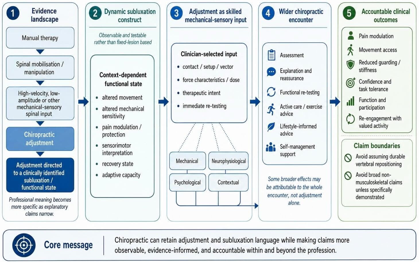
Contemporary Chiropractic adjustment within a dynamic subluxation framework. The figure locates Chiropractic adjustment within the wider manual therapy evidence landscape while distinguishing professional terminology from claim-level accountability. Subluxation is presented as a dynamic, context-dependent functional state involving observable features such as altered movement, mechanical sensitivity, pain modulation, sensorimotor interpretation, recovery state and adaptive capacity. Chiropractic adjustment is framed as a clinician-selected mechanical-sensory input whose effects may involve mechanical, neurophysiological, psychological and contextual pathways. The wider chiropractic encounter, including assessment, explanation, functional re-testing, active care, lifestyle-informed advice and self-management support, may also contribute to patient outcomes. The conceptual relationships shown are grounded in literature on subluxation as a testable construct, WHO adjustment and manipulation terminology, manual therapy mechanisms and contextual effects, spinal manipulative therapy evidence, and evidence boundaries for non-musculoskeletal claims. (3, 4, 7, 8, 15, 16, 23, 24, 25, 31) The framework encourages clinicians and researchers to distinguish adjustment-specific effects from whole-encounter effects and to link claims to observable outcomes such as pain modulation, movement access, reduced guarding, confidence, function, participation and re-engagement with valued activity
Mechanism evidence also supports restraint. Bialosky and colleagues proposed that manual therapy may initiate neurophysiological responses through interaction among mechanical stimulus, peripheral mechanisms, spinal mechanisms, supraspinal mechanisms and contextual factors. (15)
A later modelling paper argued that manual therapy mechanisms should be understood through interacting pathways rather than one isolated biomechanical correction. (16)
Keter and colleagues’ living review reported that manual therapy mechanisms are complex and distributed across body systems and contexts. (23) None of these mechanism models requires a fixed subluxation lesion as the explanatory object.
The anatomical correction claim has particularly limited support. Young and colleagues systematically reviewed immediate anatomical structural or positional changes after manually delivered high-velocity, low-amplitude spinal manipulation. (22)
They found a small and heterogeneous literature and concluded that the most credible evidence concerned immediate changes such as facet joint space or spinal stiffness rather than durable vertebral repositioning. (22) This evidence supports the possibility of short-term mechanical change. It does not support broad claims that adjustment permanently corrects a hidden lesion.
Neurophysiological evidence supports only a limited inference. Haavik and Murphy reviewed work suggesting that spinal manipulation may influence sensorimotor integration and motor control. (26) Haavik-Taylor and Murphy reported immediate somatosensory evoked-potential changes after cervical spine manipulation. (27) Niazi and colleagues reported short-term changes in H-reflexes and V-waves after spinal manipulation. (28) Christiansen and colleagues reported immediate changes in strength and cortical drive after a single session of spinal manipulation in athletes. (29) These studies measured short-term or immediate surrogate neurophysiological outcomes after spinal manipulation rather than patient-important clinical outcomes, independent subluxation diagnosis, objective subluxation correction or retained correction. (26, 27, 28, 29)
A systematic review evaluating whether spinal manipulation affects brain function found low-to-medium methodological quality, inconsistent findings and unknown clinical relevance, and concluded that promoting spinal manipulation as a treatment to improve brain function was premature. (30) Therefore, this literature supports mechanistic plausibility only. (26, 27, 28, 29, 30) It is not evidence that a reliably diagnosed subluxation has been corrected, because the subluxation construct remains insufficiently established as a validated clinical lesion. (3, 4) It is not evidence that correction is retained or that patient-important clinical outcomes necessarily improve, because the cited neurophysiological studies measured short-term surrogate outcomes. (26, 27, 28, 29, 30) It is not evidence that general non-musculoskeletal health effects follow, because the Global Summit review did not support spinal manipulative therapy for selected non-musculoskeletal disorders. (31)
The strongest boundary condition comes from non-musculoskeletal claims. Côté and colleagues, reporting the Global Summit systematic review, found that spinal manipulative therapy was not superior to sham interventions in acceptable or high-quality randomised trials for selected non-musculoskeletal disorders. (31) That evidence challenges the claim that treating spinal dysfunction through manipulation or adjustment produces reliable physiological benefits for organ function or general health. (31) A dynamic subluxation model should therefore avoid using adjustment language to imply broad non-musculoskeletal effects unless such effects are specifically demonstrated. (31) This does not preclude future evidence showing adjustment-specific features or effects. It means the burden rests with Chiropractic researchers and proponents to demonstrate those features rather than to claim them by definition.
A fair conclusion is that adjustment and manipulation should not be conflated simplistically, but neither should they be separated by definition alone. Adjustment may be a profession-specific, clinically targeted and sometimes more refined form of spinal manipulative or mechanical-sensory intervention. (7, 8) Its professional meaning may be distinctive. Its clinical claims should build on the wider spinal manipulation evidence while also encouraging more precise research into Chiropractic procedures, reasoning, dose, force characteristics, outcomes and safety. (15, 16, 23, 24, 25) Anatomical correction claims should remain careful because evidence for immediate structural or positional findings after HVLA manipulation is limited and heterogeneous. (22) Generalised non-musculoskeletal claims should be made only where condition-specific evidence supports them. (31) Table 1 summarises constructive ways to describe the relationship between chiropractic adjustment and spinal manipulation without either collapsing the terms or separating them by definition alone (Table 1).
Chiropractic adjustment as skilled mechanical-sensory input
The proposed alternative is not anti-adjustment. It makes the adjustment more scientifically accountable. A Chiropractic adjustment can be described as a skilled, salient mechanical-sensory input delivered to a person whose response is influenced by tissue state, pain sensitivity, sensorimotor prediction, expectation, confidence and recovery capacity. This framing is consistent with Bialosky's model of manual therapy mechanisms because it allows mechanical input to initiate peripheral, spinal, supraspinal and contextual responses. (15) It is also consistent with the broader living-review evidence that manual therapy mechanisms are multidimensional rather than reducible to one tissue event. (23)
Table 1: Constructive ways to describe the relationship between chiropractic adjustment and spinal manipulation. The wording in this table is informed by WHO terminology, manual therapy mechanism models, spinal manipulative therapy evidence, immediate structural-change evidence and evidence boundaries for non-musculoskeletal claims. (7, 8, 15, 16, 22, 23, 24, 25, 31) Note. SMT = spinal manipulative therapy; HVLA = high-velocity, low-amplitude.
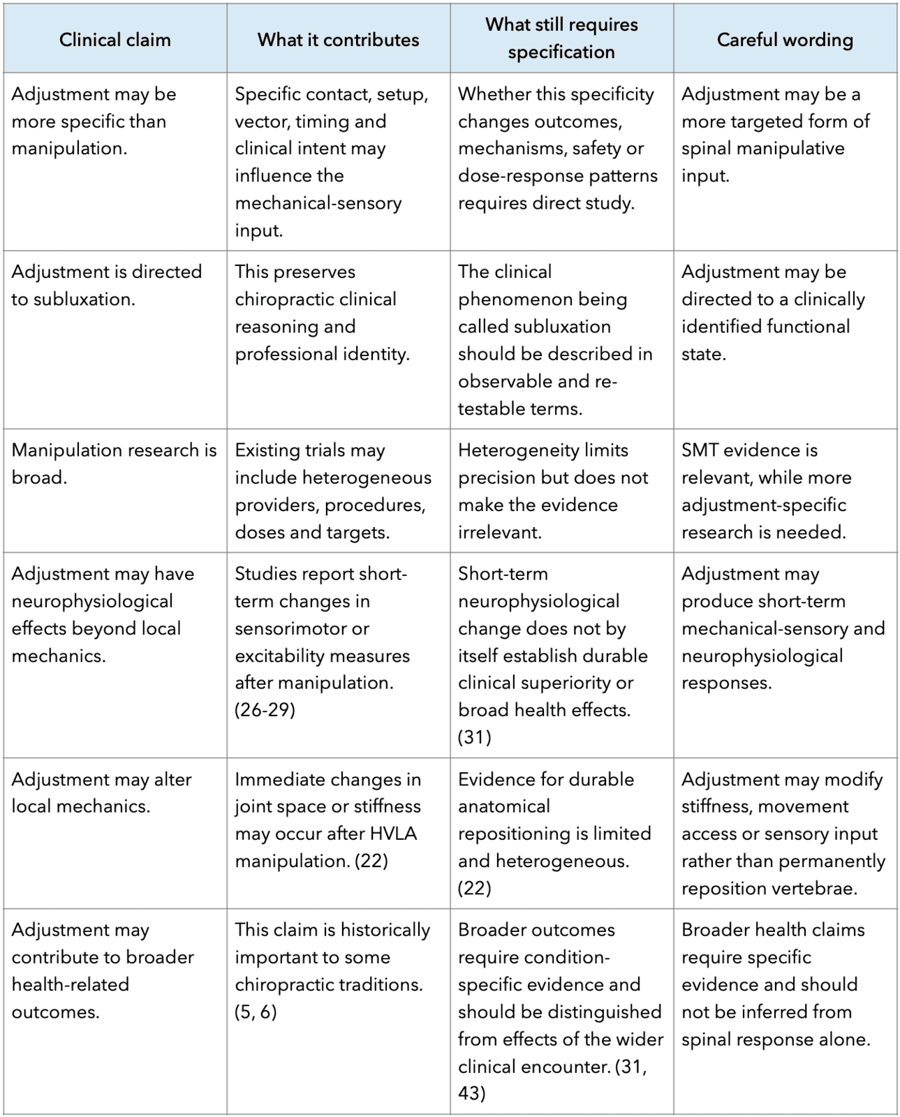
The clinical value of adjustment should therefore be tested through observable change. Relevant outcomes include pain modulation, perceived stiffness, movement access, protective tone, range of motion, confidence, task tolerance, patient-specific function and capacity to re-engage with valued activity. This is consistent with guideline-level emphasis on function, self-management and activity rather than passive correction alone. (2)
A favourable immediate response can be clinically meaningful. It should not be over-interpreted. Immediate improvement after adjustment may indicate altered sensitivity, changed movement willingness, contextual response, expectation effect or reduced guarding. (15, 16, 20) It may also reflect short-term mechanical change. (22) It does not by itself prove that a fixed lesion existed or that it was permanently corrected.
Operational rules for contemporary subluxation language
- Define the clinical phenomenon before naming it. If subluxation is used, the record should specify whether the relevant finding is painful spinal movement, altered mechanical sensitivity, restricted functional range, guarding, impaired proprioceptive control, reduced confidence, poor recovery or reduced tolerance to ordinary demand;
- Match the explanation to the evidence. It is reasonable to say that spinal manual care may provide mechanical-sensory input that can influence pain, movement and sensorimotor responsiveness. (15, 26) It is less defensible to claim that a hidden lesion has been objectively corrected unless the lesion, mechanism and outcome can be independently specified and re-tested; (3, 4)
- Document change in patient-centred terms. The record should include the patient's functional problem, relevant spinal or movement findings, the manual input used, immediate response, and next functional behaviour to test. Implementation science supports the value of clearly specified intervention processes and context when translating complex interventions into practice. (32) Process evaluation guidance similarly emphasises mechanisms, implementation and contextual influences rather than assuming that a complex intervention works through one simple pathway; (32) and
- Communicate in language that supports confidence. The clinician can explain that the spine and nervous system are receiving a precise physical input, that the aim is to reduce protective sensitivity or improve movement access, and that the effect is best retained by tolerable use of the body. This is more consistent with evidence on expectations and fear-avoidance than messages implying that the spine is fragile, unstable or chronically out of place. (20, 21)
Translating the model into practice language
For the framework to influence practice, the dynamic definition must be usable in clinical speech. Table 2 connects scientific terms with practitioner language, observable cues, patient-facing explanations and phrases to avoid. The table is intentionally operational. Its purpose is to prevent subluxation from functioning as a magic word that ends clinical reasoning (Table 2)
Table 2: Translating the dynamic subluxation model into practice language. The examples are informed by evidence on best-practice musculoskeletal care, manual therapy mechanisms, patient expectations, fear-avoidance and the need for observable, testable subluxation language. (2. 3, 4, 15, 16, 20, 21, 23) Note. Patient-facing phrases are illustrative and should be adapted to the patient, presentation, consent process and clinical context.
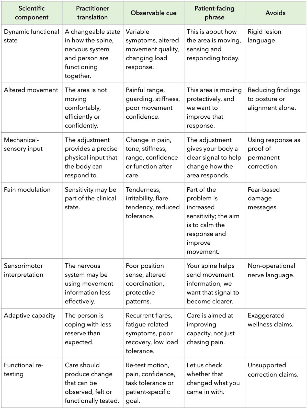
State modifiers: recovery, disuse, prior injury and load change
A dynamic subluxation construct becomes more useful when it includes state modifiers. Patient response to adjustment may be influenced by recent activity exposure, de-conditioning, prior injury, sleep, recovery, confidence, tissue irritability, stress and changes in load. These factors do not need to be treated as separate subluxations, nor as effects uniquely corrected by adjustment. Rather, they are modifiers of the person’s current capacity to tolerate ordinary demands and to respond to mechanical-sensory input. Short-term disuse can rapidly alter muscle and metabolic physiology; Dirks and colleagues found that one week of bed rest produced muscle atrophy and whole-body insulin resistance in healthy young men. (33)
Prior injury is another relevant modifier. Toohey and colleagues found in a systematic review and meta-analysis that previous lower-limb injury was associated with subsequent lower-limb injury in athletic populations. (34) This does not make recurrence inevitable. It supports the clinical need to consider prior exposure, tissue capacity, movement confidence and load tolerance.
Pain trajectories also matter. Low back pain trajectory research supports recurrent and fluctuating patterns rather than a simple acute-chronic dichotomy. (35) These findings support a state-based explanation in which recovery, recurrence and adaptive capacity are monitored over time.
Load change should also be considered. The International Olympic Committee consensus statement emphasised that both under-loading and over-loading can be problematic depending on recovery, capacity and context. (36) In Chiropractic practice, this means adjustment response should be interpreted alongside recent changes in work, sport, sleep, stress and activity exposure.
Sleep is a clinically relevant state modifier. Santos and colleagues found bidirectional associations between chronic musculoskeletal pain and sleep-related problems in a systematic review and meta-analysis. (37) These data support discussing recovery and sleep as modifiers of response without blaming the patient or converting chiropractic into an unsupported lifestyle cure.
Lifestyle medicine, contextual care and the wider chiropractic encounter
Lifestyle medicine should be integrated into this framework at the level of the Chiropractic encounter and longitudinal care model rather than as an intrinsic effect of the adjustment. Lifestyle medicine is commonly organised around evidence-informed behavioural domains including nutrition, physical activity, restorative sleep, stress management, avoidance of risky substances and social connection. (38) Musculoskeletal service redesign literature has likewise argued for patient-centred, team-based and lifestyle-focused care as a foundation for non-operative musculoskeletal management. (39)
This integration is not foreign to Chiropractic. A systematic review of primary prevention in Chiropractic practice found that many Chiropractors report positive attitudes toward prevention and offer some preventive or wellness-oriented services, while also noting that patients rarely consult specifically for primary prevention. (40) A later systematic review of clinician-based surveys found that a large majority of practising chiropractors report engaging with physical activity promotion, although the underlying survey evidence had moderate-to-high risk of bias. (41) The World Federation of Chiropractic (WFC) has also described a public-health agenda in which Chiropractors may contribute to health promotion and prevention initiatives, particularly where these are connected to musculoskeletal health. (42)
Chiropractic branding, however, has often presented the profession through an adjustment-centred public identity, even though everyday Chiropractic practice commonly includes a broader clinical encounter. Pollard has argued that professional identity and branding should better reflect the contemporary chiropractic profile, including the range of populations seen, practice interests, post-professional education, special interest groups and management approaches used in practice. (46) This distinction matters because the public, other health professionals, regulators and healthcare systems may judge Chiropractic by its simplified external message rather than by the fuller range of care that commonly occurs in clinical practice.
The critical distinction is causal attribution. Broader effects of Chiropractic care should be investigated as effects of the whole clinical encounter: assessment, explanation, reassurance, physical activity advice, sleep and recovery discussion, load management, behavioural support, referral and follow-up. They should not be attributed to the adjustment alone unless the adjustment-specific mechanism and outcome have been demonstrated.
This distinction is supported by the systematic review by Goncalves and colleagues, which found no evidence that Chiropractic treatment produced effects in primary or early secondary prevention for disease in general. (43) A modernised Chiropractic model can therefore include active care and lifestyle-informed constructs without converting them into unsupported subluxation-correction or adjustment-specific claims. (43)
A contemporary subluxation framework should therefore avoid reducing Chiropractic to adjustment alone, while also avoiding the opposite error of minimising the adjustment's professional and clinical importance. Chiropractic care may include assessment, explanation, reassurance, skilled touch, adjustment or manipulation, mobilisation, soft tissue care, activity advice, exercise recommendation, self-management support and lifestyle-informed discussion. These elements may interact to influence pain, confidence, movement behaviour, recovery expectations, participation and adaptive capacity.
Some benefits historically attributed to adjustment alone may therefore be more appropriately studied as effects of the wider Chiropractic encounter, within which adjustment remains a distinctive and potentially important mechanical-sensory input.
This distinction also gives a constructive role to contextual factors. Contextual effects are often discussed as though they weaken manual therapy claims or reduce treatment to placebo. A more clinically useful view is that context, when used ethically and transparently, is part of responsible care. Explanation, consent, therapeutic alliance, expectation, touch, confidence-building language, functional re-testing and advice may help patients interpret symptoms less threateningly and engage more successfully with activity. The relevant question is not whether contextual factors are present, because they always are. The question is whether they are used responsibly to support patient capability, self-management and participation rather than dependency.
Testable propositions and limits
A modernised subluxation framework should generate testable propositions. First, adjustment should be studied as a mechanical-sensory input whose effects can be measured through pain, movement access, perceived stiffness, confidence, sensorimotor measures and functional re-testing. This proposition follows directly from manual therapy mechanism models and does not require a fixed-lesion assumption. (15, 16, 23)
Second, the relationship between adjustment and manipulation should be studied rather than asserted. Trials or pragmatic studies would need to prospectively define target selection, contact, force-time characteristics, dose, practitioner expertise, active-care integration and comparator interventions. Without those specifications, claims of clinical or mechanistic distinctiveness remain underdetermined.
Third, subluxation communication should be studied as a clinical behaviour. Records should become clearer if clinicians translate subluxation into observable descriptors and re-testable outcomes. Process-evaluation guidance is suitable for studying how mechanisms, implementation and context influence a complex intervention. (32) Implementation-science frameworks are suitable for examining whether revised clinical language changes documentation, clinical reasoning, patient-centred outcomes and integration with active care or lifestyle-informed advice in practice. (44)
Fourth, limits must be stated plainly. The framework does not deny fracture, infection, inflammatory disease, progressive neurological disease or other conditions requiring medical recognition. It also does not prove that manual therapy mechanisms are specific, uniform or technique-dependent. Similar outcomes across different interventions may arise from overlapping mechanisms, contextual effects or limitations in measurement. (45)
A further limitation is terminological variation. WHO Chiropractic guidance uses both adjustment and manipulation within Chiropractic training and safety documents. (7, 8) Historical Chiropractic technique literature shows that subluxation concepts and technique systems vary across periods and systems. (9, 10) Some Chiropractors reserve ‘adjustment’ for HVLA thrust procedures, whereas others include low-force, instrument-assisted, tonal or response-based procedures within adjustment language. (7, 8, 9, 10) For this reason, any claim about adjustment must specify the procedure, target, dose, comparator and outcome rather than relying on the professional term alone.
Conclusion
The issue with subluxation is not only whether it exists as a discrete entity. The more important problem is that the construct has often named a clinical phenomenon while relying on explanations drawn from early Chiropractic science. A more defensible future is to retain subluxation as a functional clinical construct while requiring it to be observable, explainable, testable and useful.
The relationship between adjustment and manipulation should be handled with the same discipline. Adjustment may be more refined, more clinically intentional and more professionally meaningful than generic manipulation. The broader spinal manipulation literature therefore remains relevant to chiropractic adjustment, while also leaving room for more specific research into chiropractic procedures, clinical reasoning, dosing, force characteristics, active-care integration and patient-centred outcomes.
A contemporary Chiropractic explanation can therefore describe the adjustment as skilled mechanical-sensory input delivered within a dynamic human system and a broader clinical encounter. This preserves skilled manual contact, clinical judgement and chiropractic identity while making the language of subluxation more observable, testable and communicable. The aim is not to diminish Chiropractic, nor to force uniformity across its diverse traditions. It is to provide a shared explanatory platform that can support clinical precision, patient confidence, research development, regulatory clarity and more constructive participation in wider healthcare.
References
- Hartvigsen J, Hancock MJ, Kongsted A,, et al. What low back pain is and why we need to pay attention. Lancet. 2018;391(10137):2356-67.
- Lin I, Wiles L, Waller R, Goucke R, et al. What does best practice care for musculoskeletal pain look like? Eleven consistent recommendations from high-quality clinical practice guidelines: systematic review. Br J Sports Med. 2020;54(2):79-86.
- Keating JC Jr, Charlton KH, Grod JP, et al. Subluxation: dogma or science? Chiropr Osteopat. 2005;13:17.
- Mirtz TA, Morgan L, Wyatt LH, et al. An epidemiological examination of the subluxation construct using Hill's criteria of causation. Chiropr Osteopat. 2009;17:13.
- Palmer DD. The Chiropractor's Adjuster: The Science, Art and Philosophy of Chiropractic. Portland: Portland Printing House; 1910.
- Stephenson RW. Chiropractic Text Book. Davenport: Palmer School of Chiropractic; 1927.
- World Health Organization. WHO guidelines on basic training and safety in chiropractic. Geneva: World Health Organization; 2005.
- World Health Organization. Benchmarks for training in chiropractic. Geneva: World Health Organization; 2022.
- Senzon SA. The chiropractic vertebral subluxation part 1: introduction. J Chiropr Humanit. 2018;25:10-21.
- Senzon SA. The chiropractic vertebral subluxation part 7: technics and models from 1962 to 1980. J Chiropr Humanit. 2019;26:10-27.
- Humphrey JD, Dufresne ER, Schwartz MA. Mechanotransduction and extracellular matrix homeostasis. Nat Rev Mol Cell Biol. 2014;15(12):802-12.
- Martino F, Perestrelo AR, Vinarsky V, Pagliari S, Forte G. Cellular mechanotransduction: from tension to function. Front Physiol. 2018;9:824.
- Proske U, Gandevia SC. The proprioceptive senses: their roles in signaling body shape, body position and movement, and muscle force. Physiol Rev. 2012;92(4):1651-97.
- Wolpert DM, Diedrichsen J, Flanagan JR. Principles of sensorimotor learning. Nat Rev Neurosci. 2011;12(12):739-51.
- Bialosky JE, Bishop MD, Price DD, et al. The mechanisms of manual therapy in the treatment of musculoskeletal pain: a comprehensive model. Man Ther. 2009;14(5):531-8.
- Coronado RA, Bialosky JE. Unraveling the mechanisms of manual therapy: modeling an approach. J Orthop Sports Phys Ther. 2018;48(1):8-18.
- Foster NE, Anema JR, Cherkin D, et al. Prevention and treatment of low back pain: evidence, challenges, and promising directions. Lancet. 2018;391(10137):2368-83.
- Hodges PW, Tucker K. Moving differently in pain: a new theory to explain the adaptation to pain. Pain. 2011;152(3 Suppl):S90-S98.
- Hodges PW, Smeets RJEM. Linking pain and motor control: conceptualization of movement deficits in patients with pain. Phys Ther. 2022;102(4):pzab289.
- Bishop MD, Mintken PE, Bialosky JE, Cleland JA et al. Patient expectations of benefit from interventions for neck pain and resulting influence on outcomes. J Orthop Sports Phys Ther. 2013;43(7):457-465.
- Vlaeyen JWS, Crombez G, Linton SJ. The fear-avoidance model of pain. Pain. 2016;157(8):1588-1589.
- Young KJ, Leboeuf-Yde C, Gorrell L, et al. Mechanisms of manipulation: a systematic review of the literature on immediate anatomical structural or positional changes in response to manually delivered high-velocity, low-amplitude spinal manipulation. Chiropr Man Therap. 2024;32:28.
- Keter DL, Bialosky JE, Brochetti K, et al. The mechanisms of manual therapy: a living review of systematic, narrative, and scoping reviews. PLoS One. 2025;20(3):e0319586.
- Paige NM, Miake-Lye IM, Booth MS, et al. Association of spinal manipulative therapy with clinical benefit and harm for acute low back pain: systematic review and meta-analysis. JAMA. 2017;317(14):1451-1460.
- Rubinstein SM, de Zoete A, van Middelkoop M, et al. Benefits and harms of spinal manipulative therapy for the treatment of chronic low back pain: systematic review and meta-analysis of randomised controlled trials. BMJ. 2019;364:l689.
- Haavik H, Murphy B. The role of spinal manipulation in addressing disordered sensorimotor integration and altered motor control. J Electromyogr Kinesiol. 2012;22(5):768-76.
- Haavik-Taylor H, Murphy B. Cervical spine manipulation alters sensorimotor integration: a somatosensory evoked potential study. Clin Neurophysiol. 2007;118(2):391-402.
- Niazi IK, Turker KS, Flavel S, et al. Changes in H-reflex and V-waves following spinal manipulation. Exp Brain Res. 2015;233(4):1165-73.
- Christiansen TL, Niazi IK, Holt K, et al. The effects of a single session of spinal manipulation on strength and cortical drive in athletes. Eur J Appl Physiol. 2018;118(4):737-49.
- Meyer AL, Amorim MA, Schubert M, et al. Unravelling functional neurology: does spinal manipulation have an effect on the brain? A systematic literature review. Chiropr Man Therap. 2019;27:60.
- Côté P, Hartvigsen J, Axen I, et al. The global summit on the efficacy and effectiveness of spinal manipulative therapy for the prevention and treatment of non-musculoskeletal disorders: a systematic review of the literature. Chiropr Man Therap. 2021;29:8.
- Moore GF, Audrey S, Barker M, et al. Process evaluation of complex interventions: Medical Research Council guidance. BMJ. 2015;350:h1258.
- Dirks ML, Wall BT, van de Valk B, et al. One week of bed rest leads to substantial muscle atrophy and induces whole-body insulin resistance. Diabetes. 2016;65(10):2862-75.
- Toohey LA, Drew MK, Cook JL, et al. Is subsequent lower limb injury associated with previous injury? A systematic review and meta-analysis. Br J Sports Med. 2017;51(23):1670-8.
- Kongsted A, Kent P, Axen I, et al. What have we learned from ten years of trajectory research in low back pain? BMC Musculoskelet Disord. 2016;17:220.
- Soligard T, Schwellnus M, Alonso JM, et al. How much is too much? International Olympic Committee consensus statement on load in sport and risk of injury. Br J Sports Med. 2016;50(17):1030-41.
- Santos M, Gabani FL, de Andrade SM, et al. The bidirectional association between chronic musculoskeletal pain and sleep-related problems: a systematic review and meta-analysis. Rheumatology. 2023;62(9):2951-62.
- Lippman D, Stump M, Veazey E, et al. Foundations of lifestyle medicine and its evolution. Mayo Clin Proc Innov Qual Outcomes. 2024;8(1):97-111.
- Artz KE, Phillips TD, Moore JM, et al. Redesigning the care of musculoskeletal conditions with lifestyle medicine. Mayo Clin Proc Innov Qual Outcomes. 2024;8(5):418-30.
- Goncalves G, Le Scanff C, Leboeuf-Yde C. Primary prevention in chiropractic practice: a systematic review. Chiropr Man Therap. 2017;25:9.
- Fernandez M, Young A, Milton K, et al. Physical activity promotion in chiropractic: a systematic review of clinician-based surveys. Chiropr Man Therap. 2022;30:55.
- Maiers MJ, Agaoglu M, Brown R, et al. Chiropractic in global health and wellbeing: a white paper describing the public health agenda of the World Federation of Chiropractic. Chiropr Man Therap. 2018;26:26.
- Goncalves G, Le Scanff C, Leboeuf-Yde C. Effect of chiropractic treatment on primary or early secondary prevention: a systematic review with a pedagogic approach. Chiropr Man Therap. 2018;26:10.
- Damschroder LJ, Reardon CM, Widerquist MAO, et al. The updated Consolidated Framework for Implementation Research based on user feedback. Implement Sci. 2022;17:75.
- Cook CE, George SZ, Keefe F. Different interventions, same outcomes? Here are four good reasons. Br J Sports Med. 2018;52(15):951-2.
- Pollard H. Does chiropractic branding reflect current chiropractic practice? Chiropr J Aust. 2022;49:1-11.
- Good CJ. The great subluxation debate: a centrist's perspective. J Chiropr Humanit. 2010;17(1):33-9.
Clinical practice
AU 1: Gilbert Weiner DC, LST, FFCLB. Adjunct Assistant Professor, Adjunct Research Faculty Northeast College of Health Sciences; Private Practice, Bellingham MA . e: drweiner@hotmail.com
AU2: Charles L Blum DC. Director of Research, Sacro Occipital Technique Organization - USA. Private practice of Chiropractic, Santa Monica, CA
Purpose: To provide a qualitative report using peer-reviewed published articles, reports, and contemporary textbooks Allopathic, Osteopathic, and Chiropractic to demonstrate the neurological aetiology of VMR. Documenting its systemic repercussions especially as it concerns cerebral spinal fluid flow and the benefit that Chiropractic care offers.
Methods: A Narrative Analysis was undertaken of the literature expanding outwards from accepted definitions of vasomotor rhinitis to identify peer reviewed published studies. The studies concern linking VMR to its neurological aetiology and structural consequences. We will also link that aetiology to other health conditions demonstrating comorbidities and further documenting the autonomic nervous system (ANS) affect in various bodily functions. There will be discussion of the fascial and neurological patterns, the disruptions of those patterns and their ramifications in postural, spinal distortions, and cerebral spinal fluid (CSF) flow. Considering the neurochemicals and neurotransmitters, we will delve into the functions and dysfunctions many times where we find the factors that connect certain pathologies. This enables us to change our perspective from merely looking at a myriad of separate diseases, to seeing that so many have the common underlying causes and possible common resolutions.
Conclusions:VMR is more than nasal congestion. The mechanisms that provoke and maintain this “stuffed nose” collaterally produce problems that are far more detrimental to one’s health. The appearance of VMR should be considered a prodromal symptom and should alert us to examine further to observe what other health problems are simultaneously occurring. Chiropractic appears to offer an effective avenue for evaluation and treatment via ANS dysfunction or dysautonomia. Chiropractic presents a treatment with no secondary effects for VMR and the concomitant related cranial dysfunction. Further study is recommended.
Cite: Weiner G, Blum CL. Vasomotor Rhinitis (VMR) and Cerebral Spinal Fluid Flow: A Chiropractic issue. Asia-Pac Chiropr J. 2026;7.3 https://www.apcj.site/WeinerRhinitis.pdf
Take-away: '… Chiropractic offers a targeted specific resolution to many organic system malfunctions and with the minimal collateral symptomatology, side effects, or complications ….'
Introduction
Vasomotor rhinitis (VMR) is a nonallergic, non-infection condition which even the allopathic literature states is triggered by autonomic nervous system dysfunction. This occurs when nerves in the nasal mucosa overreact to environmental stimuli like temperature changes, smells, ambient airborne hydrocarbons or esters, stress, or other environmental elements.
It is characterised by an ANS imbalance typically hyperactive parasympathetic system, leading to increased blood flow (vasodilation) and excessive, clear mucus secretion, chronic nasal congestion, runny nose (rhinorrhea), and postnasal drip. This has been managed with nasal sprays and trigger avoidance. (1, 2, 3) The most common prescription is Ipratropium Bromide (Atrovent®) a prescription anticholinergic medication. However, introducing an anticholinergic, or any medication, into the nasal mucosa is a double-edged sword.
The nasal mucosa is a formidable vehicle for access to the blood stream, i.e. Narcan®. So, an anticholinergic can cause systemic reactions as well such as; swelling of the eyes, face, lips, tongue, throat, hands, feet, ankles, or lower legs, hoarseness, difficulty breathing or swallowing. Other warnings concerning this drug’s literature include prostate and bladder neck blockage. Label warnings state that patients with enlarged prostate, Narrow angle Glaucoma should ‘Use with caution’. The product literature also states that the drug may make these conditions worse: Kidney disease, Liver disease, stating ‘Use with caution’. However, might there be a safer avenue for therapeutic action, one with minimal adverse effects?
Background
VMR is the most common form of nonallergic rhinitis, comprising approximately 71% of all nonallergic rhinitis conditions. It is estimated that 14 million Americans suffer from vasomotor rhinitis, with a worldwide prevalence approaching 320 million. The most recent report being the report of the 2008 AAAAI Joint Task Force on Practice Parameters, The diagnosis and management of rhinitis: an updated practice parameter, which defines VMR (idiopathic rhinitis)(4) and states ‘that patients with vasomotor rhinitis (VMR) often suffer from concomitant gastric distress, particularly irritable bowel syndrome (IBS) or gastroesophageal reflux disease (GERD)’. Studies show that over 70% of VMR patients may have IBS, and 67% of IBS patients experience VMR, suggesting a strong etiological link between these problems and autonomic nervous system imbalances. (5, 6) This connection is suspected to be through the same ANS imbalance which affects the nasal mucosa.
Otorhinolaryngologic (ENT)ramifications of VMR have been found to be sinus headache, facial pressure, postnasal drip, coughing, and throat clearing are common symptoms shared among both allergic and nonallergic rhinitis. However, Vasomotor rhinitis (nonallergic rhinitis) can cause non-ENT symptoms like ‘brain fog’ or cognitive difficulties, including confusion, trouble concentrating, and fatigue. (7) This in addition to the GI symptoms already mentioned.
Patients with vasomotor rhinitis tend to be categorised into two groups depending on predominant symptomatology: ‘blockers’ with congestion and ‘runners’ with rhinorrhea. Patients with rhinorrhea tend to have an enhanced cholinergic (parasympathetic) response. Those with nasal obstruction tend to have nociceptive neurons with a heightened response to normally harmless stimuli. (8) Nociceptive neurons with a heightened response to harmless stimuli (a phenomenon known as allodynia) are primarily associated with the sympathetic nervous system. (9)
Neuroceptors
The receptors primarily associated with the sympathetic nervous system in these cases are Transient Receptor Potential Vanilloid 1(TRPV1) It is essential to look at the biochemical changes that occur as that is what will illuminate the etiological commonality in health conditions. These TRPV1 receptors are often acting as an upstream controller of sympathetic activity. They act as nociceptors (pain sensors) on sensory neurons, and their activation can trigger sympathetic responses, including increased heart rate, blood pressure, and thermogenesis (heat production). (10, 11, 12)
TRPV1 receptors are non-selective cation channels that function as molecular sensors for noxious stimuli, including temperatures above 43°C (109°F), acidic conditions, and chemical irritants like capsaicin in chilli peppers, that is what makes the nose water as you eat them. They are primarily found on peripheral nociceptive neurons and play a crucial role in pain sensation and modulation. Those patients with TRPV1 over-expression are those patients who often have an increased density of TRPV1 (capsaicin receptors) on their nasal sensory nerves. Patients with Nerve Growth Factors (NGF) issues demonstrate Inflammatory mediators that are known to up-regulate TRPV1 synthesis in sensory neurons and increase the transport of these receptors to nerve endings. People with altered Nerve Growth Factor (NGF) levels often experience conditions related to neurodegeneration, chronic pain, or mental health disorders, as NGF is critical for the survival, growth, and maintenance of neurons. It acts as a signalling protein, and its dysregulation has been connected to conditions like Alzheimer's disease, schizophrenia, and neuropathic pain. Environmental exposure complications are observed where there is frequent exposure to pollutants, cigarette smoke, strong odours, or ozone. This in turn can lead to increased TRPV1 expression, contributing to chronic airway hyper-responsiveness. (13)
There is also a Reduced Activation Threshold (RAT) in these patients. When normally innocuous stimuli, such as a drop in temperature or a strong perfume, which under normal circumstances do not activate normal neurons, will activate these hypersensitive ‘pain-sensing’ neurons in the presence of RAT. What is seen also is something called Antidromic Neuropeptide Release. A process where action potentials travel backward along sensory nerves from the axon branches toward the peripheral terminal, rather than toward the spinal cord. This triggers the local release of neuropeptides. This mechanism drives neurogenic inflammation, causing vasodilation, plasma leakage, and immune cell activation. (14, 15)
They also release neuropeptides like Substance P (SP) and Calcitonin Gene-Related Peptide (CGRP) directly into the nasal tissue. Calcitonin Gene-Related Peptide, is a 37-amino acid neuropeptide that acts as a potent vasodilator and neurotransmitter, playing a key role in pain signalling and neurogenic inflammation. It is widely distributed in both the central and peripheral nervous systems, with elevated levels heavily linked to the activation of the trigeminovascular system in migraine headaches. (16) This demonstrates a clinically established link between VMR and migraine that needs to be explored in more detail. Vasomotor reactivity, the capacity of cerebral blood vessels to adapt to changes in pressure or CO2 levels, is frequently altered during migraine attacks. Migraines cause complex hemodynamic dysregulation and share a well-documented connection with both vestibular symptoms and certain types of vasomotor rhinitis. (17, 18) Another problem well documented is Neurogenic Oedema. When these neuropeptides cause rapid vasodilation (blood vessel widening) and increased vascular permeability it has found to lead to the ‘boggy’” swollen nasal lining typical of VMR. (19, 20, 21, 22, 23, 24)
Pathophysiology
The pathophysiology of nonallergic rhinitis (VMR) is complex, with still much to be discovered. As we search for aetiology it is observed that it appears to be firmly based on the imbalance between parasympathetic and sympathetic stimuli on the nasal mucosa. (25, 26) It is continually observed that mucous secretion is regulated primarily by the parasympathetic nervous system, whereas the sympathetic nervous system controls vascular tone. (27, 28)
There have been studies investigating transient receptor potential (TRP) cation channels that emerged as important players in type 2 upper airway inflammatory disorders; those being chronic inflammatory conditions driven by a specific immune response (involving interleukins 4, 5, and 13) that often coexist and affect the nose, sinuses, and throat. Key disorders include chronic rhino-sinusitis with nasal polyps (CRSwNP), allergic rhinitis, and eosinophilic laryngitis. These frequently present with congestion, facial pain, loss of smell, and runny nose. (29, 30) Clinically, Maspero’s study found that 100% of patients with rhinitis demonstrate this ANS dysfunction pattern, as well as 55% to 70% of severe asthmatics. Multiple clinical studies demonstrate these same observations in ANS dysfunction. (31, 32, 33)
ANS
In the Parasympathetic nervous system (PNS) Acetylcholine (ACh) is a primary stimulant of mucus production and secretion, particularly in the airways, acting through parasympathetic nerves and non-neuronal sources. It binds to muscarinic receptors mainly M3 on submucosal glands and surface epithelial cells, inducing rapid secretion and increasing mucus gland activity. (34) Acetylcholine acts through muscarinic receptor signalling, an increase in intracellular calcium Ca2, and causes the fusion of secretory granules with the cell membrane. This leads to rapid mucus exocytosis. The excessive mucus secretion in the airway can at times produce symptoms as severe as chronic obstructive pulmonary disease (COPD) and asthma when left unchecked. (35)
In addition to muscarinic receptors, Alpha 7 nicotinic acetylcholine receptors (nAChRs) are critical in mediating airway mucus production, potentially playing a role in mucus metaplasia (an increase in mucus-producing cells). In chronicity one finds that beyond nerve release, inflammatory cells and airway epithelial cells can produce acetylcholine on their own, providing an autocrine/paracrine mechanism for mucus production during inflammation. This local reaction feeds into the chronic loop of nasal congestion. (36, 37)
The sympathetic nervous system (SNS) in general promotes decrease in overall nasal mucus secretion primarily by causing vasoconstriction of nasal blood vessels, which reduces tissue swelling and congestion. While it can stimulate mucin output from submucosal glands via adrenergic receptors in some instances, its dominant effect is that of reducing congestion and drying the mucosa. (38, 39, 40) Sympathetic effects on mucus production can be seen as vasoconstriction (Decongestion). Sympathetic nerves release norepinephrine, which constricts blood vessels (especially in the nose), reducing interstitial oedema and decreasing the volume of the nasal mucosa. In contrast to the parasympathetic nervous system, which causes heavy, runny mucus production, sympathetic stimulation results in less but often thicker mucus, allowing for better airflow.
The Alpha-adrenoceptor agonists (which mimic sympathetic nervous system activation) can decrease the frequency of the mucociliary wave. (41, 42, 43, 44) The mucociliary wave, or wave-like motion of cilia, is the coordinated beating movement of microscopic hair-like structures (cilia) throughout the respiratory tract. These cilia act as a ‘metachronal wave’, a sweeping motion that acts to move mucus, pathogens, and inhaled particles upward to the throat to be cleared out. In essence, the mucociliary wave is the primary innate defence mechanism on the lung. (45) The repercussions of disrupting the mucociliary wave mechanism is that it is not only the respiratory system that utilises this wave motion. The ciliary wave mechanism is also utilised in the spinal canal for the movement of CSF. (46, 47) The sympathetic nervous system acts as a natural decongestant both mechanical and ANS.
In the Parasympathetic nervous system (PNS) Acetylcholine (ACh) is a primary stimulant of mucus production and secretion, particularly in the airways, acting through parasympathetic nerves and non-neuronal sources. It binds to muscarinic receptors mainly M3 on submucosal glands and surface epithelial cells, inducing rapid secretion and increasing mucus gland activity. (48) Acetylcholine acts through muscarinic receptor signalling, an increase in intracellular calcium Ca2, and causes the fusion of secretory granules with the cell membrane. This leads to rapid mucus exocytosis. The excessive mucus secretion in the airway can at times produce symptoms as severe as chronic obstructive pulmonary disease (COPD) and asthma when left unchecked. (49)
In addition to muscarinic receptors, Alpha 7 nicotinic acetylcholine receptors (nAChRs) are critical in mediating airway mucus production, potentially playing a role in mucus metaplasia (an increase in mucus-producing cells). In chronicity one finds that beyond nerve release, inflammatory cells and airway epithelial cells can produce acetylcholine on their own, providing an autocrine/paracrine mechanism for mucus production during inflammation. This local reaction feeds into the chronic loop of nasal congestion. (50, 51)
The sympathetic nervous system (SNS) in general promotes decrease in overall nasal mucus secretion primarily by causing vasoconstriction of nasal blood vessels, which reduces tissue swelling and congestion. While it can stimulate mucin output from submucosal glands via adrenergic receptors in some instances, its dominant effect is that of reducing congestion and drying the mucosa. (52, 53, 54) Sympathetic effects on mucus production can be seen as vasoconstriction (Decongestion). Sympathetic nerves release norepinephrine, which constricts blood vessels (especially in the nose), reducing interstitial oedema and decreasing the volume of the nasal mucosa. In contrast to the parasympathetic nervous system, which causes heavy, runny mucus production, sympathetic stimulation results in less but often thicker mucus, allowing for better airflow.
The Alpha-adrenoceptor agonists (which mimic sympathetic nervous system activation) can decrease the frequency of the mucociliary wave. (55, 56, 57, 58) The mucociliary wave, or wave-like motion of cilia, is the coordinated beating movement of microscopic hair-like structures (cilia) throughout the respiratory tract. These cilia act as a ‘metachronal wave’, a sweeping motion that acts to move mucus, pathogens, and inhaled particles upward to the throat to be cleared out. In essence, the mucociliary wave is the primary innate defence mechanism on the lung. (59) The repercussions of disrupting the mucociliary wave mechanism is that it is not only the respiratory system that utilises this wave motion. The ciliary wave mechanism is also utilised in the spinal canal for the movement of CSF. (60, 61) The sympathetic nervous system acts as a natural decongestant both mechanical and significant. (62) This demonstrates the connection between VMR, HRV, and other ANS dysfunction conditions.
Figure 1: ANS. Source and copyright 2019 Dysautonomia International (DI)
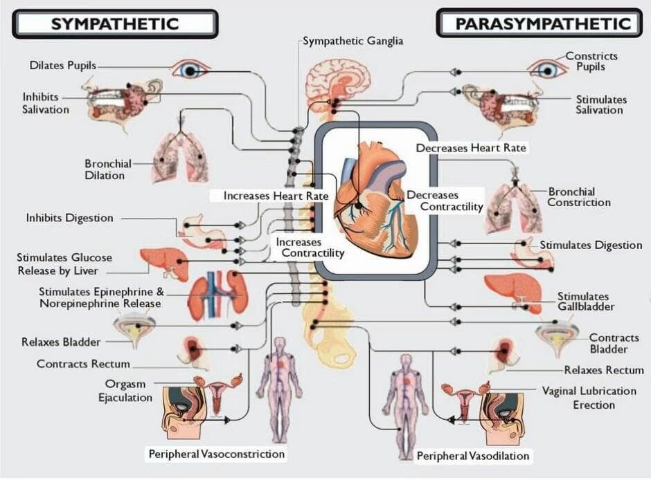
The latest studies using a cardiovascular testing laboratory convincingly demonstrated that asthmatics with gastro-oesophageal reflux demonstrate evidence of autonomic dysfunction and that these patients tended to have VMR. This testing methodology has also been used to study various disorders such as small fibre neuropathy, orthostatic imbalance or syncope, and degenerative dysautonomia such as Shy-Drager Syndrome. (63) We can now understand that ANS dysfunction exists as a spectrum of conditions. This spectrum might have simple pain and VMR on the lower end with such diseases as Shy-Drager syndrome (SDS) at the extreme high end. In this extreme case we observed an ANS imbalance causing severe organ and system collapse. This brings new appreciation for ANS–function connection. (64, 65, 66)
Structure
Considering components involved with neurological dysfunction it is interesting what else has been documented in Allopathic literature. It has been found that Vasomotor rhinitis (VMR) (nonallergic rhinitis) can occur following a cervical spine injury causing dysfunction of the autonomic nervous system that controls the nasal mucosa. (67, 68) Harlor stated that his study found ‘a traumatic injury to the neck can damage the sympathetic nerves, leading to an imbalance where the parasympathetic system becomes overactive, with depression of the sympathetic system resulting in symptoms like chronic, watery, or mucoid nasal discharge’. (69 ,70) Petersen’s study found that ‘the great majority of patients who suffer from chronic sinus problems have lost the normal curve in their neck and have spondylotic changes in the cervical spine’. Petersen documented that 83.9% of chronic rhinitis patients in the study reported neck pain prior to onset of sinus congestion and pain and presented loss of cervical lordosis.
‘Neck pain and cervical musculoskeletal dysfunction are common among persons with sinus congestion and may be a comorbid feature or contributing factor to headaches attributed to rhino-sinusitis’. (71)
While plain sinus X-rays or computer tomography of the sinus show little to no pathology in cases of vasomotor rhinitis, cervical spine X-rays in fact do. ‘Vasomotor rhinitis and cervical curve degeneration (such as the loss of cervical lordosis) are linked to mechanical stress or instability in the upper neck that irritate the cervical sympathetic chain and/or vagus nerve, disrupting autonomic control and triggering nasal congestion or rhinorrhea’. (72 )The cervical spine injuries can interrupt the sympathetic nerve pathways, leading to parasympathetic overactivity in the nasal cavity. (73)
Dicpinigaitis et al reported in their study concerning cervical spinal injury that bronchial hyperresponsiveness observed in the test population most likely reflects the loss of sympathetic airway innervation and resultant unopposed cholinergic bronchoconstrictor tone. ‘Blockade of methacholine hyperresponsiveness with ipratropium bromide® suggests a muscarinic receptor-mediated phenomenon’. (74) In a clinical report there was documentation of a patient developing persistent left-sided rhinorrhea for up to 1.5 years after a motor vehicle accident that fractured the left carotid canal, injuring the sympathetic plexus. (75)
Misalignments (subluxations) in the upper cervical spine can cause irritation to the sympathetic nervous system, which in turn causes the sinus membranes to produce excessive mucus. This condition manifests as chronic, watery nose running (rhinorrhea) and nasal congestion that often gets worse with temperature changes, stress, or even bending over. (76, 77) It appears that structurally when the neck curvature is lost it creates a posture where tensile stress on the spinal cord influences the brainstem where the 7th Cranial nerve nuclei is located. In response to the loss in curve of the neck, also the suboccipital muscles located at the back of the head become spastic interfering with sympathetic flow. (78)
Neurology
Discussing the underlying neurology, in the case of the sinus membranes, these tissues are parasympathetically controlled by the 7th Cranial nerve, better known as the facial nerve. The sympathetic innervation originates from the upper thoracic spinal cord (T1-T4), ascends the cervical sympathetic chain, and synapses in the superior cervical ganglion. Post-ganglionic fibres then form the deep petrosal nerve, which passes through the Vidian nerve (nerve of the pterygoid canal) to the pterygopalatine ganglion to regulate blood flow and reduce nasal congestion. (79)
Observing the ascending fibres, they travel up the cervical sympathetic trunk to the superior cervical ganglion (located at C2-C3 level). They then synapse in the superior cervical ganglion which acts as the source for sympathetic nerves to the head. There the post-ganglionic fibres form a plexus around the internal carotid artery (Carotid Plexus). Those fibres then leave the plexus as the deep petrosal nerve. The deep petrosal nerve joins the greater petrosal nerve (parasympathetic) to form the Vidian nerve (mixed nerve) (nerve of the Pterygoid canal).
The trigeminal network and the pterygopalatine ganglion (located adjacent to the pterygoid canal) share pathways. As a result, hypertension in the pterygoid muscles can trigger referred pain or autonomic symptoms (such as sinus pressure, nasal congestion, or eye tearing) that mimic Vidian nerve irritation. This can be caused by temporomandibular imbalance. (80, 81, 82) These fibres then pass through the canal where the nerve can be compromised before it reaches the pterygopalatine ganglion. There it continues without synapsing and distributes throughout the nasal cavity to supply blood vessels. The superior alveolar (anterior, middle, and posterior) nerves, branches of the maxillary nerve provide sensory innervation. This system works in opposition to the parasympathetic system, which causes vasodilation and increased secretion. (83)
As we stated, there may be local stimulation in the chronic stage. Sensory neuropeptides and nociceptive type C fibres of the trigeminal nerve contribute to mast cell degranulation and stimulate the itching/sneezing reflexes. (84) Mast cell degranulation causes itching and sneezing due to rapid immune process where mast cells release inflammatory mediators, most notably histamine which is stored in cytoplasmic granules. Triggered by allergens (via IgE antibodies), infections, or physical stimuli, this process causes immediate allergic reactions, inflammation, and symptoms like itching, flushing, and vascular changes. (85) There is also neurogenic dysfunction when the condition involves a malfunction of nerve signals between the brain and nasal tissue, which is sometimes linked to broader autonomic disorders. (86)
There is another mechanism which merits mentioning as it causes brainstem and spinal cord irritation, the myodural bridge. (87) (Figure 2) The myodural bridge (MDB) functions as a mechanical pump that facilitates the circulation and drainage of cerebrospinal fluid (CSF), which directly impacts the clearance of metabolic waste through the cribriform plate. (88) By connecting the suboccipital muscles to the spinal dura mater, the MDB transmits head movements into pressure changes that drive fluid toward major exit routes like the nasal lymphatics. (89)
Figure 2: ANS. Myodural bridge
The myodural bridge. Sagittal section of the myodural bridge and its connections to the dura and spine. The posterior atlantooccipital membrane (1) extends from the occiput and coalesces with the dura mater at the cerebrospinal junction. The superior myodural bridge (2) merges with the superior vertebrodural ligament (3) of the atlas and fuses with the PAOM at the level of the atlantooccipital interspace. The inferior myodural bridge comprised of the rectus capitis posterior major fascia (5a) and obliquus capitis inferior fascia (5b) courses between the atlantoaxial ligamentum flavum (4) as bundles of dense fibers. The inferior myodural bridge fuses with the PAOM. The nuchal bridge (6) merges with the inferior vertebrodural bridge (7) and attaches to the PAOM. The PAOM terminates at the level of C3 after this transition point (*). The dura mater (DM) continues as an independent structure after that. O, occiput; C1, atlas; C2, axis; C3, third cervical vertebra; NL, nuchal ligament; PAOM, posterior atlantooccipital membrane. Adapted from Scali et al. .Source: Scali F, Ohno A, Enix D, Hassan S. The Posterior Atlantooccipital Membrane: The Anchor for the Myodural Bridge and Meningovertebral Structures. Cureus. 2022;14(5):e25484. Published May 30, 2022. DOI 10.7759/cureus.25484
Glymphatic system and CSF
There is an intimate connection between the nasal lymphatics, congestion via vasomotor rhinitis, foggy brain and dull mental function that so many VMR patients complain of. This brain fog or mental confusion has always been attributed to lack of adequate oxygen to the brain due to respiratory issues. However, a new study utilising neuro-imaging biomarkers such as the diffusion tensor imaging along the perivascular space (DTI-ALPS) index and choroid plexus volume (CPV) has described glymphatic congestion as the aetiology. (90) This connection is now explained via the Glymphatic System which we will see is intertwined with nasal health. (91) The glymphatic system clears metabolic waste (such as amyloid beta) from the brain, moving them into the cervical lymph nodes and to peripheral drainage sites. A major route for this CSF outflow is alongside olfactory nerves as they pass through the cribriform plate into the nasal cavity. (Figure 3)
Figure 3: CSF flow and the cribriform plate
You can edit text on your website by double clicking on a text box on your website. Alternatively, when you select a text box a settings menu will appear. your website by double clicking on a text box on your website. Alternatively, when you select a text box
When there exists a naso-lymphatic dysfunction studies show that this drainage pathway is blocked or impaired, it can lead to, or be associated with, chronic rhinitis and olfactory dysfunction. When the CSF cannot drain efficiently through the nasal mucosa (due to obstruction or congestion), it can impair the entire brain's clearance system, potentially contributing to further neuro-inflammation (, , , , , ). Phillips states that, ‘nasal turbinate vasodilation and resultant blood pooling lead to the obstruction of normal nasal lymphatic clearance of cerebrospinal fluid and its waste products from the brain’. (98) The cribriform plate is a vital, sieve-like subcomponent of the ethmoid bone that forms this horizontal boundary. The ethmoid is directly related to the Vomer and the sphenoid bone. In SOT, motion dysfunction of the sphenoid is paramount, and the ethmoid moves in conjunction with the anterior aspect of the sphenoid. (99) (Figure 4) Whedon et al demonstrated that physiologic factors affecting the normal circulation of CSF include cardiovascular, respiratory, and vasomotor influences. Physiologic impedance or cessation of CSF flow may occur commonly in the absence of degenerative changes or pathology and may compromise the normal physiologic functions of the CSF. Their paper found that therapies directed at affecting CSF flow include osteopathic care (especially cranial manipulation), Sacro Occipital Technique, chiropractic adjustment of the spine and cranium, were all successful in restoration of CSF flow. (100, 101)
Considering the glymphatic system, attention must be given to Alpha 7 nicotinic acetylcholine receptors (alpha7nAChR) which we mentioned in relation to VMR, as we observe their affects here as well. The connection with the glymphatic system via neuro-immune regulation is noted through alpha7nAChR activation which reduces neuro-inflammation and maintains the astrocytic function that is so critical for cerebrospinal fluid (CSF) flow and amyloid-beta clearance. By regulating microglia and astrocytes, alpha7nAChR signalling directly supports the efficiency of the glymphatic system in clearing waste from the brain. (98)
The importance of the glymphatic system as a ‘pseudo-lymphatic’ network in the brain is that it flushes out metabolic waste, such as amyloid beta, primarily during deep sleep. The term glymphatic system was only introduced in 2012 to describe a glia-dependent, perivascular clearance pathway in the brain. And yet, everything it refers to, including perivascular spaces, astrocyte end-feet, cerebrospinal fluid flow, and interstitial clearance, was already well documented. ‘They were just not interconnected … this new label did something powerful. It brought anatomy, physiology, sleep biology, and neurodegeneration into a single framework, “Channelopathy” … suddenly, CSF clearance is not just a plumbing issue but a potential key factor affecting health and disease’.
Helbig further stated that ‘neurodegeneration research reframed protein accumulation as a drainage problem’. Ion channels do not only set membrane potentials. They also influence development, synaptic release, dendritic integration, and network wiring. (Ingo Helbig neurologist and epilepsy genetics researcher working at the Children’s Hospital of Philadelphia (CHOP), USA)
Figure 4: In SOT, motion dysfunction of the sphenoid is paramount, and the ethmoid moves in conjunction with the anterior aspect of the sphenoid.
SOT / Chiropractic
These statements by. Helbig has opened a new window for therapies that can affect CSF flow. In dealing with the glymphatic system we recall that the waste-filled fluid exits the brain and must pass through the meningeal lymphatic vessels at the base of the skull before entering the cervical lymph nodes in the neck. (99, 100, 101) The ‘Neck Bottleneck’ of poor posture causes tension, physical obstruction in the cervical region which will impair this outflow, potentially contributing to ‘brain fog’ or neurodegenerative decline.
SOT Therapy, as low-force mobilisation of the cranial sutures and suboccipital region (base of the skull), is used to assist the glymphatic-lymphatic continuum. (102) SOT and Chiropractic can affect this entire system as documented by various studies concerning its effect on HRV and hence validating the Chiropractic - ANS -Organ system connection. (103, 104, 105) We know that HRV is an indicator of sympathetic – parasympathetic balance. We further know that the mechanism of VMR is ANS sympathetic-parasympathetic imbalance. Finally, we have shown here that VMR is intimately related to failure of lymphatic and glymphatic drainage and other forms of neurogenic inflammation which are responsible for causative factors in many disease conditions. A case report by Bloink and Blum in the Journal of Chiropractic Education (2020) explored the use of Sacro Occipital Technique (SOT) and cranial procedures for a patient with dysautonomia suspected to be caused by vagus nerve entrapment at the jugular foramen. The intervention aimed to alleviate structural tension between the occiput and temporal bones, it resulted with the patients demonstrating improvement and reduction of the dysautonomia. (106, 107)
In SOT, cranial manipulation exerts an effect on the pterygopalatine plates, part of the sphenoid bone's pterygoid process. This serves as a crucial anatomical anchor and spatial landmarks for the pterygoid muscles of mastication. (Figure 5)
The lateral pterygoid plate, in particular, provides the major site of origin for both the medial and lateral pterygoid muscles. (108) Their primary mechanical effect on these plates is the exertion of continuous tensile and traction forces during mastication, which can influence bone remodelling, temporomandibular joint biomechanics, and long-term skeletal disorders and effect the Vidian nerve (109) Chiropractic and Osteopathic cranial manipulative medicine view the sphenoid bone as the keystone of the cranium. Again, the Vidian nerve passes through the pterygoid canal in the sphenoid body. Chiropractic and Osteopathic theory suggest that restricted mobility or spatial strains of the sphenoid can compress the nerve, potentially impairing autonomic function. (110) Upper Cervical Alignment concentrating on C1 (atlas) and C2 (axis) vertebrae can also ensure the major circulation pathways for brain detox are not restricted. (111, 112) This was shown in various study with asymptomatic control groups.
Figure 5: Cranial relationships of relevance to SOT.
As we place all the focus on the cranium and upper spine we must remember CSF produced in the brain flows through the subarachnoid space and down the spinal canal to the lumbosacral area. Pressure management such as from obstructions in the spinal column, i.e. spinal stenosis, tumors such as Schwannomas, (113) or mechanical tension (subluxation), can impair this flow, leading to metabolic waste buildup. The Sacral Pump utilised in SOT models, acts as a ‘pump’ for CSF; rhythmic sacral movement during breathing helps drive fluid upward toward the brain and downward for drainage. Sphenoidal movement with respiration provides a similar pumping mechanism. The sphenoid and occipital bones flex and extend at the sphenobasilar junction. (114, 115, 116) (Figure#6)
This continuous rocking action acts as a mechanical pump to force CSF down the spine. Rationale and research have been presented in various papers to explain the basis behind Dural Port Therapy. Blum states that ‘This Sacro-Occipital Technique (SOT) procedure, which uses the sacrum as a lever to influence and balance the spine and cranium by way of the meningeal system, is an excellent method of affecting the craniospinal system and its relationship to the meninges’. (117, 118) Mechanical Influence is obvious where manipulating the lumbosacral segments can relieve mechanical ‘traction’ on the dura mater, potentially reducing ‘metabolic tension’ in the brain's drainage pathways. (119, 120, 121) The therapeutic role of lumbosacral manipulation for meningeal normalisation (122) will aid in glymphatic drainage helping to restore mobility, targeting restricted (hypomobile) segments in the lumbar spine, like L4/L5. (123)
This will normalise the mechanical activity of the spinal cord. There will also be a symbiotic effect with C2 slope unloading the pressure at the brainstem through various fascial and reflexive avenues. (124) Therapeutically SOT enhances CSF outflow even during upright posture and movement. It enhances CSF outflow in the lumbosacral region via gravitational adaptations. SOT also reduces dural tension, addresses sacral trauma or lumbar stiffness and can transmit positive mechanical changes through the ‘fascial continuum’ to the base of the skull enhancing the siphoning effect. It has been documented that Manual therapies are observed to create a ‘system-wide siphoning effect’ that helps move fluids through the brain and body. (125) This has a huge effect on VMR and glymphatic drainage.
The prestigious Cleveland Clinic recommends Chiropractic (SOT) including self-massage, acupressure, and techniques targeting the vomer bone, that can provide immediate, drug-free relief for nasal congestion by promoting sinus drainage, reducing fascial pressure, and relaxing tense muscles often bringing relief within seconds to minutes. (Cleveland Clinic Health-Sinus) (126)
Figure 6: Source, Blum. (116)
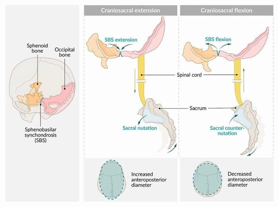
Continuing to examine the connections we observe that the sphenoid articulates with the occiput at the sphenobasilar synchondrosis (SBS). During inhalation, the SBS exhibits an upward movement (flexion), and during exhalation, it moves downward (extension). (127) (Figure 6) In the Flexion Phase (Inhalation), The sphenoid body rotates such that the anterior portion moves superiorly and anteriorly, while the greater wings move inferiorly and anteriorly. The midline of the cranium widens. While in the Extension Phase (Exhalation): The movement reverses. The sphenoid rotates in the opposite direction, and the midline narrows. (128) The sphenoid moves in conjunction with the Vomer and Ethmoid bones. As the sphenoid rotates during inhalation, the ethmoid rotates in the opposite direction to maintain balanced tension on the reciprocal tension membranes (such as the falx cerebri) and promote proper intercranial CSF fluid motion. (129) So, we can see how normalising motion of the occiput and upper spine can contribute to proper CSF flow.
Conclusion
Through this report we believe we have demonstrated the effects that Chiropractic can have on organ function in general and on vasomotor rhinitis in particular. In correcting VMR and restoring normal nasal drainage we have demonstrated the concomitant effect that will have on glymphatic drainage and hence, CSF flow. The information we shared was from a broad spectrum of therapeutic sciences all pointing to an overall aetiology of autonomic nervous system dysfunction, neurogenic inflammation and those biochemical changes that they cause in the human body. We have demonstrated that chiropractic can offer a targeted specific resolution to many organic system malfunctions and with the minimal collateral symptomatology, side effects, or complications. It is the hope of the authors that further clinical studies are performed to complement our conclusions.
References
1. Aldawas, G. and I. N. Bhat (2025). "Vasomotor Rhinitis: Current Concepts and Emerging Therapies." Cureus 17(10): e95631
2. Pattanaik, D. and P. Lieberman (2010). "Vasomotor rhinitis." Curr Allergy Asthma Rep 10(2): 84-91.
3. Geiger (2026). Nonallergic Rhinitis (Vasomotor Rhinitis). StatPearls. Treasure Island (FL).
4. Settipane, R. A. (2009). "Epidemiology of vasomotor rhinitis." World Allergy Organ J 2(6): 115-118
5. Jaradeh, S. S., et al. (2000). "Autonomic nervous system evaluation of patients with vasomotor rhinitis." Laryngoscope 110(11): 1828-31.
6. Lauriello M, Eibenstein A, Angelone AM, et al. Association between vasomotor rhinitis and irritable bowel syndrome. Allergy Rhinol (Providence). 2016 Jan 1;7(4):249-255. DOI 10.2500/ar.2016.7.0184. PMID: 28683253; PMCID: PMC5244286
7. Geiger (2026). Nonallergic Rhinitis (Vasomotor Rhinitis). StatPearls. Treasure Island (FL).
8. ibid
9. Cui X, Zhang Z, Xi H, Liu K, Zhu B, Gao X. Sympathetic-Sensory Coupling as a Potential Mechanism for Acupoints Sensitization. J Pain Res. 2023 Aug 30;16:2997-3004. DOI 10.2147/JPR.S424841. PMID: 37667684; PMCID: PMC10475306.
10. Phan, T. X., et al. (2020). "TRPV1 expressed throughout the arterial circulation regulates vasoconstriction and blood pressure." J Physiol 598(24): 5639-5659
11. Hudson, A. S., et al. (2016). "Involvement of the TRPV1 channel in the modulation of spontaneous locomotor activity, physical performance and physical exercise-induced physiological responses." Braz J Med Biol Res 49(6): e5183
12. Mohammed, M., et al. (2018). "Activation of TRPV1 in nucleus tractus solitarius reduces brown adipose tissue thermogenesis, arterial pressure, and heart rate." Am J Physiol Regul Integr Comp Physiol 315(1): R134-R143
13. Van Gerven, L., et al. (2014). "Capsaicin treatment reduces nasal hyperreactivity and transient receptor potential cation channel subfamily V, receptor 1 (TRPV1) overexpression in patients with idiopathic rhinitis." J Allergy Clin Immunol 133(5): 1332-1339, 1339 e1331-1333
14. Sorkin, L. S., et al. (2018). "Origins of antidromic activity in sensory afferent fibers and neurogenic inflammation." Semin Immunopathol 40(3): 237-247
15. Szolcsanyi, J. (1988). "Antidromic vasodilatation and neurogenic inflammation." Agents Actions 23(1-2): 4-11.
16. Russell FA, King R, Smillie SJ, Kodji X, Brain SD. Calcitonin gene-related peptide: physiology and pathophysiology. Physiol Rev. 2014 Oct;94(4):1099-142. doi: 10.1152/physrev.00034.2013. PMID: 25287861; PMCID: PMC4187032
17. Ozturk, B. and O. Karadas (2020). "Cerebral Hemodynamic Changes During Migraine Attacks and After Triptan Treatments." Noro Psikiyatr Ars 57(3): 192-196
18. Liu, Y., et al. (2021). "Association between migraine and vasomotor rhinitis." International Forum of Allergy & Rhinology 11: 1378 - 1380
19. Backaert W, Steelant B, Hellings PW, Talavera K, Van Gerven L. A TRiP Through the Roles of Transient Receptor Potential Cation Channels in Type 2 Upper Airway Inflammation. Curr Allergy Asthma Rep. 2021 Mar 18;21(3):20. doi: 10.1007/s11882-020-00981-x. PMID: 33738577; PMCID: PMC7973410
20. Leader P, Geiger Z. Vasomotor rhinitis. StatPearls. Updated July 10, 2023. Accessed March 5, 2026. https://www.ncbi.nlm.nih.gov/books/NBK547704/
21. Van Gerven, L., et al. (2014). "Capsaicin treatment reduces nasal hyperreactivity and transient receptor potential cation channel subfamily V, receptor 1 (TRPV1) overexpression in patients with idiopathic rhinitis." J Allergy Clin Immunol 133(5): 1332-1339, 1339 e1331-1333
22. Dykewicz, M. S., et al. (2020). "Rhinitis 2020: A practice parameter update." J Allergy Clin Immunol 146(4): 721-767
23. Baraniuk, J. N. (1992). "Sensory, parasympathetic, and sympathetic neural influences in the nasal mucosa." J Allergy Clin Immunol 90(6 Pt 2): 1045-1050
24. Lindh, Elfvin, L. G., et al. (1993). "The chemical neuroanatomy of sympathetic ganglia." Annu Rev Neurosci 16: 471-507
25. Yan, C. H. and P. H. Hwang (2018). "Surgical Management of Nonallergic Rhinitis." Otolaryngol Clin North Am 51(5): 945-955
26. Safira,D. (2023) Vasomotor Rhinitis: Etiology, Prevalence, Risk Factors, and Management.MEDULA ( Medical Professiom Jour. Of Lampung.Vol. 13 No. 4.1 (2023): Medula - Special Edition (Special Sense)
27. Geiger (2026). Nonallergic Rhinitis (Vasomotor Rhinitis). StatPearls. Treasure Island (FL).
28. Preston, E. and C. Heath (1972). "Depression of the vasomotor system in rabbits poisoned with an organophosphate anticholinesterase." Arch Int Pharmacodyn Ther 200(2): 245-54.
29. Maspero, J., et al. (2022). "Type 2 inflammation in asthma and other airway diseases." ERJ Open Research 8(3): 00576-02021
30. Backaert W, Steelant B, Hellings PW, Talavera K, Van Gerven L. A TRiP Through the Roles of Transient Receptor Potential Cation Channels in Type 2 Upper Airway Inflammation. Curr Allergy Asthma Rep. 2021 Mar 18;21(3):20. doi: 10.1007/s11882-020-00981-x. PMID: 33738577; PMCID: PMC7973410
31. Sánchez-Manso JC, Gujarathi R, Varacallo MA. Autonomic Dysfunction. [Updated 2023 Aug 4]. In: StatPearls [Internet]. Treasure Island (FL): StatPearls Publishing; 2026 Jan-. Available from: https://www.ncbi.nlm.nih.gov/books/NBK430888
32. Chen D, Zhou Z, Zhou Z, Zhao L, Chen Y. Autonomic nervous system dysfunction in chronic obstructive pulmonary disease: mechanistic insights and therapeutic implications. Ann Med. 2025 Dec;57(1):2596426. DOI 10.1080/07853890.2025.2596426. Epub 2025 Dec 7. PMID: 41355150; PMCID: PMC12687893.
33. Hamrefors V. The Autonomous Nervous System: A Novel and Potentially Modifiable Risk Factor for Chronic Obstructive Pulmonary Disease in the Population? Ann Am Thorac Soc. 2023 Oct;20(10):1402-1403. DOI 10.1513/AnnalsATS.202307-618ED. PMID: 37772941; PMCID: PMC10559132.
34. Kistemaker, L. E. and R. Gosens (2015). "Acetylcholine beyond bronchoconstriction: roles in inflammation and remodelling." Trends Pharmacol Sci 36(3): 164-71.
35. Shah, B. K., et al. (2023). "Mucus Hypersecretion in Chronic Obstructive Pulmonary Disease and Its Treatment." Mediators Inflamm 2023: 8840594
36. Fatemikia, H., et al. (2022). "Roles of alpha-7 nicotinic acetylcholine receptors and spleen in the lung injury induced by a repeated saline lavage in rat." BMC Pulm Med 22(1): 367.
37. Williams, O. W., et al. (2006). "Airway mucus: From production to secretion." Am J Respir Cell Mol Biol 34(5): 527-36.
38. Baraniuk, J. N. (1992). "Sensory, parasympathetic, and sympathetic neural influences in the nasal mucosa." J Allergy Clin Immunol 90(6 Pt 2): 1045-50.
39. Pack, R. J., et al. (1988). "The functional significance of the sympathetic innervation of mucous glands in the bronchi of man." J Physiol 403: 211-19.
40. Gluckman, Jack L. and Stegmoyer, Robert. Nonallergic Rhinitis. In: Paparella, Michael M., Shumrick, Donald A., Meyerhoff, William, eds., Otolaryngology, Volume III, Head and Neck. W. B. Saunders Co., 1991, pp.1889-98.
41. Baraniuk, J. N. (1992). "Sensory, parasympathetic, and sympathetic neural influences in the nasal mucosa." J Allergy Clin Immunol 90(6 Pt 2): 1045-50.
42. Nadel, J. A., et al. (1979). "Control of mucus secretion and ion transport in airways." Annu Rev Physiol 41: 369-81.
43. Mercke, U., et al. (1982). "Parasympathetic and sympathetic influences on mucociliary activity in vivo." Rhinology 20(4): 201-4.
44. Pack, R. J., et al. (1988). "The functional significance of the sympathetic innervation of mucous glands in the bronchi of man." J Physiol 403: 211-19.
45. Bustamante-Marin, X. M. and L. E. Ostrowski (2017). "Cilia and Mucociliary Clearance." Cold Spring Harb Perspect Biol 9(4).
46. Grimes, D. T., et al. (2016). "Zebrafish models of idiopathic scoliosis link cerebrospinal fluid flow defects to spine curvature." Science 352(6291): 1341-4.
47. Weiner G, Blum C. Genetic aetiology of adolescent idiopathic scoliosis (AIS): Chiropractic’s role. Asia-Pac Chiropr J. 2022;3.1 apcj.net/papers-issue-3-1/#WeinerAIScoliosis
48. Kistemaker, L. E. and R. Gosens (2015). "Acetylcholine beyond bronchoconstriction: roles in inflammation and remodeling." Trends Pharmacol Sci 36(3): 164-71.
49. Shah, B. K., et al. (2023). "Mucus Hypersecretion in Chronic Obstructive Pulmonary Disease and Its Treatment." Mediators Inflamm 2023: 8840594
50. Fatemikia, H., et al. (2022). "Roles of alpha-7 nicotinic acetylcholine receptors and spleen in the lung injury induced by a repeated saline lavage in rat." BMC Pulm Med 22(1): 367
51. Williams, O. W., et al. (2006). "Airway mucus: From production to secretion." Am J Respir Cell Mol Biol 34(5): 527-36.
52. Baraniuk, J. N. (1992). "Sensory, parasympathetic, and sympathetic neural influences in the nasal mucosa." J Allergy Clin Immunol 90(6 Pt 2): 1045-50.
53. Pack, R. J., et al. (1988). "The functional significance of the sympathetic innervation of mucous glands in the bronchi of man." J Physiol 403: 211-9.
54. Gluckman, Jack L. and Stegmoyer, Robert. Nonallergic Rhinitis. In: Paparella, Michael M., Shumrick, Donald A., Meyerhoff, William, eds., Otolaryngology, Volume III, Head and Neck. W. B. Saunders Co., 1991, pp.1889-98.
55. Baraniuk, J. N. (1992). "Sensory, parasympathetic, and sympathetic neural influences in the nasal mucosa." J Allergy Clin Immunol 90(6 Pt 2): 1045-50.
56.. Nadel, J. A., et al. (1979). "Control of mucus secretion and ion transport in airways." Annu Rev Physiol 41: 369-81.
57. Mercke, U., et al. (1982). "Parasympathetic and sympathetic influences on mucociliary activity in vivo." Rhinology 20(4): 201-4.
58. Pack, R. J., et al. (1988). "The functional significance of the sympathetic innervation of mucous glands in the bronchi of man." J Physiol 403: 211-9.
59. Bustamante-Marin, X. M. and L. E. Ostrowski (2017). "Cilia and Mucociliary Clearance." Cold Spring Harb Perspect Biol 9(4).
60. Grimes, D. T., et al. (2016). "Zebrafish models of idiopathic scoliosis link cerebrospinal fluid flow defects to spine curvature." Science 352(6291): 1341-4.
61. Weiner G, Blum C. Genetic aetiology of adolescent idiopathic scoliosis (AIS): Chiropractic’s role. Asia-Pac Chiropr J. 2022;3.1 apcj.net/papers-issue-3-1/#WeinerAIScoliosis
62. Vayisoglu, Y., et al. (2006). "Autonomic nervous system evaluation using heart rate variability parameters in vasomotor rhinitis patients." J Otolaryngol 35(5): 338-42.
63. Fealey, R. D., et al. (1989). "Thermoregulatory sweating abnormalities in diabetes mellitus." Mayo Clin Proc 64(6): 617-28.
64. Freeman, R., et al. (1990). "Autonomic function and human immunodeficiency virus infection." Neurology 40(4): 575-80.
65. Suarez, G. A., et al. (1994). "Idiopathic autonomic neuropathy: clinical, neurophysiologic, and follow-up studies on 27 patients." Neurology 44(9): 1675-82.
66. Conrad, L. A., et al. (2021). "Increased Heart Rate Variability Response Among Infants with Reported Rhinorrhea and Watery Eyes: A Pilot Study." J Asthma Allergy 14: 1349-54.
67. Scarupa, M. D. and M. A. Kaliner (2009). "Nonallergic rhinitis, with a focus on vasomotor rhinitis: clinical importance, differential diagnosis, and effective treatment recommendations." World Allergy Organ J 2(3): 20-5.
68. Sarin, S., et al. (2006). "The role of the nervous system in rhinitis." J Allergy Clin Immunol 118(5): 999-1016.
69. Harlor, E. J., et al. (2012). "Traumatic unilateral vasomotor rhinitis." Ear Nose Throat J 91(11): E4-6
70. Guimarães,R. (2003) Vasomotor rhinitis post-operative: differential diagnosis of cerebral spinal fluid rhinorrhea. Brazilian J. of Otorynolaryngology Vol. 69 pg. 252-255 Ed. 2- (16º)
71. Petersen, S. M., et al. (2019). "Self-reported sinus headaches are associated with neck pain and cervical musculoskeletal dysfunction: a preliminary observational case control study." J Man Manip Ther 27(4): 245-52.
72. Hauser RA, Matias D, Rawlings BR. Cervicovagopathy: ligamentous cervical instability and dysstructure as a potential etiology for vagus nerve dysfunction in the cause of human symptoms and diseases. Front Neurol. 2025 Jul 2;16:1572863. DOI 10.3389/fneur.2025.1572863. PMID: 40672458; PMCID: PMC12263383.
73. Petersen, S. M., et al. (2019). "Self-reported sinus headaches are associated with neck pain and cervical musculoskeletal dysfunction: a preliminary observational case control study." J Man Manip Ther 27(4): 245-52.
74. Dicpinigaitis, P. V., et al. (1994). "Bronchial hyperresponsiveness after cervical spinal cord injury." Chest 105(4): 1073-6.
75. Harlor, E. J., et al. (2012). "Traumatic unilateral vasomotor rhinitis." Ear Nose Throat J 91(11): E4-6. Vasomotor rhinitis (VMR) is a commonly encountered entity that may be difficult to diagnose
76. Dicpinigaitis, P. V., et al. (1994). "Bronchial hyperresponsiveness after cervical spinal cord injury." Chest 105(4): 1073-6.
77. Guimarães,R.(2003) Vasomotor rhinitis post-operative: differential diagnosis of cerebral spinal fluid rhinorrhea. Brazilian J. of Otorynolaryngology Vol. 69 pg. 252-255 Ed. 2 - (16º)
78. Petersen, S. M., et al. (2019). "Self-reported sinus headaches are associated with neck pain and cervical musculoskeletal dysfunction: a preliminary observational case control study." J Man Manip Ther 27(4): 245-52.
79. Sobiesk, J. L. and S. Munakomi (2026). Anatomy, Head and Neck, Nasal Cavity. StatPearls. Treasure Island (FL).
80. Chen, J. and J. Xiao (2015). "Morphological study of the pterygoid canal with high-resolution CT." Int J Clin Exp Med 8(6): 9484-90.
81. Shao, Y. X., et al. (2012). "Microsurgical anatomy of the greater superficial petrosal nerve." World Neurosurg 77(1): 172-82.
82. Lundy, J. A. and T. McNary (2026). Neuroanatomy, Pterygopalatine Ganglion. StatPearls. Treasure Island (FL).
83. Beule, A. G. (2010). "Physiology and pathophysiology of respiratory mucosa of the nose and the paranasal sinuses." GMS Curr Top Otorhinolaryngol Head Neck Surg 9: Doc07
84. Geiger (2026). Nonallergic Rhinitis (Vasomotor Rhinitis). StatPearls. Treasure Island (FL).
85. Li, D., et al. (2025). "Mast cell-neuron axis as a core mechanism in chronic pruritus of atopic dermatitis: from mechanistic insights to therapeutic targets." Front Immunol 16: 1645095
86. Xiong L, Leung TWH. Autonomic dysfunction in neurological disorders. Aging (Albany NY). 2019 Apr 9;11(7):1903-1904. doi: 10.18632/aging.101896. PMID: 30969941; PMCID: PMC6503877.
87. Weiner G. The Leg Length Check and its importance to the complete practice of Chiropractic. Asia-Pac Chiropr J. 2026;6.4. www.apcj.net/papers-issue-6-4/#WeinerDerifield
88. Xu, Q., et al. (2021). "Head-nodding: a driving force for the circulation of cerebrospinal fluid." Sci Rep 11(1): 14233.
89. Yang, H.(2023) The relationship between myodural bridge, atrophy and hyperplasia of the suboccipital musculature, and cerebrospinal fluid dynamics. Sci Rep. Nov 2;13:18882. DOI 10.1038/s41598-023-45820-x
90. Jia, L., et al. (2025). "The glymphatic system in neurodegenerative diseases and brain tumors: mechanistic insights, biomarker advances, and therapeutic opportunities." Acta Neuropathol Commun 14(1): 19.
91. Phillips WT, Schwartz JG. Nasal lymphatic obstruction of CSF drainage as a possible cause of Alzheimer's disease and dementia. Front Aging Neurosci. 2024 Oct 21;16:1482255. DOI 10.3389/fnagi.2024.1482255. PMID: 39497786; PMCID: PMC11532075.
92. Chae, J., et al. (2024). "The nasal lymphatic route of CSF outflow: implications for neurodegenerative disease diagnosis and monitoring." Anim Cells Syst (Seoul) 28(1): 45-54.
93. Goodman, B. The Glymphatic System and its Potential Role in Sjögren's. The Glymphatic System and its Potential Role in Sjögren's | Sjögren’s Foundation
94. Ding Z, Fan X, Zhang Y, Yao M, Wang G, Dong Y, Liu J, Song W. The glymphatic system: a new perspective on brain diseases. Front Aging Neurosci. 2023 Jun 15;15:1179988. DOI 10.3389/fnagi.2023.1179988. PMID: 37396658; PMCID: PMC10308198.
95. Rasmussen,M. (2022). Fluid transport in the brain. Physiol Rev Apr 1;102(2):1025-1151. DOI 10.1152/physrev.00031.2020. Epub 2021 May 5
96. Xue, Y., et al. (2022). "Sustained glymphatic transport and impaired drainage to the nasal cavity observed in multiciliated cell ciliopathies with hydrocephalus." Fluids Barriers CNS 19(1): 20
97. Plog, B. A. and M. Nedergaard (2018). "The Glymphatic System in Central Nervous System Health and Disease: Past, Present, and Future." Annu Rev Pathol 13: 379-94.
98. Conejero-Goldberg C, Davies P, Ulloa L. Alpha7 nicotinic acetylcholine receptor: a link between inflammation and neurodegeneration. Neurosci Biobehav Rev. 2008;32(4):693-706. DOI 10.1016/j.neubiorev.2007.10.007. Epub 2007 Nov 28. PMID: 18180036; PMCID: PMC2895566.
99. Reddy OC, van der Werf YD. The Sleeping Brain: Harnessing the Power of the Glymphatic System through Lifestyle Choices. Brain Sci. 2020 Nov 17;10(11):868. DOI 10.3390/brainsci10110868. PMID: 33212927; PMCID: PMC7698404.
100. Plog, B. A. and M. Nedergaard (2018). "The Glymphatic System in Central Nervous System Health and Disease: Past, Present, and Future." Annu Rev Pathol 13: 379-94.
101. Chachaj, A., et al. (2023). "The Lymphatic System In The Brain Clearance Mechanisms - New Therapeutic Perspectives For Alzheimer's Disease." Curr Neuropharmacol 21(2): 380-91.
102. Upledger, J. E. (1995). "Craniosacral therapy." Phys Ther 75(4): 328-30.
103. Benner C. Effects of sacro occipital technique (SOT) chiropractic and cranial therapy on dizziness in three patients 68, 72, and 75-years-old: A case series [Abstract]. Asia-Pac Chiropr J. 2025;6.2. www.apcj.net/SOT-Abstracts-2025/#BennerCaseSeries
104. Steinberg B, Clodgo-Gorden K, Postlethwaite R, McIvor C. Improved Heart Rate Variability, mood, reproductive health and gut function, and decreased eczema symptoms in a 30-year-old female under chiropractic care: A case report. Asia-Pac Chiropr J. 2023;4.2. apcj.net/papers-issue-4-2/#SteinbergWell-Being
105. Luu J, Atkinson B, Postlethwaite R, McIvor C. Restored heart function and reduced recovery time in a 36-year-old female patient following a Pulmonary Embolism and induced coma: A case report. Asia-Pac Chiropr J. 2024;4.4. apcj.net/papers-issue-4-4/#LuuPE
106. emeta, Esther & Blum, Charles. (2012). Chiropractic Sacro Occipital Technique (SOT) and Cranial Treatment Model for Traumatic Brain Injury Along with Monitoring and Supplementing for Neurotransmitter Balance: A Case Report. Journal of Head Trauma Rehabilitation. 27. E34-E34
107. Park J, Benitez L, Hamanaka A, Abbas GH, Faluade E, Pouwels S, Eller J. Optimizing Airway Function Through Craniofacial and Cervical Manipulations and Emergency-Anesthesia Maneuvers: Applications in Airway Function Enhancement, Pneumonia, and Asthma-Narrative Review. J Clin Med. 2025 Jun 25;14(13):4494. DOI 10.3390/jcm14134494. PMID: 40648867; PMCID: PMC12249762
108. Cappello ZJ, Arbor TC, Potts KL. Anatomy, Pterygopalatine Fossa. [Updated 2024 Apr 20]. In: StatPearls [Internet]. Treasure Island (FL): StatPearls Publishing; 2026 Jan-. Available from: https://www.ncbi.nlm.nih.gov/books/NBK513269
109. Guerrero,M. (2015) Can pterygoid plate asymmetry be linked to temporomandibular joint disorders? Imaging Sci Dent, Jun 19;45(2):89–94. DOI 10.5624/isd.2015.45.2.89
110. Bordoni, B. and R. E. Sina (2026). Osteopathic Principles and Practices: 5 Diaphragm Procedure. StatPearls. Treasure Island (FL).
111. Welch A., Boone R. Sympathetic and parasympathetic responses to specific diversified adjustment to chiropractic vertebral subluxations of the cervical and thoracic spine. J Chiropr Med. 2008;7(3):86–93. DOI 10.1016/j.jcm.2008.04.001.
112. Win, N. N., et al. (2015). "Effects of Upper and Lower Cervical Spinal Manipulative Therapy on Blood Pressure and Heart Rate Variability in Volunteers and Patients With Neck Pain: A Randomized Controlled, Cross-Over, Preliminary Study." J Chiropr Med 14(1): 1-9.
113. Bayazit, Y. A., et al. (2009). "Management of cerebrospinal fluid leak following retrosigmoid posterior cranial fossa surgery." ORL J Otorhinolaryngol Relat Spec 71(6): 329-33.
114. Koren,T.(2006) The Sphenoid Pattern. The American Chiropractor May1.
115. Blum, CL, "Biodynamics of the Cranium: A Survey," The Journal of Craniomandibular Practice, Mar/May 1985: 3(2):, 164-71.
116. Blum, CL, Curl, DD, "The Relationship Between Sacro-occipital Technique and Sphenobasilar Balance. Part One: the Key Continuities," Chiropractic Technique, Aug 1998, Vol. 10, No. 3.
117. Farmer JA, Blum CL. Dural port therapy. J Chiropr Med. 2002 Spring;1(2):54-61. DOI 10.1016/S0899-3467(07)60003-2. PMID: 19674561; PMCID: PMC2646921
118. Bordoni, B. and A. R. Escher (2023). "Rethinking the Origin of the Primary Respiratory Mechanism." Cureus 15(10): e46527.
119. Hou, Y., et al. (2026). "The spinal lymphatic system: an emerging pathway bridging fluid homeostasis, immunity, and disease." Bone Res 14(1).
120. Lee, D. S., et al. (2020). "Brain Glymphatic/Lymphatic Imaging by MRI and PET." Nucl Med Mol Imaging 54(5): 207-23.
121. Bordoni, B. and R. E. Sina (2026). Osteopathic Principles and Practices: 5 Diaphragm Procedure. StatPearls. Treasure Island (FL).
122. Pick M. Immediate changes to lumbosacral dural regions upon simultaneous bilateral compression to the parietooccipital (asterion) cranial region: A dissection study. Asia-Pac Chiropr J. 2022;31. www.apcj.net/papers-issue-3-1/#PickDural
123. Waqqar, S., et al. (2016). "McKenzie treatment versus mulligan sustained natural apophyseal glides for chronic mechanical low back pain." Pak J Med Sci 32(2): 476-9.
124. Weiner,G., Blum,C. (2026) Is the C2 tilt a reliable indicator of decompensated spinopelvic alignment: A Sacro Occipital Technique perspective SOT RESEARCH CONFERENCE-2026.
125. Hou, Y., et al. (2026). "The spinal lymphatic system: an emerging pathway bridging fluid homeostasis, immunity, and disease." Bone Res 14(1).
126. Omar, B (2020) Non-Allergic Rhinitis with Osteopathic Treatment Techniques. Osteopathic Family Physician | Volume 12, No. 2 | March/April, 2020
127. Childs B, Hammes C. Dr. William Garner Sutherland (1873-1954): The Founder of Cranial Osteopathy. Cureus. 2025 Feb 7;17(2):e78705. DOI10.7759/cureus.78705. PMID: 40062177; PMCID: PMC11890225.
128. Park J, Benitez L, Hamanaka A, Abbas GH, Faluade E, Pouwels S, Eller J. Optimizing Airway Function Through Craniofacial and Cervical Manipulations and Emergency-Anesthesia Maneuvers: Applications in Airway Function Enhancement, Pneumonia, and Asthma-Narrative Review. J Clin Med. 2025 Jun 25;14(13):4494. DOI 10.3390/jcm14134494. PMID: 40648867; PMCID: PMC12249762.
129. Childs B, Hammes C. Dr. William Garner Sutherland (1873-1954): The Founder of Cranial Osteopathy. Cureus. 2025 Feb 7;17(2):e78705. DOI 10.7759/cureus.78705. PMID: 40062177; PMCID: PMC11890225.
AU: Charles L Blum DC. Director of Research, Sacro Occipital Technique Organization - USA. Private practice of Chiropractic, Santa Monica, CA. e: drcblum@aol.com
Narrative: Healthy aging depends on sustained physical activity that preserves function, independence, and quality of life. This narrative review examines principles for developing individualised exercise programmes integrating aerobic activity, resistance training, flexibility, and balance. Drawing on published literature and clinical considerations,
I describe the cardiovascular, metabolic, cognitive, and musculoskeletal benefits of regular exercise, including maintenance of muscle mass, bone health, joint mobility, and capacity for activities of daily living. Particular emphasis is placed on recognising asymmetries in strength and flexibility and modifying exercise to address individual limitations rather than applying uniform routines. Obesity and generalised joint hypermobility illustrate the need to adjust exercise priorities: aerobic activity supports metabolic health and weight management, whereas hypermobility warrants greater attention to strength, neuromuscular control, and joint stability. Gradual progression, sustained participation, varied activities, and cautious stretching are advocated to support safe adaptation.
Periodic reassessment enables programmes to evolve with changing health status and functional goals. An individualised, adaptable approach to exercise offers a practical strategy for maintaining resilience and promoting healthy ageing throughout life.
Indexing terms: Chiropractic; aging; exercise; health; fitness.
Cite: Blum CL. Developing an exercise program for healthy aging. Asia-Pac Chiropr J. 2026;7.3 https://www.apcj.site/BlumHealthyAging.pdf
Introduction
Developing a healthy lifestyle is important for healthy aging. The choices we make today will affect our health, capacity to function, and ability to enjoy our lives as we age. While there are many aspects of a healthy lifestyle (not smoking cigarettes, having a healthy weight, limiting alcohol intake, partaking in a healthy diet and lifestyle, etc.) this article will focus on one of the most important of these, exercise. (1)
Biological aging takes place regardless of what we do or don’t do, however we can minimise the biological affects of aging with regular exercise. Regular exercise can ‘increase active life expectancy by limiting the development and progression of chronic disease and disabling conditions’. (2) There is also emerging evidence for significant psychological and cognitive benefits accruing from regular exercise participation by older adults. Ideally, exercise prescription should include aerobic exercise, muscle-strengthening exercises, flexibility exercises, and balance training. For the purposes of this article, activities of daily living (ADLs) refer to essential tasks such as walking, climbing stairs, dressing, bathing, carrying objects, and maintaining balance during everyday activities.
Together, these forms of exercise help preserve cardiovascular health, muscular strength, mobility, and functional independence throughout the aging process. Being physically active in younger years translates into a higher likelihood of being both physically and mentally healthy as we age into our 80’s, 90’s, and even into our 100’s. Ultimately this means as we age our quality of life and activities of daily living do not have to precipitously decline. (3, 4, 5, 6)
There are four important factors when considering the development of a healthy exercise lifestyle, and these include:
- Performing aerobic, muscle strengthening, flexibility, and balance exercises;
- Developing symmetry in muscle use and function-strength and flexibility;
- Understanding your body type and what proportion of aerobic, strengthening, flexibility, and balance exercises should be performed based on individual strengths and weaknesses; and
- Periodically reassessing your exercise program as your body, health status, and goals change over time.
Performing aerobic, muscle strengthening, and flexibility exercises
The evidence supports that participation in regular physical activity (both aerobic and strength exercises) elicits a number of favourable responses that contribute to healthy aging. Ideally the benefits associated with regular exercise and physical activity contributes to a healthier independent lifestyle, greatly improving the functional capacity and quality of our lives. (7) It is fairly evident that cardiovascular fitness is associated with decreases in cardiovascular disease risk. (8, 9) Along those lines, research demonstrates that regular physical activity can help reduce the cognitive decline associated with aging. (4, 6) More and more studies are supporting the notion that there is ‘a strong biological basis for the role of aerobic fitness in maintaining and enhancing central nervous system health and cognitive functioning in older adults’. (5)
Key takeaways for aerobic exercise
- Supports cardiovascular health and endothelial function (the ability of blood vessels to dilate and function properly);
- Helps maintain cognitive function and brain health;
- Assists with weight management and metabolic health; and
- Contributes to independence and quality of life as we age.
It is commonly understood that calcium and vitamin D supplementation along with weight bearing exercise are important for post-menopausal women to prevent osteoporosis. Currently the research supports that exercise as a means of preventing osteoporosis needs to be performed both frequently and sustained over the course of a lifetime. (10) The studies are showing that being consistent with nutritional supplementation and exercise can ‘go far in preventing the development of this disorder’. (11)
Osteoporosis and age-related muscle loss (sarcopenia) frequently occur together because bone and muscle function as an interconnected system. Muscles provide mechanical loading to bones during movement and exercise, helping stimulate bone maintenance and remodelling. As muscle mass and strength decline with age, bone density may also decrease, increasing the risk of osteoporosis, falls, and fractures.
While men are usually not concerned with osteoporosis as they age, skeletal muscle decreases in both men and women, particularly after the age of 50-60. While aerobic activity helps improve endothelial function, the ability of blood vessels to relax, dilate, and maintain healthy circulation, as we age and prevents cardiovascular diseases, regular strength training prevents the loss of muscle mass as we age. (12) Therefore sustaining an exercise friendly lifestyle that incorporates strength training improves muscle function and functional activities in men and women, even in subjects over 75 years old. (13)
Key takeaways for strength training
- Helps preserve muscle mass and strength;
- Supports bone health and osteoporosis prevention;
- Improves balance, stability, and functional capacity;
- Enhances the ability to perform activities of daily living; and
- Remains beneficial even when initiated later in life.
While strength and aerobics are important for health, the third part of this equation is flexibility. Some studies note that building strength can be accomplished in weeks to months whereas building flexibility takes months to years of regular stretching. Stretching of muscles is not something that can be performed quickly and adequate time is needed with stretching to develop the needed flexibility to help increase ranges of motion. (14, 15) Creating long term flexibility in muscles can be quite challenging. For instance one study found that ‘long-term stretching (three weeks) increased joint range of motion as a result of a change in stretch tolerance rather than in the passive properties’. (16) This means that while the subjects could tolerate the stretching the actual length of the muscle had not changed over the three-week trial period.
Clinically, long-term improvements in flexibility appear to require regular, sustained stretching performed over months and years rather than days or weeks. While strength and cardiovascular adaptations may occur relatively quickly, flexibility improvements often develop more gradually. Therefore, patience, consistency, and appropriate progression are essential components of an effective flexibility program.
Along with aerobic and strength exercises ‘flexibility training should be incorporated into one’s musculoskeletal program, as well as into the overall fitness program’. (2) Just as strength training serves to preserve, protect and develop abilities related to the successful completion of activities of daily living, flexibility training is beneficial in that it develops and maintains one’s ranges of motion. (2)
Key takeaways for flexibility training
- Maintains joint mobility and range of motion;
- May help preserve movement efficiency and comfort;
- Requires long-term consistency for meaningful change; and
- Should emphasise muscles and joints that demonstrate limitations rather than simply stretching all regions equally.
While it’s important to perform aerobic, muscle strengthening, and flexibility exercises for our long term health, how we perform these activities is essential in order to gain the optimal effects.
Developing symmetry in muscle use and function – strength and flexibility
It is not uncommon for someone to say ‘this is my stronger side’ or ‘this is my more flexible side’. Asymmetry of strength from one side to the other is commonly found since we all tend to have a dominant side, either right or left. Sometimes body use or imbalance will also cause asymmetry in flexibility from one side to the other or even greater flexibility in the flexor compartment of a limb (e.g. hamstring) versus the extensor (e.g. quadriceps). When attending exercise classes it is common that a standard group of exercises are taught with the goal of spending the same time on all muscles, with the goal of not favouring one side of the body. While this seems like a logical approach in general, we need to consider how this approach can lead to the perpetuating of our personal body asymmetries while exercising.
To optimise our exercise program it starts with us becoming aware of our own bodies, this can be aided by answering the following questions:
- Assess asymmetry of our strength by asking ‘Is one side of my body stronger than the other’?
- Assess asymmetry of our flexibility by asking ‘Is one side of my body more flexible than the other’?
- Assess asymmetry of the body from top to bottom by asking ‘Are my arms stronger or more flexible than my legs’? and
- Assess asymmetry of the body from front to back by asking a question such as, “Are the biceps in my arm as strong or flexible as the triceps in my arm?”
Developing an awareness of our own body can help us modify our strengthening and stretching exercise program to fit what our body needs. We may need to strengthen our weaker side by performing more repetitions or stretch a tighter side by extending the time we stretch that side. This asymmetry of exercise activity will help build better symmetry in how our body functions. Along with classes or guidance that focuses on working the body equally, it is good to also do the extra asymmetrical strengthening or stretching as a means to build better symmetry and ultimately healthy aging.
Building healthy exercise programs should include not only aerobic, strength, and flexibility training, but also balance training and symmetrical dexterity practices. Balance is a critical component of healthy aging because it contributes to fall prevention, confidence during movement, and the ability to safely perform daily activities. Balance exercises can range from simple single-leg standing activities to more advanced dynamic movements and can often be incorporated into existing strengthening and flexibility routines.
Balance training
Balance training can easily be incorporated into an exercise program of strengthening and stretching. To help improve our symmetrical dexterity we can utilise lifestyle modifications, such as brushing our teeth with the opposite hand, using the opposite hand to hold a utensil, etc. Becoming aware of our body’s preferences can help us develop personal exercise and lifestyle modifications that can enhance our body’s function as we age. (17)
Understanding your body type and whether amounts of aerobic, muscle strengthening, and flexibility exercises are evenly distributed or unequally weighted.
How might the individuality of our body type affect our aerobic, strength, and flexibility exercises? What body types dictate the type, amount, and proportion of exercise activities we should optimally perform?
Obesity and health
While there can be many factors utilised for directing the development of an exercise program, two major issues that should be considered involve conditions related to obesity or generalised hypermobility syndrome (GHS). (18) Understanding your body type can help determine how to balance the optimal percentages of aerobic, strengthening, and flexibility exercises.
‘Obesity is a multifactorial disease that results from a combination of both physiological, genetic, and environmental inputs’ (19) and ‘has become a major public health concern in the United States and the rest of the world ’. Obesity is associated with adverse health consequences, including type two diabetes mellitus, cardiovascular disease, musculoskeletal disorders, obstructive sleep apnea, and many types of cancer. (20, 21, 22, 23)
Even recent research is finding an adverse relationship between obesity and long-term brain function and cognition. (24) The good news is that the ‘probability of developing adverse health outcomes can be decreased with maintained weight loss of 5% to 10% of current body weight’. (25)
While we may be aware that weight loss is important for our health, it is not an easy task to lose weight since there can be many psychological factors making the process challenging. This is usually the case with dietary modification so in some instances it may be easier to initially focus on exercise when starting a weight loss program since exercise may be a more important factor for weight loss than dietary control. (26)
Obesity and mitochondrial function
The mitochondria are the part of our body’s cells associated with energy production and utilisation and are vital for maintaining metabolic homeostasis. (27) Current research ‘strongly suggests that the lower mitochondrial capacity associated with obesity, type 2 diabetes and aging is not an irreversible lesion. However, weight loss does not appear to affect this response, even when the weight loss is extreme. In contrast, increasing physical activity improves mitochondrial content and perhaps the function of individual mitochondrion’. (28) Advancing age and increased adipose tissue contribute to the development of sarcopenic obesity, a condition characterised by the coexistence of excess body fat and reduced muscle mass and function.
Weight loss solely related to dietary control does not tend to have an affect on sarcopenic obesity, whereas exercise does. (29) It is crucial to understand when performing the necessary moderate-intensity aerobic exercise program to facilitate weight loss that the process will take approximately six to twelve months to induce a modest reduction in weight and waist circumference. (30)
Benign joint hypermobility syndrome
Generalised joint hypermobility (GJH) is a hereditary connective tissue condition characterised by increased joint mobility beyond normal ranges, often accompanied by musculoskeletal symptoms, pain, instability, or recurrent injury. The syndrome has been under-recognised and has only recently been taken more seriously. (31, 32, 33) The research suggests that GJH may be found as a normal variant in 20% of the population with a greater prevalence in women than men. Those with GHJ have a tendency to prefer flexibility exercises (e.g., yoga) whereas those without commonly prefer strengthening exercises (e.g., weight lifting). An easy way to determine if you have GJH is if you give a positive answer to any two questions in the following five-part questionnaire: (18)
- Can you now [or could you ever] place your hands flat on the floor without bending your knees?
- Can you now [or could you ever] bend your thumb to touch your forearm?
- As a child, did you amuse your friends by contorting your body into strange shapes or could you do the splits?
- As a child or teenager, did your kneecap or shoulder dislocate on more than one occasion?
- Do you consider yourself ‘double-jointed’?
Individuals with generalised joint hypermobility should generally prioritise muscular strength, neuromuscular control, joint stability, and balance training. Flexibility exercises should be used judiciously and directed only toward areas that demonstrate true movement restrictions rather than routinely attempting to increase mobility throughout the body.
Flexibility training
Flexibility training involves important considerations such as which joints are best to focus on with our stretching? The type of joint and its function gives clues to this answer:
- Ball and socket joints such as the hip and shoulder generally need to maintain good flexibility for walking, balance, and upper body use;
- Hinge joints such as the knee and elbow usually are not a region we will need to focus for flexibility; and
- Saddle joints like the ankle and wrist are a bit complex due to the ankle joint being an important weight-bearing joint, whereas the wrist is not.
Most commonly we want to maintain adequate ankle forward bending (dorsiflexion) as a means to keep our Achilles tendon flexible for walking. Ideally we need to focus on maintaining good hip ranges of motion, good shoulder ranges of motion (particularly at the front of the shoulder), and good ankle dorsiflexion.
The spinal column is also an important region of the body to maintain flexibility but it is crucial to maintain a high degree of exercise awareness and caution in the process. With all spinal movements that involve flexion and extension and right and left lateral flexion or rotation the first concept with stretching should be to focus upon the feeling of our body lengthening. It is important that before we stretch the spine we initiate any motion by first attempting to lengthen our spine and then move as if we are using the whole spine, from head to tailbone, and not focusing solely on one segment or portion.
When considering a stretching program it is important to focus on the joints to determine where to best focus our attention and how to maintain a manner of stretching that will optimise the body and spinal column’s health. But while joints are one region of focus for stretching, another is the muscles. There are many muscles of the body that need attention when developing a flexibility based exercise program and each body has its own patterns as well as symmetry issues. One important factor is that when stretching muscles it is important to feel the stretch in the muscle and not within the joint. For instance, when stretching the hamstring we don’t want to over stretch the knee and hyperextend, or when we are attempting to stretch the hip joint we don’t want to inadvertently overstretch the weight-bearing sacroiliac joint.
Strength training
Developing a general strengthening and stability exercise program is especially important for post-menopausal women and anyone with GHS, but also of value for healthy functioning in the general population. One important caution is lifting weights in a manner that overloads our sacroiliac joint, vertebral discs, and even the vertebral bodies. When beginning a strengthening program it is usually best to start with low weights and build up over time. This gives both your muscles, joints, and bones the ability to adapt to the new loads. It is also helpful to use activities that utilise your own body’s weight or incorporate springs or stretch bands for resistance. One key factor to consider is that it is better to have an eclectic exercise program that uses different parts of the body in different ways so that the body is challenged differently each day.
When developing an exercise program it is important to determine if you need to focus on weight loss and/or a program that involves more stability versus flexibility training. If obesity is an issue then aerobics will be an essential part of a lifestyle exercise program. If GHS is a factor then you would want to limit excessive stretching and focus more on strengthening and stability training. On the other hand if you have reduced flexibility and ranges of motion you may want to limit your strength or stability exercises and focus more on stretching or flexibility training. Whatever type of training you find best for your body it is always a good idea to vary your activities so you are stimulating your myofascial, skeletal and neurological systems in a manner that will not cause repetitive trauma to your body over time.
Exercise priorities for healthy aging
Although exercise programs should be individualised, several general principles apply to most adults:
- Perform regular aerobic exercise to support cardiovascular, metabolic, and cognitive health;
- Engage in strength training to preserve muscle mass, bone health, and physical function;
- Maintain flexibility through consistent stretching focused on areas of restriction;
- Incorporate balance training to reduce fall risk and improve confidence in movement;
- Address asymmetries in strength and flexibility whenever possible;
- Modify exercise emphasis according to personal factors such as obesity, hypermobility, injury history, and mobility limitations; and
- Periodically reassess your program and adjust it as your body and health status change.
Conclusion
Exercise is an important part of a healthy lifestyle, which can be used for disease Exercise is one of the most powerful lifestyle interventions available for promoting healthy aging. Regular participation in aerobic, strength, flexibility, and balance activities can help prevent disease, preserve physical and cognitive function, and maintain independence throughout life. However, exercise programs are most effective when they are individualised to address personal asymmetries, strengths, weaknesses, health conditions, and functional goals.
As our bodies change with age, our exercise programs should also evolve. Through ongoing self-assessment and thoughtful adjustment, exercise can serve not only as a means of maintaining health, but also as a lifelong strategy for maximising function, resilience, and quality of life.
References
1. Peel NM, McClure RJ, Bartlett HP. Behavioral determinants of healthy aging. Am J Prev Med. 2005 Apr;28(3):298-304.
2. Hartman-Stein PE, Potkanowicz ES. Behavioral determinants of healthy aging: good news for the baby boomer generation. Online J Issues Nurs. 2003;8(2):6.
3. American College of Sports Medicine, Chodzko-Zajko WJ, Proctor DN, Fiatarone Singh MA, Minson CT, Nigg CR, Salem GJ, Skinner JS. American College of Sports Medicine position stand. Exercise and physical activity for older adults. Med Sci Sports Exerc. 2009 Jul;41(7):1510-30.
4. Paillard T. Preventive effects of regular physical exercise against cognitive decline and the risk of dementia with age advancement. Sports Med Open. 2015;1(1):4.
5. Colcombe SJ, Erickson KI, Scalf PE, Kim JS, Prakash R, McAuley E, Elavsky S, Marquez DX, Hu L, Kramer AF. Aerobic exercise training increases brain volume in aging humans. J Gerontol A Biol Sci Med Sci. 2006 Nov;61(11):1166-70.
6. Brinke LF, Bolandzadeh N, Nagamatsu LS, Hsu CL, Davis JC, Miran-Khan K, Liu-Ambrose T. Aerobic exercise increases hippocampal volume in older women with probable mild cognitive impairment: a 6-month randomised controlled trial. Br J Sports Med. 2015 Feb;49(4):248-54.
7. Mazzeo RS, Cavanagh P, Evans WJ, Fiatarone M, Hagberg J, McAuley E, Startzell J. Exercise and physical activity for older adults. Medicine and Science in Sports and Exercise. 1998; 30(6), 992-1008.
8. Wilson MG, Ellison GM, Cable NT. Basic science behind the cardiovascular benefits of exercise. Postgrad Med J. 2015 Dec;91(1082):704-11.
9. Lavie CJ, Arena R, Swift DL, Johannsen NM, Sui X, Lee DC, Earnest CP, Church TS, O'Keefe JH, Milani RV, Blair SN. Exercise and the cardiovascular system: clinical science and cardiovascular outcomes. Circ Res. 2015 Jul 3;117(2):207-19.
10. Kemmler W, von Stengel S, Kohl M. Exercise frequency and bone mineral density development in exercising postmenopausal osteopenic women. Is there a critical dose of exercise for affecting bone? Results of the Erlangen Fitness and Osteoporosis Prevention Study. Bone. 2016 Aug;89:1-6.
11. Hingorjo MR, Syed S, Qureshi MA. Role of exercise in osteoporosis prevention--current concepts. J Pak Med Assoc. 2008 Feb;58(2):78-81.
12. Hollmann W, Strüder HK, Tagarakis CV, King G. Physical activity and the elderly. Eur J Cardiovasc Prev Rehabil. 2007 Dec;14(6):730-9.
13. Capodaglio P, Capodaglio Edda M, Facioli M, Saibene F. Long-term strength training for community-dwelling people over 75: impact on muscle function, functional ability and life style. Eur J Appl Physiol. 2007 Jul;100(5):535-42.
14. Matsuo S, Suzuki S, Iwata M, Banno Y, Asai Y, Tsuchida W, Inoue T. Acute effects of different stretching durations on passive torque, mobility, and isometric muscle force. J Strength Cond Res. 2013 Dec;27(12):3367-76.
15. Halbertsma JP, van Bolhuis AI, Göeken LN. Sport stretching: effect on passive muscle stiffness of short hamstrings. Arch Phys Med Rehabil. 1996 Jul;77(7):688-92.
16. Magnusson SP. Passive properties of human skeletal muscle during stretch maneuvers. A review. Scand J Med Sci Sports. 1998 Apr;8(2):65-77.
17. Halder K, Chatterjee A, Pal R, Tomer OS, Saha M. Age related differences of selected Hatha yoga practices on anthropometric characteristics, muscular strength and flexibility of healthy individuals. Int J Yoga. 2015 Jan;8(1):37-46.
18. Hakim AJ, Grahame R. A simple questionnaire to detect hypermobility: an adjunct to the assessment of patients with diffuse musculoskeletal pain. Int J Clin Pract. 2003 Apr;57(3):163-6.
19. Skolnik NS, Ryan DH. Pathophysiology, epidemiology, and assessment of obesity in adults. J Fam Pract. 2014 Jul;63(7 Suppl):S3-S10.
20. Shamseddeen H, Getty JZ, Hamdallah IN, Ali MR. Epidemiology and economic impact of obesity and type 2 diabetes. Surg Clin North Am. 2011 Dec;91(6):1163-72, vii.
21. Brunner EJ, Shipley MJ, Ahmadi-Abhari S, Tabak AG, McEniery CM, Wilkinson IB, Marmot MG, Singh-Manoux A, Kivimaki M. Adiposity, obesity, and arterial aging: longitudinal study of aortic stiffness in the Whitehall II cohort. Hypertension. 2015 Aug;66(2):294-300.
22. Horvath S, Erhart W, Brosch M, Ammerpohl O, von Schönfels W, Ahrens M, Heits N, Bell JT, Tsai PC, Spector TD, Deloukas P, Siebert R, Sipos B, Becker T, Röcken C, Schafmayer C, Hampe J. Obesity accelerates epigenetic aging of human liver. Proc Natl Acad Sci U S A. 2014 Oct 28;111(43):15538-43.
23. Canning KL, Brown RE, Jamnik VK, Kuk JL. Relationship between obesity and obesity-related morbidities weakens with aging. J Gerontol A Biol Sci Med Sci. 2014 Jan;69(1):87-92.
24. Bischof GN, Park DC. Obesity and Aging: Consequences for Cognition, Brain Structure, and Brain Function. Psychosom Med. 2015 Jul-Aug;77(6):697-709.
25. Alpert MA, Lavie CJ, Agrawal H, Aggarwal KB, Kumar SA. Obesity and heart failure: epidemiology, pathophysiology, clinical manifestations, and management. Transl Res. 2014 Oct;164(4):345-56.
26. Swift DL, Johannsen NM, Lavie CJ, Earnest CP, Church TS. The role of exercise and physical activity in weight loss and maintenance. Prog Cardiovasc Dis. 2014 Jan-Feb;56(4):441-7.
27. Boudina S, Graham TE. Mitochondrial function/dysfunction in white adipose tissue. Exp Physiol. 2014 Sep;99(9):1168-78.
28. Toledo FG, Goodpaster BH. The role of weight loss and exercise in correcting skeletal muscle mitochondrial abnormalities in obesity, diabetes and aging. Mol Cell Endocrinol. 2013 Oct 15;379(1-2):30-4.
29. Vincent HK, Raiser SN, Vincent KR. The aging musculoskeletal system and obesity-related considerations with exercise. Aging Res Rev. 2012 Jul;11(3):361-73.
30. Thorogood A, Mottillo S, Shimony A, Filion KB, Joseph L, Genest J, Pilote L, Poirier P, Schiffrin EL, Eisenberg MJ. Isolated aerobic exercise and weight loss: a systematic review and meta-analysis of randomized controlled trials. Am J Med. 2011 Aug;124(8):747-55.
31. Mathews SL, Miller J. [Joint hypermobility syndrome] [case report]. J Acad Chiropr Orthoped (JACO). 2010 Mar;7(1): 8-15.
32. Grahame R, Joint hypermobility and genetic collagen disorders: are they related? Arch Dis Child, 1999; 80:188- 91.
33. Grahame R and Hakim A. High prevalence of joint hypermobility syndrome in clinic referrals to a north London community hospital. Rheumatology 2004;43(2):ii91.
AU: David Traster DC, MS, DACNB, FABBIR, FABVR, CCSP, PAK. Private practice, Boc Raton, FL. w: https://neurologicwellnessinstitute.com/our-team/
Narrative: Carbon dioxide plays a central role in cerebral circulation, acid–base balance, respiratory drive, and autonomic regulation. This clinical exposition explores how disturbed carbon dioxide homeostasis, particularly hypocapnia associated with excessive ventilation, may contribute to neurological and autonomic symptoms. It examines the relationship between restricted thoracic and rib cage mobility, dysfunctional breathing patterns, and ventilatory inefficiency.
Chiropractic adjusting, combined with targeted diaphragmatic and nasal breathing exercises, is proposed as a potential adjunct to multidisciplinary management. The paper emphasises matching ventilation to metabolic demand and considering respiratory mechanics alongside neurological, cardiovascular, and metabolic factors when developing comprehensive care strategies for affected patients.
Indexing terms: Chiropractic; adjustment; Carbon Dioxide; hypocapnia; hypercapnia; multidisciplinary care.
Cite: Traster D. Carbon Dioxide regulation, respiratory mechanics, and Autonomic function: A potential role for Chiropractic care. Asia-Pac Chiropr J. 2026;7.3. https://www.apcj.site/TrasterRespiratory.pdf
Take-away: '… For many patients, learning to breathe more efficiently rather than more forcefully or rapidly may be one of the most important steps toward reducing symptoms and improving neurological resilience …’
Introduction
A fundamental component of life is breathing. While most consider oxygen during breathing, carbon dioxide (CO2) may be one of the most misunderstood molecules in human physiology. CO2 is not only a waste product the body eliminates, but is a critical regulator of blood flow, nervous system function, acid-base balance, breathing drive, and cerebral circulation.
In many neurological and autonomic disorders, including dysautonomia, postural orthostatic tachycardia syndrome (POTS), vestibular disorders, chronic fatigue, post-concussion syndrome, and chronic anxiety states, abnormal CO₂ regulation may play a significant role in symptom generation. Understanding how CO₂ influences the brain and body can provide valuable insight into why patients experience dizziness, air hunger, brain fog, fatigue, headaches, and exercise intolerance.
Regarding breathing mechanics, chiropractors are uniquely positioned to help patients improve respiratory function through the mobilisation of the rib cage and thoracic spine. By restoring mobility to these structures and combining it with targeted breathing exercises that emphasise diaphragmatic activation and efficient breathing patterns, chiropractors may play an important role in supporting patients with autonomic nervous system disorders. Improved thoracic and rib cage mobility can enhance respiratory mechanics, optimise ventilatory efficiency, and potentially influence autonomic regulation through better integration of respiratory and nervous system function.
Understanding Carbon Dioxide
Carbon dioxide is produced continuously by every cell in the body during metabolism. As cells utilise oxygen to generate energy within the mitochondria, CO₂ is created as a byproduct. This CO₂ enters the bloodstream and is transported to the lungs, where it is exhaled. Most of the body’s CO₂ is not transported as a gas. Instead, it is converted into bicarbonate through a reversible chemical reaction involving water and carbonic acid. This bicarbonate system serves as one of the body’s primary buffering mechanisms for maintaining blood pH. As a result, CO₂ is intimately connected to acid-base regulation, vascular control, and nervous system function.
Normal arterial carbon dioxide pressure (PaCO₂) generally ranges from approximately 35 to 45 mmHg. Values below 35 mmHg are referred to as hypocapnia, meaning low carbon dioxide levels. Values above 45 mmHg are referred to as hypercapnia, meaning elevated carbon dioxide levels. Even relatively small changes in CO₂ can significantly alter blood vessel diameter, cerebral blood flow, autonomic function, and symptoms. The brain is particularly sensitive to changes in CO₂ because it uses carbon dioxide levels as a major signal to regulate blood flow and breathing.
Carbon Dioxide and the brain
Carbon dioxide acts as a potent vasodilator in the brain. When CO₂ levels rise, cerebral blood vessels expand and increase blood flow. When CO₂ levels fall, cerebral blood vessels constrict and blood flow decreases. A drop in CO₂ of only a few millimetres of mercury can reduce cerebral blood flow by as much as 20-40%. This highlights one of the intimate relationships between breathing and cerebral blood flow.
Low CO₂ most commonly occurs from excessive ventilation, also known as hyperventilation. Importantly, hyperventilation does not necessarily mean breathing rapidly. Many individuals hyperventilate by breathing too deeply, taking frequent sighs, overusing accessory respiratory muscles, or unconsciously breathing through the mouth. Because excessive breathing removes CO₂ faster than it is produced, blood levels begin to fall. Common causes of low CO2 include, but are not limited to, anxiety/panic disorders, chronic stress, dysautonomia, chronic pain, concussion/TBI, vestibular disorders, over-breathing during exercise, sleep-disordered breathing, and chronic mouth breathing.
Elevated CO₂ usually results from inadequate ventilation. In these situations, the lungs are unable to adequately eliminate CO₂, causing levels to rise. While hypercapnia can cause symptoms, many neurological and autonomic patients more commonly struggle with hypocapnia. Common causes include, but are not limited to, chronic obstructive pulmonary disease (COPD), asthma, neuromuscular disorders, obesity hypoventilation syndrome, sleep apnea, sedative medications, respiratory muscle weakness, and advanced pulmonary disease.
Exploring the Chiropractic role in Carbon Dioxide homeostasis
Many patients assume they need more oxygen when they feel these symptoms. They may need more carbon dioxide. Individuals with low CO₂ levels may experience symptoms of brain fog, difficulty concentrating, dizziness, lightheadedness, visual disturbances, fatigue, head pressure/headaches, feelings of discomfort, air hunger, palpitations, tingling of the face and/or hands, exercise intolerance, sleep disturbances, and cognitive slowing. Improving rib cage mechanics and breathing patterns may help address hypocapnia, a condition characterised by abnormally low levels of carbon dioxide (CO₂) in the blood.
When the rib cage and thoracic spine are restricted, individuals frequently compensate by relying on shallow, rapid upper-chest breathing rather than efficient diaphragmatic breathing. This pattern can increase respiratory rate and ventilation beyond the body's metabolic needs, causing excessive CO₂ elimination. By restoring mobility to the rib cage and thoracic spine, patients are often able to take deeper, more efficient breaths with less effort. When combined with breathing exercises that emphasise diaphragmatic activation, slower respiratory rates, nasal breathing, and improved breath control, ventilation can become better matched to metabolic demand. This may reduce excessive CO₂ loss and help normalise carbon dioxide levels.
Understanding the physiology of carbon dioxide shifts the focus from simply increasing oxygen intake to restoring the proper balance between oxygen, carbon dioxide, blood flow, and autonomic regulation. For many patients, learning to breathe more efficiently rather than more forcefully or rapidly may be one of the most important steps toward reducing symptoms and improving neurological resilience.
However, when the thoracic cage and rib cage are restricted in mobility, normal breathing mechanics may be compromised, limiting both respiratory efficiency and the effectiveness of breathing exercises. This creates a unique opportunity for Chiropractors to co-manage patients by improving thoracic and rib cage mobility, thereby supporting more efficient breathing patterns and potentially contributing to improvements in autonomic function, cardiovascular regulation, and pulmonary symptoms. This relationship further emphasises the importance of a multidisciplinary, team-based approach to healthcare.
Conclusion
Many patients present with complex signs, symptoms, and comorbidities that span multiple physiological systems. By addressing mechanical, neurological, respiratory, cardiovascular, and metabolic factors simultaneously, healthcare providers may be better positioned to identify contributing factors and develop more comprehensive treatment strategies.
Bibliography
Banzett, R. B., O’Donnell, C. R., Guilfoyle, T. E., et al. (2021). Multidimensional dyspnea profile: An instrument for clinical and laboratory research. European Respiratory Journal, 58(2), 2002936.
Courtney, R. (2009). The functions of breathing and its dysfunctions and their relationship to breathing therapy. International Journal of Osteopathic Medicine, 12(3), 78-85.
Davenport, P. W., & Vovk, A. (2009). Cortical and subcortical central neural pathways in respiratory sensations. Respiratory Physiology & Neurobiology, 167(1), 72-86.
Edvinsson, L., & Krause, D. N. (Eds.). (2002). Cerebral blood flow and metabolism. Lippincott Williams & Wilkins.
Ide, K., Eliasziw, M., & Poulin, M. J. (2003). Relationship between middle cerebral artery blood velocity and end-tidal PCO₂ in the hypocapnic-hypercapnic range in humans. Journal of Applied Physiology, 95(1), 129-37.
Jonely, H., Brismée, J. M., Sizer, P. S., James, C. R., & Dedrick, G. S. (2023). Changes in pulmonary function following thoracic spine manipulation. Journal of Manipulative and Physiological Therapeutics, 46(5), 325-33.
Jung, S. H., Kim, Y. N., & Lee, D. K. (2022). Does mobilisation of the thoracic spine improve diaphragmatic excursion and respiratory function in individuals with thoracic hyperkyphosis? Healthcare, 10(6), 1050.
Lee, Y. J., Lee, J. H., & Kim, S. Y. (2023). Effects of thoracic mobilization and respiratory muscle endurance training on diaphragm thickness and respiratory function in patients with COVID-19. Healthcare, 11(11), 1600.
Shin, D. C., & Lee, M. M. (2016). The immediate effects of spinal thoracic manipulation on respiratory function. Journal of Physical Therapy Science, 28(10), 2917-19.
Wearing, J., Beaumont, S., Forbes, D., Brown, B., & Engel, R. (2016). The use of spinal manipulative therapy in the management of chronic obstructive pulmonary disease: A systematic review. Journal of Alternative and Complementary Medicine, 22(2), 108-24.
AU: Richard M. Burger, DC, CCSP, DIBAK. Clinic Director, San Diego Sports Medicine Chiropractic Clinic, San Diego CA. e: rmbdc@earthlink.net
Narrative: It has been found by this author that mandibular torsion may be responsible for recurring cranial fault patterns and their attendant symptoms and that correction of this pattern can yield lasting correction in sometimes difficult and challenging cases. The challenge and therapy localisation of this condition are similar in nature to that found for a pelvic Category I.
Indexing terms: Chiropractic; jaw; TMJ; Category I; jaw torsion; cranial fault; mandibular torsion.
Cite: Burger RM. Mandibular Torsion: Category I of the Jaw. Asia-Pac Chiropr J. 2026;7.3. http://www.apcj.site/BurgerMandibularTorsion.pdf
Introduction
Any doctor who has been treating patients with cranial faults for any length of time has come across resistant, recurrent or difficult cases that do not respond as expected or hoped for with resolution of symptoms and improved or restored function.
In the treatment of these patients with jaw or head pain, or recurring cranial faults and their associated far-reaching effects, one may look to the dura, the pelvis, the feet, and anywhere in between to find the solution. These cases may be particularly frustrating to the doctor and the patient due to the less than desired for response.
In dealing with just such a case, I recalled that in making the correction for the Pineal Cranial Fault which was first described by Goodheart in 1979, (1) it was necessary to correct the mandible by spreading, in order to prevent it from re-compressing the sphenoid. It occurred to me, that if the mandible could be compressed medially and effect the cranium, then perhaps it could be torqued and have a similar detrimental cranial effect.
The patient in question had unremitting and recurring trigeminal neuralgia in the V2 and V3 and occasionally in the V1 dermatomal areas which had not responded to usual cranial techniques. I challenged the mandible for torque by pressing inferior on the left while simultaneously pressing superior on the right, causing a clockwise (my view) torsion around the menton.
There was an immediate weakening of the indicator muscle. Following the ‘rebound challenge’ rule of the cranium and axial skeleton, I attempted correction using firmer pressure with my gloved left thumb on the left lower molars pressing inferiorly and my right hand contact under the right ramus pressing superiorly, enhanced by inspiration, four or five times until I sensed resilience. Immediately, the intractable pain was gone, as were all of the previously identified and recurring cranial faults. Happily for the patient and me the correction was lasting.
Subsequent to that initial observation, I have found the problem of mandibular torsion to be fairly common and to resolve a number of persistent cranial faults.
Method
The challenge for this Mandibular Torsion fault is to press inferiorly on the bony ridge of the inferior aspect of the mandible, approximately 2.5 cm anterior to the angle of the mandible on one side, while simultaneously pressing superiorly from under the same landmark on the opposite side, using the pressure one would normally use for cranial challenge (0.5 lb. or less).
It is important that the teeth not be clenched and that the jaw is in a relaxed state during challenge. A positive response is indicated by a change in the indicator or previously associated weak muscle. So far, I have only observed the positive torsion challenge to be in a clockwise direction, even though I consistently screen for counter-clockwise as well if I do not find it in the clockwise direction. This may have to do with handedness, as my subjects to this point have all been right-handed. It may also have to do with the same common patterns of dural torsion which cause the Universal Cranial Fault to be predominantly one direction as well.
Therapy Localisation will be positive with the patient’s index fingers over both TMJs simultaneously, one on each side, and over only one side when done separately. The TL is also positive with one index finger over the positive side TMJ and the other index finger on top of the nail of that finger. Once again, the pattern has demonstrated only on the left in the patients I have found it in so far, but variation cannot be ruled out. This pattern is reminiscent of that found in the Category I fault of the pelvis, which is also due to torsion of the pelvic structure, and this is why I have referred to this pattern of mandibular torsion as ‘Category I of the jaw’.
Correction is achieved by using a gloved thumb over the mandibular molars on the side which challenged inferiorly, applying approximately three to four pounds of pressure inferiorly, while simultaneously applying similar pressure in a superior direction from under the opposite mandible with the intent to cause torsion around the menton. This is repeated three to five times on inspiration, or until resilience is palpable.
Discussion
It has been well documented that malocclusion can be an important factor in cranial dysfunction, and dental equilibration has been used both successfully and unsuccessfully to attempt correction.(2) The technique discussed in this paper has been found useful for resolution of recurring cranial faults, particularly the rotational faults (internal and external frontals, temporal bulge, parietal decent and universal cranial faults).(3)
Using the same logic applied with the need to separate the mandible in order to gain lasting correction for a compressed sphenoid (Pineal Cranial Fault), it has been found that torsion of the mandible must be corrected to gain lasting correction for the cranium. The challenge and therapy localisation are similar in nature to that found for the Category I of the pelvis, and as Goodheart has been heard to say, ‘the jaw is the pelvis of the skull’, I feel comfortable with the ‘Jaw Category I’ reference and designation.
Of interest is that I have also seen resolution of a Pelvic Category I after this procedure, even though no corrections were made to the pelvis. The body truly is an integrated structure and everything is connected to everything.
References
- Goodheart G. 1979 Applied Kinesiology Manual. p. 13.
- Walther D. Applied Kinesiology, Volume II, Head Neck and Jaw Pain and Dysfunction- The Stomatognathic System. Pueblo: Systems DC; 1983. p. 270-271.
- Walther D. Applied Kinesiology, Advanced Approach in Chiropractic. Pueblo: Systems DC; 1976. p. 148.
AU1: Angelo LM Battiston BSc, DC, MSc, PG Cert (Med Ed), ICSSD, FEAC, CCEP, FHEA. HSU (Health Sciences University), AECC university college, Parkwood Campus Bournemouth. e: angelobattiston@hotmail.com
AU2: Nik R Nunn BSc, MSc, DC, FRCC (ortho). HSU (Health Sciences University), AECC university college, Parkwood Campus Bournemouth.
AU3: Sheila Breeze DC, BSc (Hons), MSc, PG Cert (Anat), CCEP. HSU (Health Sciences University), AECC university college, Parkwood Campus Bournemouth.
Au4: Joy Harrison MChiro, DC, PgDip (pain Mgmnt). HSU (Health Sciences University), AECC university college, Parkwood Campus Bournemouth.
Background: Cervical spinal adjustment or manipulation (CeSAM) is widely used for musculoskeletal conditions and is generally considered safe and effective. However, rare neurovascular complications have been reported, particularly following CeSAM. The affected demographic commonly includes young adults who would otherwise be considered healthy. Several risk factors are linked to blood vessel integrity. One such risk factor is heavy sympathomimetic drug use, including cocaine use of approximately 1 g/day, which is strongly associated with vascular pathology, including vasospasm, thrombosis, and arterial dissection leading to strokes. Other sympathomimetic substances, including amphetamines and methylphenidate, have also been associated with adverse vascular effects.
Discussion: This narrative review examines the literature relating to sympathomimetic drug-associated vascular pathology and adverse vascular events reported in association with CeSAM. Heavy cocaine use has been shown to contribute to endothelial dysfunction, vasospasm, hypertension, hyper-coagulability and structural vascular pathology. Although direct evidence linking sympathomimetic drug use, CeSAM and cervical artery dissection is lacking, a biologically plausible mechanism exists whereby pre-existing vascular pathology may increase susceptibility to an adverse vascular event. Cervical manipulation could theoretically contribute to thrombus or embolus displacement in an already compromised cervical artery. Clinicians considering CeSAM should therefore recognise heavy sympathomimetic drug use as a potentially important vascular risk factor and, where clinically appropriate, regard active heavy use as a relative contraindication while prioritising conservative management approaches.
Conclusion: Heavy use of cocaine, amphetamines and methylphenidate has been associated with vascular pathology and arterial injury. These exposures should therefore be considered potentially modifiable risk factors when assessing patients before cervical spinal adjustment or manipulation. Appropriate history-taking, individualised risk assessment and consideration of conservative treatment alternatives are warranted when significant sympathomimetic drug use is identified.
Indexing terms: Chiropractic; Cocaine; methamphetamine; methylphenidate; cervical spinal manipulation; cervical artery dissection.
Cite: Battiston ALM, Nunn N, Breeze S, Harrison J. Chiropractic spinal adjustments in patients with heavy sympathomimetic drug use. Asia-Pac Chiropr J. 2026;7.3 https://www.apcj.site/BattistonDrugUse.pdf
Take-away: '… Epidemiological evidence and case reports demonstrate that cocaine use is associated with both haemorrhagic and ischaemic stroke, while heavy or prolonged exposure has been linked to vascular dysfunction …’
What is already known on this topic
Cervical artery dissection (CeAD), which can result in ischaemic intra-cerebral stroke, is an uncommon vascular pathology that affects approximately 1 to 3 individuals per 100,000 asymptomatic, otherwise healthy young adults annually. 2:1 female: male ratio with age <50
The literature has proposed a range of potential risk factors for cervical artery dissection (CeAD), informed by emerging epidemiological and clinical evidence.
Certain pharmacological agents have been implicated in vascular injury, leading to inflammation (vasculitis), luminal narrowing or constriction, and structural weakening of arterial walls. Notably, substances such as cocaine, methamphetamines and methylphenidate, collectively known as sympathomimetic drugs, are recognised for their capacity to induce pronounced vasoconstriction. Chronic exposure may result in irreversible vascular injury, thereby elevating the risk of hypertension, stroke, and other cardiovascular pathologies.
Introduction
The pathogenesis of cervical artery dissection (CeAD) is highly complex and not yet fully understood. Multiple risk factors have been shown to increase vulnerability to CeAD, as evidenced by independent associations that have been reported in the literature. (1) Among these, connective tissue disorders, particularly Ehlers-Danlos syndrome type IV, are well-documented to compromise arterial wall integrity, thereby substantially increasing the propensity for dissection. (2) Recent infection has been associated with an increased risk of CeAD, possibly through inflammatory or immune-mediated mechanisms that transiently weaken the arterial wall. (3)
Mild hyperhomocysteinaemia has similarly been implicated through mechanisms involving endothelial dysfunction and vascular injury. Elevated plasma homocysteine concentrations have been reported as a recognised risk factor for cardiovascular disease, stroke and thrombotic events. (4) Reported estimates of the incidence of CeAD associated with cervical spinal adjustment or manipulation vary substantially within the literature, ranging from approximately one event per 400,000 treatments, as reported by Dvorak and Orelli in 1985, (5) to 6.39: 10,000,000 manipulations. (6)
These figures may be considered alongside the estimated spontaneous incidence of cervical artery dissection within the general population, reported by Béjot et al. (2014) (7) to be approximately 1 - 3 cases per 100,000 people annually. This has been updated to 2 - 9 cases per 100,000 by Griffin et al. (2024), (1) who found a 4-fold increase between 2002 - 2020. The considerable variability in these estimates reflects uncertainty about the incidence and causal interpretation of these events.
Chaibi & Russell (2019) (8) proposed three possible explanations for the temporal association between CeSAM and cervical artery dissection: the association may be coincidental; the event may be directly iatrogenic; or the patient may already possess an underlying predisposition to arterial dissection, including vascular vulnerability associated with hypermobility syndromes or other conditions. In an experimental investigation, Symons et al. (2002) (9) subjected cadaveric vertebral arteries to mechanical strain and concluded that the forces typically generated during spinal manipulation were unlikely to directly damage an otherwise healthy vertebral artery. However, mechanical forces could theoretically displace a pre-existing thrombus or embolus. Thomas (2016) similarly concluded that manipulation could theoretically dislodge a pre-existing thrombus or embolus in a susceptible individual.
Common modifiable risk factors that may contribute to vascular damage include hypertension, hypercholesterolaemia, obesity, type 2 diabetes mellitus, oral contraceptive use, suboptimal dietary habits, excessive alcohol consumption and tobacco use; these have been extensively discussed by Triano and Kawchuk (2006) (11) Another modifiable risk factor that should be considered is the use of cocaine and cocaine-like substances. Cocaine is a potent sympathomimetic drug that increases concentrations of neurotransmitters including norepinephrine and serotonin and markedly increases sympathetic nervous system activity by inhibiting catecholamine re-uptake. (12) Its physiological effects include tachycardia, hypertension and widespread vasoconstriction.
Cocaine increases heart rate, blood pressure, and cardiac contractility, thereby raising myocardial oxygen demand. Second, vasoconstriction promotes platelet activity, facilitating thrombus formation, and triggers coronary spasm, collectively reducing oxygen supply. These effects are associated with substantial cardiovascular and cerebrovascular morbidity. Heavy or prolonged cocaine use (~1 g/day) has been associated with an increased risk of ischaemic stroke, arterial dissection and other serious cardiovascular complications. (13)
Other stimulant drugs warrant consideration. Hemphill et al. (2024) (14) reported an association between methamphetamine use and cerebrovascular disease in younger adults. Long-term methylphenidate therapy, commonly used in the treatment of attention-deficit/hyperactivity disorder (ADHD), has also been investigated in relation to cardiovascular risk, vascular function and cerebral blood flow. (15, 16) Although these substances differ pharmacologically from cocaine, they share sympathomimetic and vasoconstrictive properties that may contribute to vascular compromise in susceptible individuals. Vascular compromise may present with symptoms including numbness, altered sensation, or pain in the extremities, and may be mistaken for musculoskeletal symptoms. (16)
What this study adds
This study should provide insight into how sympathomimetic drugs may influence clinical practice.
Understanding the risk factors for certain sympathomimetic drugs can help identify individuals at risk for cerebrovascular and coronary artery disease and enable chiropractors to make better-informed clinical decisions, including when to refer patients for medical care.
Cervical spinal adjustment or manipulation is a recognised intervention within Chiropractic practice and is commonly used to manage cervical spine pain, dysfunction, and some headache disorders. Although most reported adverse effects following spinal manipulation are mild and transient, cervical artery dissection remains among the most serious vascular events reported in temporal association with CeSAM. A systematic review by Pankrath et al. (2024) (17) and large retrospective cohort studies by Chu et al. (2023) (18) have not demonstrated convincing evidence of a direct causal relationship between CeSAM and CeAD. Findings reported by Brown (2024) (19) and Gorrell et al. (2023) (20) further illustrate the continuing uncertainty surrounding direct causation.
Mechanisms of cocaine-induced vascular injury
Long-term cocaine use has been reported to damage the vascular endothelium, promote a prothrombotic state and accelerate atherosclerosis. Several interrelated mechanisms may contribute to these vascular effects.
Endothelial dysfunction
The vascular endothelium plays a central role in maintaining vascular homeostasis by regulating vascular tone, platelet activity, and inflammatory responses. Cocaine exposure has been associated with endothelial dysfunction, impairing these protective mechanisms and potentially contributing to progressive vascular pathology. Chronic cocaine use may also contribute to vascular fibrosis through excessive deposition of connective tissue within the vessel wall.
This process can reduce arterial elasticity and compliance, impairing blood vessels' ability to adapt to blood pressure changes. Progressive vascular stiffening may contribute to sustained hypertension and increase susceptibility to adverse cardiovascular events, including myocardial infarction and stroke. (21)
Prothrombotic effects
Cocaine promotes a prothrombotic state through mechanisms including enhanced platelet aggregation and alterations in circulating fibrinogen and von Willebrand factor concentrations. Together, these effects may facilitate intravascular clot formation and increase the risk of arterial thrombosis. (22) Arterial thrombosis may restrict or completely obstruct blood flow, potentially causing serious cardiovascular or cerebrovascular complications depending upon the vessel involved.
Vasospasm
Cocaine is a potent vasoconstrictor and may cause significant vasospasm through sustained contraction of vascular smooth muscle. The resulting reduction in arterial diameter limits tissue perfusion and the delivery of oxygen and nutrients, predisposing affected tissues to ischaemia. (23) Persistent ischaemia may subsequently result in infarction, defined as tissue death resulting from prolonged interruption of an adequate oxygen supply.
Depending on the affected vascular territory, these pathophysiological processes can cause myocardial infarction, cerebral infarction, or gastrointestinal ischaemia and infarction. (23)
Structural degeneration of the arterial wall
Experimental and clinical evidence indicates that cocaine may compromise the structural integrity of the arterial wall. Clare et al. (23) described important neurovascular effects associated with cocaine exposure, including effects on vascular cellular function. Cocaine has also been associated with processes involving apoptosis of vascular smooth muscle cells. These cells are important in maintaining the structural integrity and contractile function of arterial walls.
Cocaine use has additionally been associated with pathological changes affecting the medial layer of blood vessels. Loss of smooth muscle integrity and degeneration within the arterial wall may reduce vascular resilience and potentially increase vulnerability to rupture or dissection, particularly when combined with substantial fluctuations in blood pressure. An arterial dissection occurs when disruption of the vessel wall allows blood to enter between arterial wall layers, potentially separating those layers and compromising blood flow to downstream tissues.
Accelerated atherosclerosis
Chronic cocaine use is associated with accelerated atherosclerotic disease, including premature atherosclerotic changes within the coronary circulation. Importantly, these pathological changes may occur in younger individuals who do not necessarily possess conventional cardiovascular risk factors such as hypercholesterolaemia or longstanding hypertension. (24) Cocaine exposure may promote the accumulation of lipids, inflammatory cells and fibrous tissue within arterial walls.
Over time, atherosclerotic plaques may progressively narrow arteries and restrict blood flow. Plaque disruption may additionally promote thrombus formation and acute arterial occlusion, potentially resulting in myocardial infarction or other ischaemic events. Accelerated vascular ageing therefore represents a clinically important potential consequence of chronic cocaine exposure. (24)
Evidence of vascular injury outcomes
Cocaine-associated myocardial infarction may occur through mechanisms that differ from those typically associated with chronic coronary atherosclerosis. One important mechanism is intra-coronary thrombosis, in which thrombus formation within a coronary artery obstructs myocardial blood flow. Importantly, this may occur even in the absence of extensive underlying atherosclerosis. (25)
A second important mechanism is pronounced coronary vasospasm. Cocaine-induced constriction of the coronary arteries may substantially reduce or temporarily interrupt myocardial blood supply, resulting in ischaemia and potentially infarction. Both intra-coronary thrombosis and severe coronary vasospasm therefore contribute to cocaine-associated acute myocardial infarction, including in relatively young individuals who may otherwise appear healthy. (25, 26)
Cocaine use is also associated with both ischaemic and haemorrhagic stroke. Severe hypertension, vasospasm, thrombosis and other vascular abnormalities may contribute to these events. Cocaine-induced vasoconstriction can cause arterial narrowing and reduced cerebral perfusion, thereby increasing the risk of ischaemic stroke. Conversely, marked elevations in blood pressure may increase the risk of vascular rupture and haemorrhagic stroke. Some cocaine-associated vascular changes may remain clinically silent until a major neurological or cardiovascular event occurs. Early recognition of significant cocaine exposure and associated vascular risk may therefore be important in appropriate clinical assessment and prevention. (See table 1 and figure 1)
Figure 1: Proposed Pathophysiological Pathways Linking Cocaine, Methylphenidate (Ritalin), and Methamphetamine Use to Ischaemic and Haemorrhagic Stroke
Note. This figure illustrates the potential cardiovascular and cerebrovascular effects of cocaine, methylphenidate (Ritalin), and methamphetamine, highlighting the proposed mechanisms through which these substances may contribute to ischaemic and haemorrhagic stroke. Image: OpenAI. (2026). ChatGPT [Large language model
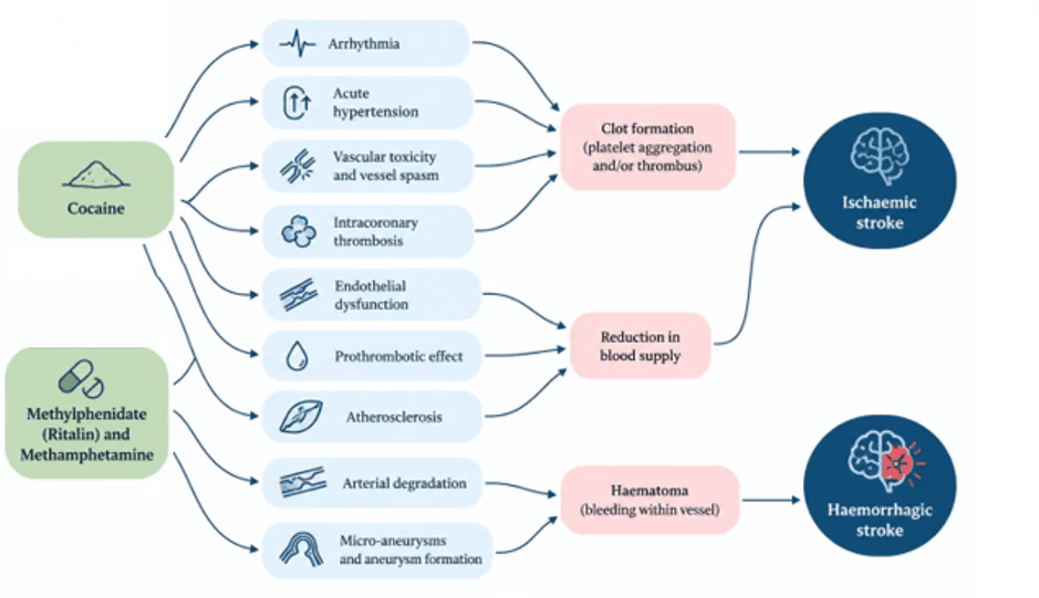
Table 1: Effects of cocaine across multiple anatomical structures and physiological systems.
Effect of Cocaine (C17H21NO4)on the heart (28)
Cardiomyopathy: This condition causes the heart muscle to become stiffer and reduces its efficiency in pumping blood.
Myocardial dysfunction and heart failure: Progressive myocardial injury may impair cardiac output and cause fatigue, dyspnoea and reduced exercise tolerance.
Cardiac arrhythmias: Cocaine may disrupt cardiac electrical conduction and precipitate potentially life-threatening arrhythmias or cardiac arrest.
Spikes vital signs: These effects may include a rapid increase in heart rate, blood pressure, and body temperature.
Increased cardiovascular activity: Sympathetic stimulation may acutely increase heart rate, blood pressure, myocardial oxygen demand and body temperature.
Effect of Cocaine (C17H21NO4) on blood vessels (28)
Increases vasoconstriction: Cocaine induces vasoconstriction by stimulating α-adrenergic receptors on vascular smooth muscle and inhibiting norepinephrine reuptake. These mechanisms cause acute, significant narrowing of arteries, including the coronary arteries that supply the myocardium.
Atherosclerosis: Accelerated atherosclerosis.
Coronary vasospasm: Rare cases of coronary vasospasm and myocardial ischaemia have been reported; these are not routine effects of prescribed use.
Damages blood vessels: Aortic injury.
Blood Clotting and Thrombosis: Platelet activation and thrombosis.
This impairment increases the risk of intravascular micro-thrombus formation within stenotic arteries.
Table 2: Effects of methylphenidate across multiple anatomical structures and physiological systems
Effect of Methylphenidate (C14H19NO2) on the heart (37; 38)
Increases Heart Rate: Heart rate can increase up to 10 beats per minute.
Rare Vascular Complications: Misuse, including crushing and injecting pills, may result in physical obstruction of blood vessels. Rare medical reports have linked stimulant use to severe conditions such as cerebral vasculitis and reversible cerebral vasoconstriction syndrome (RCVS).
Increased Cardiac Output: The heart beats slightly faster and with greater force.
Increased Blood Pressure: The majority of individuals exhibit modest increases in blood pressure, typically ranging from 1 to 5 mmHg.
Palpitations or Arrhythmias: Some individuals may have rapid, irregular, or forceful heartbeats.
Effect of Methylphenidate (C14H19NO2) on blood vessels (39; 40)
Increases vasoconstriction: Methylphenidate (Ritalin) acts as a central nervous system stimulant. It increases norepinephrine and dopamine concentrations, resulting in blood vessel constriction.
Atherosclerosis: Accelerated atherosclerosis.
Coronary artery disease: Vasoconstriction, coronary artery vasospasm, intra-coronary thrombosis.
Peripheral Circulation: Reduced blood flow to the fingers and toes may result in sensations of coldness, numbness, or pain.
Raynaud’s Phenomenon: Methylphenidate (Ritalin) has been linked to the development of peripheral vasculopathy. This includes Raynaud's syndrome, a disorder characterised by temporary colour changes in the fingers in response to specific stimuli.
Table 3: Effects of methamphetamine across multiple anatomical structures and physiological systems. Note: Note: Methamphetamine is a type of amphetamine, but they are not the same; methamphetamine is more potent and has a higher potential for abuse and addiction. While amphetamines are often prescribed for medical conditions, methamphetamine is primarily known as an illicit street drug with limited medical use
Effect of Amphetamine (C₉H₁₃N) on the heart (41)
Catecholaminergic stimulation: Amphetamine increases catecholaminergic signalling, particularly involving norepinephrine and dopamine, resulting in enhanced sympathetic activity.
Increased heart rate and contractility: Sympathetic stimulation may increase heart rate and myocardial contractility, raising cardiac workload and oxygen demand.
Elevated blood pressure:Increased cardiac activity and peripheral vascular resistance can raise blood pressure, sometimes markedly at high doses.
The effect of Amphetamine (C₉H₁₃N) on blood vessels (42)
Increases vasoconstriction: Amphetamine drugs elevate norepinephrine levels, which leads to vasoconstriction and a subsequent reduction in blood flow to specific tissues.
Vasospasm: Sudden, severe contractions of blood vessel walls can occur, restricting blood flow to the extremities, as seen in conditions such as Raynaud's syndrome.
Atherosclerosis: Accelerated atherosclerosis in young.
Coronary artery disease: Coronary vascular complications.
Clotting Risk: Thrombosis and coagulation.
Increased Blood Pressure: Blood pressure often increases markedly because of concurrent vasoconstriction and an elevated heart rate.
Vascular Permeability: High doses or prolonged exposure may alter the endothelium, the inner lining of blood vessels, increasing permeability and susceptibility to inflammation or the formation of small intercellular gaps.
Experimental studies suggest that the mechanical forces generated during cervical spinal manipulation are unlikely to routinely damage an otherwise healthy vertebral artery. Symons et al. (2002) found no evidence of routine mechanical disruption of healthy vertebral arteries under forces comparable to those associated with manipulation. (9) However, an artery already compromised by vascular pathology associated with cocaine or other sympathomimetic drug use may possess reduced structural resilience. This creates a biologically plausible hypothesis that chronic stimulant-related vascular injury could increase susceptibility to an adverse vascular event in a predisposed individual undergoing CeSAM. (43)
This distinction is important because it remains consistent with literature that has not established a direct causal relationship between cervical spinal manipulation and cervical artery dissection, while acknowledging that individual patient characteristics may materially influence clinical risk. The question becomes particularly relevant when vasoactive substances such as cocaine, amphetamines and methylphenidate are involved. These substances can affect vascular integrity through vasoconstriction, elevated blood pressure, endothelial dysfunction, and altered platelet aggregation.
Individually or collectively, these mechanisms may contribute to arterial wall injury or thrombus formation. When such pharmacological factors are present, clinical risk assessment before cervical manipulation should therefore consider the possibility of underlying vascular vulnerability, particularly when additional vascular risk factors or suspicious symptoms are present. (43)
Clinical implications
From a clinical perspective, active heavy use of cocaine, amphetamines or methylphenidate should be regarded as a potentially significant vascular risk factor. Where substantial stimulant exposure is identified, it may be appropriate to consider this a relative contraindication to cervical spinal adjustment or manipulation until an appropriate clinical assessment has been completed. (44)
Comprehensive history-taking should include appropriate enquiry regarding recreational and prescribed stimulant use, particularly cocaine, methamphetamine and other sympathomimetic substances. This information may materially affect assessment of vascular risk and subsequent clinical decision-making. Where heavy stimulant use is identified, conservative management strategies should be considered. Mobilisation, appropriate exercise therapy and soft-tissue interventions may represent alternatives for some patients and avoid the theoretical vascular stresses associated with high-velocity cervical manipulation.
Treatment selection should remain individualised and should take account of the patient's complete cardiovascular, neurological and vascular risk profile rather than stimulant exposure in isolation. Interprofessional collaboration may also be appropriate. Patients with suspected stimulant-associated vascular disease should be considered for medical assessment where clinically indicated, particularly when presenting with neurological symptoms, uncontrolled hypertension or other features suggestive of underlying cardiovascular or cerebrovascular pathology. (45) Appropriate monitoring for emerging cardiovascular or neurological symptoms remains important during the management of individuals with recognised vascular risk factors. (46)
Areas where further study is needed
Several clinically important questions remain unanswered. Prospective research is required to determine whether chronic cocaine use independently increases the risk of CeAD following cervical spinal manipulation or whether cocaine exposure should instead be understood as one component of a broader vascular risk profile. Large multi-centre observational studies may be particularly valuable in addressing this question because individual vascular events are rare and appropriately powered prospective interventional studies may be difficult to conduct.Chronic hypertension
Numerous studies have identified an association between chronic cocaine exposure and the development of hypertension. Repeated stimulation of the sympathetic nervous system may produce recurrent elevations in blood pressure through vasoconstriction and increased cardiac output. (47) Hypertension is an important consideration during vascular risk assessment before cervical adjustment because chronically elevated blood pressure may coexist with other conditions that affect vascular integrity. However, the available literature has not established a direct relationship between long-term cocaine use, cervical spinal adjustment and subsequent cervical artery dissection. Further research is needed to clarify the long-term effects of cocaine and other sympathomimetic substances on blood-pressure regulation, cervical arterial integrity and the risk of vascular events in patients undergoing cervical manual interventions.
Venous thrombosis
Although the arterial complications of cocaine use, including myocardial infarction and stroke, have been extensively investigated, its effects on the venous system are less well characterised.
Emerging evidence suggests that cocaine may contribute to an increased risk of deep-vein thrombosis and pulmonary embolism through mechanisms including endothelial injury, platelet activation and hypercoagulability. (48) These processes may promote venous thrombus formation and create potentially life-threatening complications when a thrombus embolises to the pulmonary circulation. Data regarding the prevalence and specific risk factors for deep-vein thrombosis and pulmonary embolism among cocaine users remain limited.
Further investigation is also required into the effects of other sympathomimetic drugs on cervical vascular integrity. Although such substances share some pharmacological characteristics with cocaine, their specific relationship with cervical arterial pathology has not been adequately investigated.
Future studies should also examine long-term vascular outcomes among chronic stimulant users and determine whether clinically useful risk-stratification tools can be developed to identify patients who may require modification of manual therapy techniques or referral for medical assessment before treatment. (24, 49, 50)
Cocaine- and sympathomimetic drug-related vascular disease, may possess distinctive clinical features, including earlier onset, severe presentations and occurrence in younger individuals without conventional cardiovascular risk factors. However, evidence concerning the prevalence and magnitude of any additional CeSAM-related risk in cocaine users remains limited. Further research in this area could inform more targeted screening, prevention, referral and management strategies for patients with significant stimulant exposure. (45)
Conclusion
Drug misuse represents a significant social and public-health concern. The occurrence of coronary, cerebrovascular and other vascular disease among individuals who use cocaine highlights the importance of recognising potentially modifiable vascular risk factors and improving strategies for early identification and prevention of cardiovascular and cerebrovascular morbidity.
Adverse vascular events reported in temporal association with cervical spinal adjustment or manipulation have frequently involved younger patients who may not have previously recognised arterial pathology. Epidemiological evidence and case reports demonstrate that cocaine use is associated with both haemorrhagic and ischaemic stroke, while heavy or prolonged exposure has been linked to substantial vascular dysfunction. Amphetamines and methylphenidate may also affect vascular function through mechanisms including vasoconstriction, hypertension and alterations in platelet activity.
Although the current literature does not establish a direct causal relationship between sympathomimetic drug use, cervical spinal adjustment or manipulation and cervical artery dissection, the known vascular effects of these substances provide a biologically plausible basis for increased caution. Patients who are heavy users of cocaine, amphetamines or methylphenidate may have underlying vascular pathology that could increase their susceptibility to cervical arterial complications.
Consequently, clinicians should specifically enquire about sympathomimetic drug use as part of a comprehensive clinical history and incorporate this information into individualised vascular risk assessment before undertaking CeSAM. Heavy active use should be recognised as a potentially modifiable risk factor and may constitute a relative contraindication to cervical spinal adjustment or manipulation pending appropriate clinical assessment. If significant vascular risk are identified, consider conservative management strategies and, when indicated, medical referral.
Author contributions
Angelo LM Battiston (AB): Conceptualisation, supervision, writing—review and editing, and final approval.
Nik R Nunn (NRN): Conceptualisation, methodology, data curation, investigation, writing—original draft, and final approval.
Sheila Breeze (SB): Data curation, literature review, writing—review and editing, and final approval.
Joy Harrison (JH): Critical review, manuscript editing, and final approval.
Funding
This research received no specific grant from any funding agency in the public, commercial or not-for-profit sectors.
Ethics approval and consent to participate
Ethics approval was not required because this study is a narrative review of published literature and did not involve human participants or identifiable patient data.
Competing interests
The authors declare no potential conflicts of interest concerning the research, authorship and/or publication of this article.
Copyright and Permission Statement
The authors confirm that this manuscript does not infringe copyright. Where required, permission has been obtained for copyrighted material, and all sources have been appropriately acknowledged and referenced.
Editor's note: The authors collectively retain copyright to this article and, as is Journal policy, they retain the IP.
References
- Griffin KJ, Harmsen WS, Mandrekar J, Brown RD Jr, Keser Z. Epidemiology of Spontaneous Cervical Artery Dissection: Population-Based Study. Stroke. 2024; 55(3):670-677. DOI: 10.1161/STROKEAHA.123.043647
- Debette S, Goeggel Simonetti B, Schilling S, et al. Familial occurrence and heritable connective tissue disorders in cervical artery dissection. Neurology. 2014; 83(22):2023-31. DOI: 10.1212/WNL.0000000000001027
- Guillon B, Berthet K, Benslamia L, Bertrand M, Bousser MG, Tzourio C. Infection and the risk of spontaneous cervical artery dissection: a case-control study. Stroke. 2003;34(7):e79-81. DOI: 10.1161/01.STR.0000078309.56307.5C
- Wang X, He B. Endothelial dysfunction: molecular mechanisms and clinical implications. MedComm 2024;5(8):e651. DOI: 10.1002/mco2.651
- Dvorak J, Orelli F. How dangerous is manipulation to the cervical spine? Manual Medicine. 1985; 2: 1-4.
- Coulter ID. Efficacy and Risks of Chiropractic Manipulation: What Does the Evidence Suggest? Integrative Medicine 1998; 1(2): 61-66. DOI.org/10.1016/S1096-2190(98)00015-8
- Béjot Y, Daubail B, Debette S, Durier J, Giroud M. Incidence and outcome of cerebrovascular events related to cervical artery dissection: the Dijon Stroke Registry. Int J Stroke. 2014; 9(7):879–882. DOI: 10.1111/ijs.12154
- Chaibi A, Russell MB. A risk-benefit assessment strategy to exclude cervical artery dissection in spinal manual-therapy: a comprehensive review. Ann Med. 2019; 51(2):118-127. DOI: 10.1080/07853890.2019.1590627
- Symons BP, Leonard T, Herzog W. Internal forces sustained by the vertebral artery during spinal manipulative therapy. J Manipulative Physiol Ther. 2002; 25(8):504-10. DOI: 10.1067/mmt.2002.127076
- Thomas LC. Cervical arterial dissection: An overview and implications for manipulative therapy practice. Man Ther. 2016; 21:2-9. DOI: 10.1016/j.math.2015.07.008
- Triano JJ, Kawchuk G. Current concepts of spinal manipulation and cervical arterial incidence. NCMIC Insurance Company 2006. ISBN 1-892734-14-1.
- Rendon LF, Malta S, Leung J. et al. Cocaine and Ischaemic or Haemorrhagic Stroke: A Systematic Review and Meta-Analysis of Clinical Evidence. J. Clin. Med. 2023; 12: 5207. DOI: 10.3390/jcm12165207
- Westover AN, McBride S, Haley RW. Stroke in young adults is associated with cocaine use. Arch Gen Psychiatry. 2007; 64(4):495-502. DOI: 10.1001/archpsyc.64.4.495
- Hemphill K, Tierney S, Tirschwell D and Davis AP. A review of methamphetamine use and stroke in the young. Front. Neurol. 2024; 15:1397677. DOI.org/10.3389/fneur.2024.1397677
- Liu H, Feng W, Zhang D. Association of ADHD medications with the risk of cardiovascular diseases: a meta-analysis. Eur Child Adolesc Psychiatry. 2019; 28(10):1283-1293. DOI: 10.1007/s00787-018-1217-x
- Schrantee A, Bouziane C, Bron EE, et al. Long-term effects of stimulant exposure on cerebral blood flow response to methylphenidate and behaviour in attention-deficit hyperactivity disorder. Brain Imaging Behav. 2018;12(2):402-410. DOI: 10.1007/s11682-017-9707-x
- Pankrath N, Nilsson S, Ballenberger N. Adverse Events After Cervical Spinal Manipulation - A Systematic Review and Meta-Analysis of Randomised Clinical Trials. Pain Physician. 2024; 27(4):185-201. ISSN 1533-3159. PMID: 38805524
- Chu EC, Trager RJ, Lee LY, Niazi IK. A retrospective analysis of the incidence of severe adverse events among recipients of chiropractic spinal manipulative therapy. Sci Rep. 2023; 13(1): 1254. DOI: 10.1038/s41598-023-28520-4
- Brown SP. Plausible Mechanisms of Causation of Immediate Stroke by Cervical Spine Manipulation: A Narrative Review. Cureus. 2024; 16(3): e56565. DOI: 10.7759/cureus.56565
- Gorrell LM, Brown BT, Engel R, et al. Reporting of adverse events associated with spinal manipulation in randomised clinical trials: an updated systematic review. BMJ Open. 2023; 13: e067526. DOI: 10.1136/bmjopen-2022-067526
- Manini AF, Gibson CL, Miller ML, Richardson LD, Vargas-Torres CC, Vedanthan R, Hurd YL. Biomarkers of endothelial dysfunction in cocaine overdose and overdose-related cardiovascular events. Addict Biol. 2021; 26(2):e12901. DOI: 10.1111/adb.12901.
- Sharma T, Kumar M, Rizkallah A, Cappelluti E, Padmanabhan P. Cocaine-induced Thrombosis: Review of Predisposing Factors, Potential Mechanisms, and Clinical Consequences with a Striking Case Report. Cureus. 2019; 11(5):e4700. DOI: 10.7759/cureus.4700
- Clare K, Park K, Pan Y, Lejuez CW, Volkow ND, Du C. Neurovascular effects of cocaine: relevance to addiction. Front Pharmacol. 2024; 15:1357422. DOI: 10.3389/fphar.2024.1357422
- Bachi K, Mani V, Jeyachandran D, Fayad ZA, Goldstein RZ, Alia-Klein N. Vascular disease in cocaine addiction. Atherosclerosis. 2017; 262:154-162. DOI: 10.1016/j.atherosclerosis.2017.03.019
- Rezkalla SH, Kloner RA. Cocaine-induced acute myocardial infarction. Clin Med Res. 2007; 5(3):172-6. DOI: 10.3121/cmr.2007.759
- Pergolizzi JV Jr, Magnusson P, LeQuang JAK, Breve F, Varrassi G. Cocaine and Cardiotoxicity: A Literature Review. Cureus. 2021; 13(4):e14594. DOI: 10.7759/cureus.14594
- Feller D, Giudice A, Faletra A, et al Identifying peripheral arterial diseases or flow limitations of the lower limb: Important aspects for cardiovascular screening for referral in physiotherapy. Musculoskelet Sci Pract. 2022; 61:102611. DOI: 10.1016/j.msksp.2022.102611
- Kim ST, Park T. Acute and Chronic Effects of Cocaine on Cardiovascular Health. Int J Mol Sci. 2019; 20(3):584. DOI: 10.3390/ijms20030584.
- Darke S, Kaye S, Duflou J. Comparative cardiac pathology among deaths due to cocaine toxicity, opioid toxicity and non-drug-related causes. Addict. (Abingd. Engl.). 2006; 101:1771–1777. DOI: 10.1111/j.1360-0443.2006.01601.x
- Arnold M, Kappeler L, Georgiadis D, Berthet K, Keserue B, Bousser MG, Baumgartner RW. Gender differences in spontaneous cervical artery dissection. Neurology. 2006; 67:1050–1052. DOI: 10.1212/01.wnl.0000237341.30854.6a
- Lodato M, Pini R, Porcelli, A, et al. Ten-Year Outcomes of Cervical Artery Dissection: A Retrospective Study in a Real-World Cohort. J Clin Med. 2025; 14: 6836. DOI: 10.3390/jcm14196836
- Hoffman RS. Treatment of patients with cocaine-induced arrhythmias: bringing the bench to the bedside. Br J Clin Pharmacol. 2010; 69(5):448-57. DOI: 10.1111/j.1365-2125.2010.03632.x
- Alaraj A, Wallace A, Mander N, et al. Effect of acute cocaine use on vasospasm and outcome in aneurysmal subarachnoid haemorrhage. World Neurosurg. 2010; 73(4):357–360. DOI: 10.1016/j.wneu.2010.01.011
- Aleksic I, Dzambas J, Marinkovic N, et al. Subarachnoid and intracerebral haemorrhage in cocaine abusers. Vojnosanit. Pregl. 2021; 78:1166–1172. DOI.org/10.2298/VSP191229034A
- Treadwell SD, Robinson TG. Cocaine use and stroke. Postgrad Med J. 2007; 83(980):389-94. DOI: 10.1136/pgmj.2006.055970.
- Dominic P, Ahmad J, Awwab H, et al. Stimulant Drugs of Abuse and Cardiac Arrhythmias. Circ Arrhythm Electrophysiol. 2022; 15(1):e010273. DOI: 10.1161/CIRCEP.121.010273.
- Schelleman H, Bilker WB, Kimmel SE, et al. Methylphenidate and risk of serious cardiovascular events in adults. Am J Psychiatry. 2012; 169(2):178-85. DOI: 10.1176/appi.ajp.2011.11010125
- Jackson JW. The cardiovascular safety of methylphenidate. BMJ. 2016; 353:i2874 DOI: 10.1136/bmj.i2874
- Cai W, Giacobini M, Österholm C. et al. Effects of methylphenidate on the human vascular endothelium. Transl Psychiatry. 2026; 16: 369. DOI.org/10.1038/s41398-026-04237-6
- Zhang L, Li L, Andell P. et al. Attention-deficit/hyperactivity disorder medications and long-term risk of cardiovascular diseases. JAMA Psychiatry. 2024; 81:178-187. DOI: 10.1001/jamapsychiatry.2023.4294
- Böttner J, Fischer-Schaepmann T, Werner S, Knauth S, Jahnke HG, Thiele H, Büttner P. Amphetamine increases vascular permeability by modulating endothelial actin cytoskeleton and NO synthase via PAR-1 and VEGF-R. Sci Rep. 2024; 14(1):3596. DOI: 10.1038/s41598-024-53470-w
- Simonds SE, Pryor JT, Lam BYH, Dowsett GK, Mustafa T, Munder A, Elysee K, Balland E, Cowley LO, Yeo GSH, Lawrence A, Spanswick DC, Cowley MA. The metabolic and cardiovascular effects of amphetamine are partially mediated by the central melanocortin system. Cell Rep Med. 2025; 6(2):101936. DOI: 10.1016/j.xcrm.2025.101936
- Middlekauff HR, Cooper ZD, Strauss SB. Drugs of Misuse: Focus on Vascular Dysfunction. Can J Cardiol. 2022; 38(9):1364-1377. DOI: 10.1016/j.cjca.2022.04.011
- Cheng YC, Ryan KA, Qadwai SA, et al. Cocaine Use and Risk of Ischaemic Stroke in Young Adults. Stroke. 2016; 47(4):918-22. DOI: 10.1161/STROKEAHA.115.011417
- Gliedt JA, Reynolds M, Moonaz S, Long CR, Russell R, Schneider MJ. Identifying and addressing patient substance use: a survey of chiropractic clinicians. Chiropr Man Therap. 2023; 31(1):18. DOI: 10.1186/s12998-023-00490-4
- Havakuk O, Rezkalla SH, Kloner RA. The Cardiovascular Effects of Cocaine. J Am Coll Cardiol. 2017; 70(1):101-113. DOI: 10.1016/j.jacc.2017.05.014.
- Alzghoul BN, Abualsuod A, Alqam B, Innabi A, Palagiri DR, Gheith Z, Amer FN, Meena NK, Kenchaiah S. Cocaine Use and Pulmonary Hypertension. Am J Cardiol. 2020; 125(2):282-288. DOI: 10.1016/j.amjcard.2019.10.008
- Griffin D, Cha S. Cocaine: A Provoking Risk Factor in Venous Thromboembolism. Cureus. 2019; 11(12):e6520. DOI: 10.7759/cureus.6520
- Jacobs W, Wright DS, Pohlman K, Rosenbaum R, Hutten BA, Scholten-Peeters GG, Haldeman S, Ket JC, Rubinstein SM. Risk factors for cervical artery dissection: a systematic review with meta-analysis. Stroke Vasc Neurol. 2026; 11(3):275-285. DOI: 10.1136/svn-2025-004186
- Gaban GLNA, Vægter HB, Vivaldini MRS et al. Acute and long-term effect of specific and non-specific exercises in patients with chronic neck pain: A protocol for a randomized controlled trial. Exp Physiol. 2025; 110(1):58-67. DOI: 10.1113/EP091907
AU: Scott Cuthbert BA, DC. Chiropractor, Dauin, The Philippines; Associate Editor. e: cranialdc@hotmail.com
Abstract: The gluteus maximus is the largest muscle of the human beings we serve as Chiropractor. It is distinctive muscle in terms of size, anatomy and function compared to apes and other non-human primates. The gluteus maximus is one of the principal muscles responsible for controlling the human trunk and lower limb during powerful locomotion. In essence, unless Chiropractors correct this muscle’s functional capacity, our patients will fail to recover from walking impairments and low back pain. The gluteus maximus is the principal extensor of the hip and an important contributor to pelvic and lower-limb function.
Despite its biomechanical importance, standardised manual muscle testing (MMT) of the gluteus maximus is not consistently emphasised as a core examination procedure in chiropractic education. The available literature provides a meaningful evidentiary foundation for standardised gluteus maximus and hip-extensor MMT. Studies have demonstrated clinically meaningful inter-examiner agreement, while controlled investigations have shown that manual grades of hip-extensor strength correspond with objectively measured torque. A 2025 systematic review specifically examining MMT in Applied Kinesiology identified standard MMT, without additional non-musculoskeletal challenge procedures, as potentially useful and reported moderate-to-very-strong reliability for several muscles, including the gluteus maximus.
This paper argues that standardised gluteus maximus MMT should be considered a fundamental Chiropractic examination competency, not because MMT replaces dynamometry, electromyography, imaging, or neurological examination, but because it provides a rapid, inexpensive, repeatable bedside assessment of hip-extensor performance. Proper testing requires appropriate patient positioning, pelvic stabilisation, consistent examiner contact, consistent resistance, and careful interpretation. A weak result should be interpreted as a finding of impaired force production or motor performance, not, by itself, proof of a specific pathology.
Indexing Terms: gluteus maximus; manual muscle testing; chiropractic; Applied Kinesiology; reliability; validity; hip extension; pelvic stability; low back pain; neuromusculoskeletal examination.
Cite: Cuthbert S. The Gluteus Maximus Manual Muscle Test in Chiropractic clinical practice: A critical review of reliability, validity, clinical utility, and its importance as a core Chiropractic examination competency. Asia-Pac Chiropr J. 2026;7.3. https://www.apcj.site/CuthbertGluteMaxMMT.pdf
Introduction
The gluteus maximus occupies a uniquely important position in the ensemble of human movement. It is the largest of the gluteal muscles and a major hip extensor, contributing to propulsion, stair climbing, rising from a seated position, running, jumping, lifting, and control of the pelvis and trunk.
Consequently, assessment of gluteus maximus performance has implications extending beyond the hip itself.
Figure 1: The basic rationale for the manual muscle test -- for life and movement
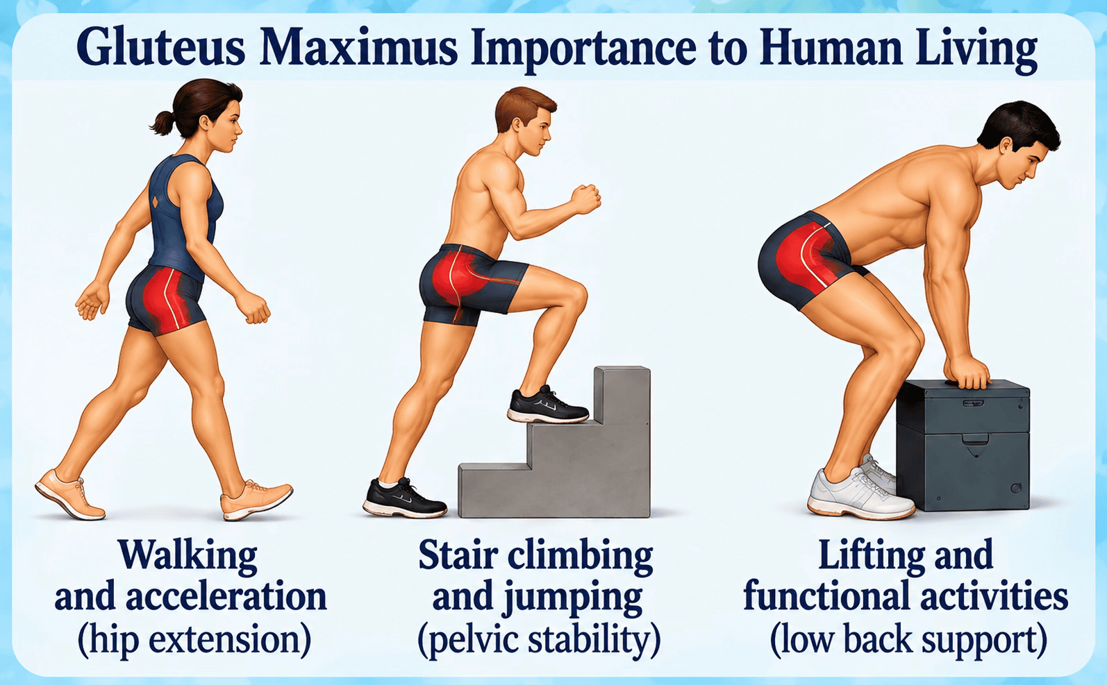
For Chiropractors this is particularly relevant because common clinical presentations involve the interaction of the lumbar spine, pelvis, sacroiliac region, hip, and lower extremity. A clinician examining these regions needs methods capable of identifying meaningful functional impairments rather than relying exclusively on structural imaging or symptom location. Manual muscle testing offers one such method.
The scientifically useful question is not simply whether ‘muscle testing works’, but whether a specific, standardised gluteus maximus MMT provides reproducible and clinically meaningful information about hip-extensor performance. The literature reviewed here indicates that it can.
The Gluteus Maximus as a target of Chiropractic examination
The gluteus maximus produces powerful hip extension and contributes to control of the pelvis and lower extremity. During functional activities, hip-extensor performance is especially important when the body must generate propulsion or control the trunk over the lower extremities.
A 2024 systematic review of hip biomechanics in people with low back pain reported differences in hip muscle performance and identified reduced hip-extensor and abductor strength in affected populations across several forms of assessment. (Perry, 2004) These findings support the clinical relevance of assessing hip-extensor function as one component of a broader lumbopelvic examination.
Standardised, reliable and validated Gluteus Maximus manual muscle testing:
The Chiropractic profile
The traditional prone gluteus maximus test is performed with the patient prone and the knee flexed approximately 90°. The examiner stabilises the pelvis while the patient attempts hip extension against downward resistance applied to the posterior distal thigh. (Conroy et al, 2022; Schmitt, Cuthbert, 2008)
- Patient position: prone.
- Knee: flexed approximately 90 degrees.
- Pelvis: stabilised to minimise lumbar extension and pelvic rotation.
- Action: patient actively extends the hip.
- Resistance: examiner applies downward resistance toward the table against the posterior distal thigh.
- Observation: assess the patient's ability to maintain hip extension against the applied resistance.
- Comparison: where appropriate, compare right and left sides and document the grade or qualitative response.
- Repeat testing: use the same position, contact point, resistance direction, stabilisation, and examiner mechanics during reassessment.
Gluteus maximus (after Walther, with permission, 2000)
Origin: posterior gluteal line of ilium, tendon of sacrospinalis, dorsal surface of sacrum and coccyx, and sacrotuberus ligament.
Insertion: gluteal tuberosity of femur and iliotibial tract of fascia lata.
Action: extends hip, assists in externally rotating the thigh.
Test: The supine patient flexes the knee and extends the hip. The knee flexion is necessary to help take the hamstrings out of the test. The examiner directs pressure on the distal one-third of the femur in a direction of hip flexion. Observe for adequate pelvic fixation to the trunk by the trunk extensors and oblique abdominal musculature.
Nerve supply: inferior gluteal, L4, 5, Sl, 2.
Neurolymphatic:
Anterior: anterolateral thigh.
Posterior: between posterior superior iliac spine and L5 spinous.
Neurovascular: on the lambdoidal suture midway between lambda and asterion.
Nutrition: vitamin E, male or female endocrine concentrates or nucleoprotein extracts.
Meridian association: circulation sex.
Organ/gland association: reproductive organs or glands.
Body Language of Inhibition:
Testing position: Much information is gained as the patient goes into the testing position. IN the presence of inhibition, she may attempt to rotate the pelvis around the vertical axis, lifting it from the table on the side of the test to aid in lifting the thigh from the table. Another indication of inhibition or malfunction is a change of knee flexion by the patient. Even though the tester may have placed the knee in proper flexion, the patient may attempt to extend it to recruit synergistic action in the hamstrings. Inhibition may be evident by an inability to reach or maintain the testing position in the first place.
During test: Continues improper effort, such as that used in an attempt to hold the resting position, will be observed. The patient will attempt to straighten the leg from the flexed position, and to rotate the pelvis.
Movement aberrations: Difficulty in arising from a chair, or in arising from the standing hip flexion position (Adams orthopedic test).
Postural imbalance: Anterior rotation of the pelvis, or the patient carries the weight of the trunk posteriorly on a posteriorly rotated pelvis in an attempt to compensate for the weakness. In either the standing or the prone position, an inhibited gluteus maximus may be observed by the atonic appearance of the muscle. In the prone position, there is a lack of firmness and roundness of the gluteus maximus. In the standing position, the gluteus maximus appears to sag.
Alternate Testing Methods: In the presence of a short rectus femoris, it is necessary to test the gluteus maximus with less knee flexion. The shortened rectus femoris is observed when the thigh cannot be raised from the table with knee flexion; it can raise adequately when less flexion is present. An improvement in the length of the rectus femoris can often be obtained by fascial release or percussion on the origin or insertion or myofascial trigger points of the muscle.
Note: The gluteus maximus is frequently tested in the weight-bearing position to evaluate for an upper cervical or foot dysfunction which appears to affect this muscle only when weight-bearing. The test is generally accomplished against an upright hi-lo table, and it is done in a manner similar to the prone test. If a hi-lo table is not available, the test can be done stabilising against a wall; however, it is more difficult to obtain precise information.
Figure 2: Glute Max MMT
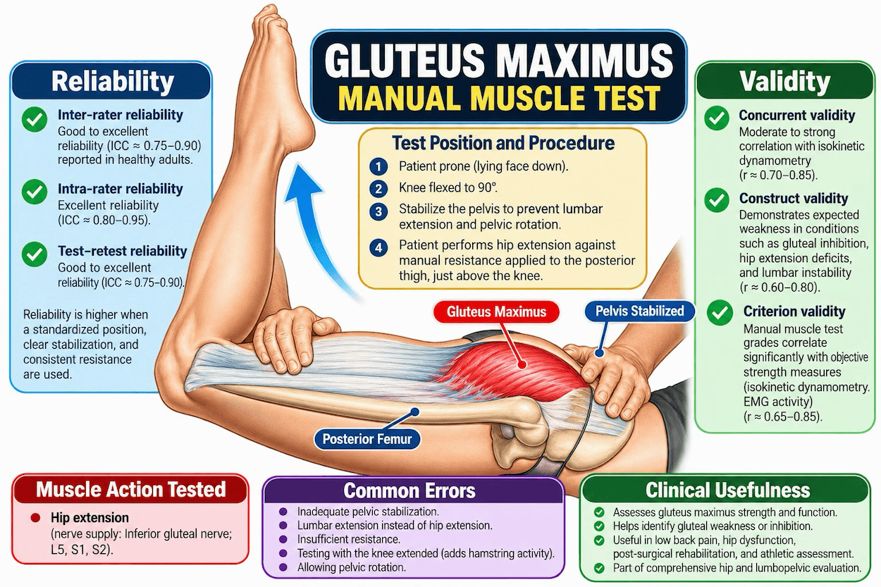
Common spinal fixation complex associated with bilaterally inhibited gluteus maximus MMT
When a fixation complex is present in the spinal column, it has a specific bilateral muscle weakness in Applied Kinesiology. The bilateral weakness observed by Goodheart (Goodheart, 1973) reveals many vertebral fixations that would otherwise be overlooked.
Fixation levels associated with bilaterally inhibited gluteus maximus muscles are:
An upper cervical fixation: bilateral gluteus maximus muscles. This has been found repeatedly and for decades many tens of thousands of Chiropractors around the world since Goodheart first recognised the correlation.
Figures 3, 4: Upper cervical fixation correction for bilaterally inhibited gluteus maximus muscles - frequently found upon Chiropractic examination. The viscerosomatic reflexes for the gluteus maximus muscle in Applied Kinesiology Chiropractic [Walther, 2000, 1994]
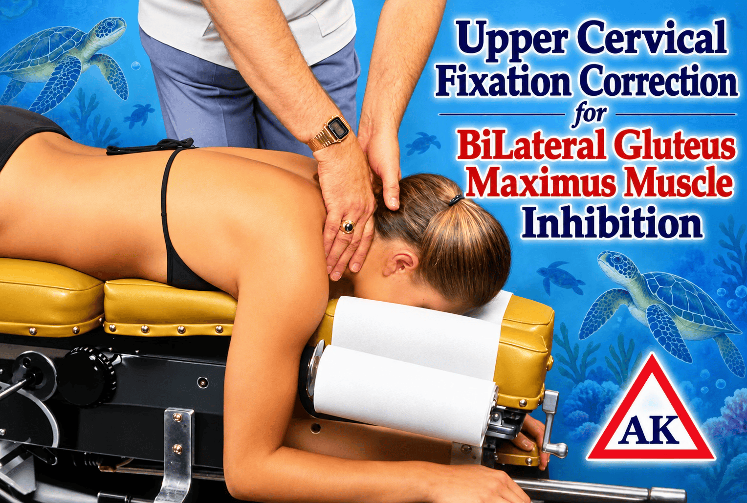
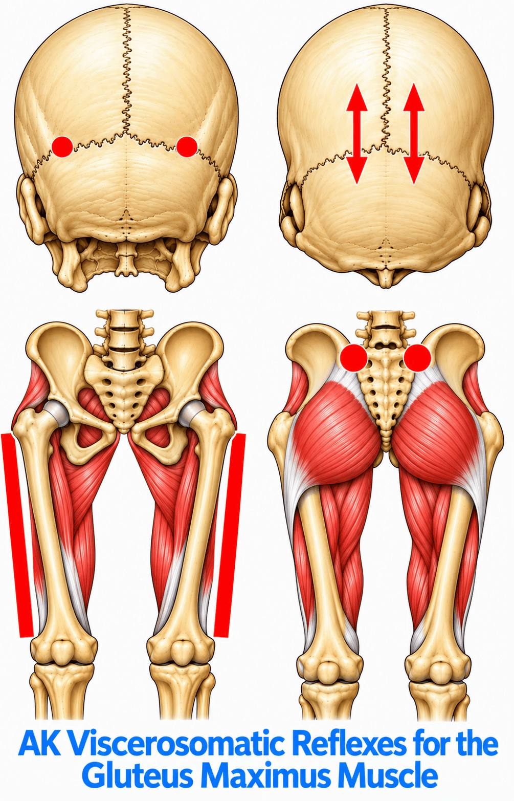
The specific reflex-to-gluteus-maximus relationships have not been adequately tested with EMG, H-reflexes, TMS, dynamometry, autonomic measurements, sham controls, and blinded examiners. However, the widely reported in-office clinical trials by Chiropractors and other AK-users around the world have given considerable outcomes information on this subject.
Frank Chapman described tender reflex areas that he believed were associated with visceral and lymphatic dysfunction. Goodheart subsequently observed that stimulating particular Chapman reflexes appeared to change the result of specific muscle tests and incorporated this into AK as the neurolymphatic reflex system.
The proposed viscerosomatic pathway is approximately:
visceral/lymphatic disturbance
↓
somatic/autonomic afferent input
↓
spinal cord/brainstem integration
↓
alteration of α-motor-neuron excitability
↓
altered muscle recruitment
↓
MMT changes
The important part is that the general neurophysiological architecture of these discoveries are neuro-physiologically plausible.
The importance of standardisation cannot be overstated. Manual muscle testing is affected by starting position, direction and timing of force, contact point, examiner mechanics, stabilisation, and other procedural variables. The objective is not merely to observe whether the leg moves backward, but to assess hip-extension force while minimising substitution by lumbar extension and pelvic rotation. (Schmitt, Cuthbert, 2008)
Reliability
Can different examiners obtain similar results?
Reliability is essential to any clinical examination. If one Chiropractor identifies a gluteus maximus as strong and another using the same procedure identifies it as weak, the test has limited value. Available evidence indicates that standardised gluteus maximus MMT can produce meaningful examiner agreement.
Pollard and colleagues examined inter-examiner reliability of lower-limb MMT while examiners were ‘blinded’, attempting to minimise visual, verbal, and nonverbal cues. For the gluteus maximus, the study reported approximately 75.6% agreement between examiners. The authors concluded that MMT demonstrated fair-to-substantial agreement across the muscles examined, while noting the need for further research regarding validity for specific clinical conditions.
Clinical meaning
The test is useful primarily as a low-cost clinical examination of hip-extensor performance, side-to-side differences, gross weakness, and change over time. With the addition of Applied Kinesiology diagnostic tests (challenge and therapy localisation), the operative factors contributing to the muscle inhibition found can be precisely and quickly diagnosed by the clinician. (Cuthbert and Walther, 2014)
Figure 5: Detailed illustration of the standardised standing gluteus maximus MMT, emphasising the multiplicity of functional assessment developed in Applied Kinesiology MMT assessment. This image emphasises the importance of gluteus maximus function in the evolution of homo sapiens sapiens. (Lieberman et al, 2006)
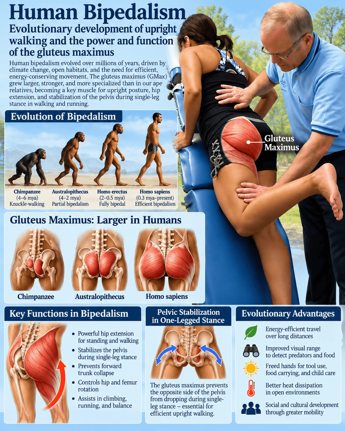
Figure 6: Standing gluteus maximus MMT illustration demonstrating an alternative clinical approach. This figure is presented as an illustrative variation; evidence for one testing configuration should not automatically be generalised to another.
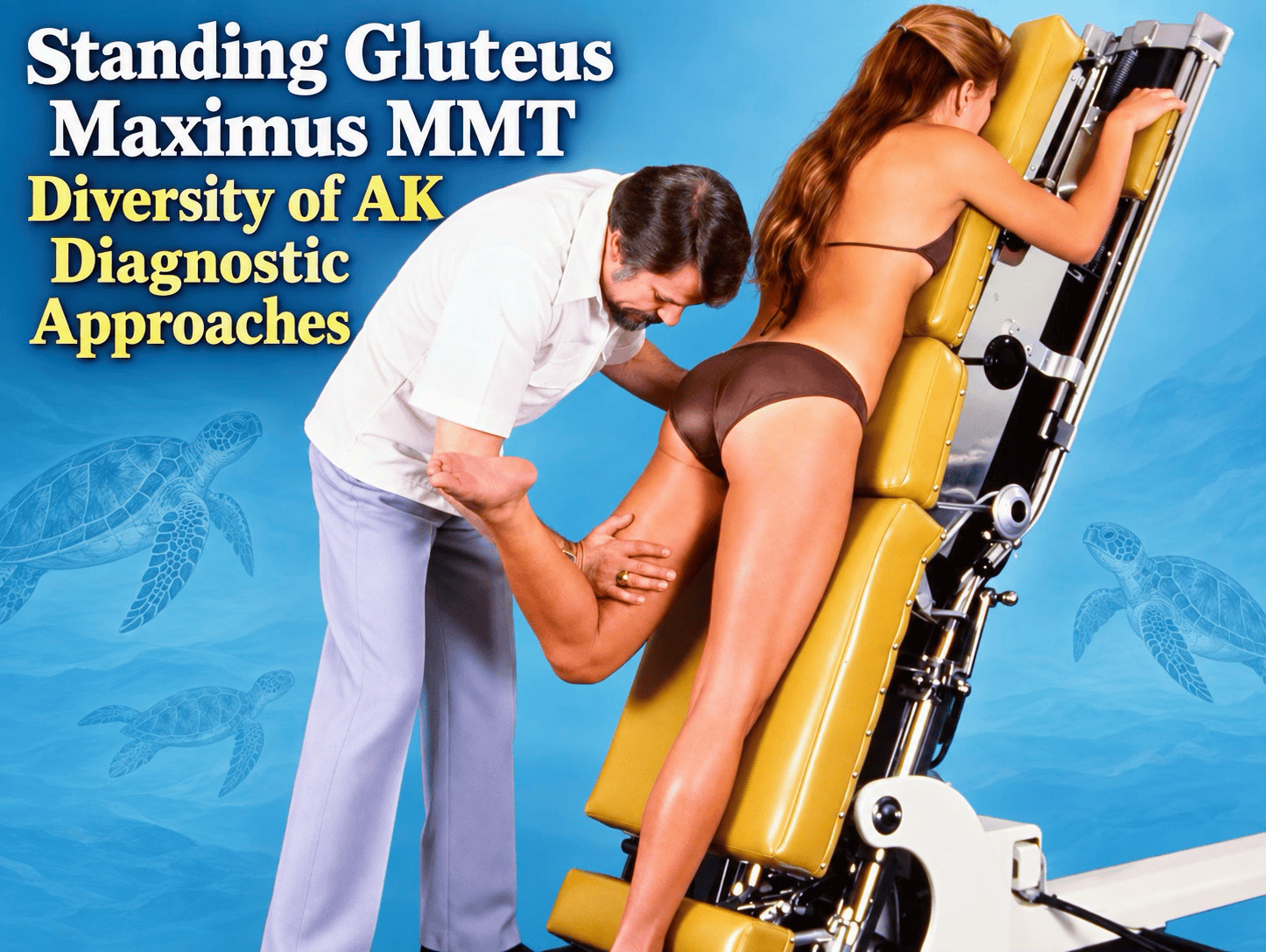
The importance of standardisation cannot be overstated. Manual muscle testing is affected by starting position, direction and timing of force, contact point, examiner mechanics, stabilisation, and other procedural variables. The objective is not merely to observe whether the leg moves backward, but to assess hip-extension force while minimising substitution by lumbar extension and pelvic rotation. (Schmitt, Cuthbert, 2008)
Reliability is essential to any clinical examination. If one chiropractor identifies a gluteus maximus as strong and another using the same procedure identifies it as weak, the test has limited value. Available evidence indicates that standardised gluteus maximus MMT can produce meaningful examiner agreement.
Pollard and colleagues examined inter-examiner reliability of lower-limb MMT while examiners were “blinded”, attempting to minimise visual, verbal, and nonverbal cues. For the gluteus maximus, the study reported approximately 75.6% agreement between examiners. The authors concluded that MMT demonstrated fair-to-substantial agreement across the muscles examined, while noting the need for further research regarding validity for specific clinical conditions.
Clinical meaning
The test is useful primarily as a low-cost clinical examination of hip-extensor performance, side-to-side differences, gross weakness, and change over time. With the addition of Applied Kinesiology diagnostic tests (challenge and therapy localization), the operative factors contributing to the muscle inhibition found can be precisely and quickly diagnosed by the clinician. (Cuthbert and Walther, 2014)
Reliability evidence for the gluteus maximus MMT
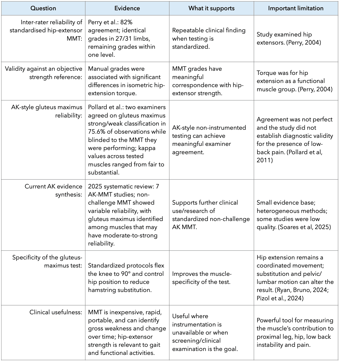
Figure 7: Reliability and Validity of the Gluteus Maximus MMT
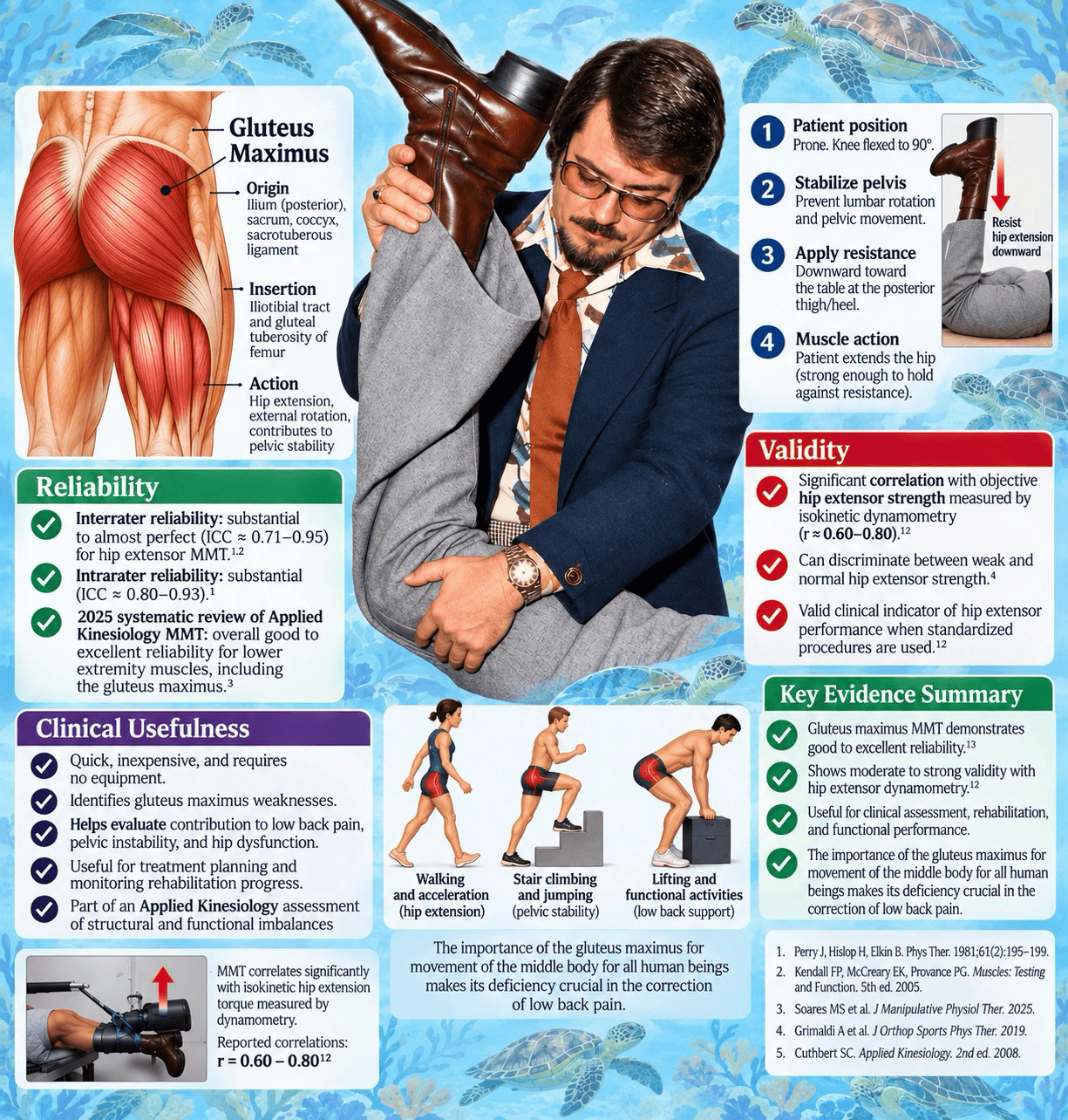
Validity
Does MMT correspond to actual hip-extensor force?
Reliability alone is insufficient: a test can be reproducible yet fail to measure the construct of interest. The validity question is whether manual grades correspond with objective measures of hip-extensor force.
Perry, Weiss, Burnfield, and Gronley investigated the hip-extensor MMT by comparing manual muscle grades with objectively measured isometric hip-extension torque. Interrater agreement was approximately 82%, and different MMT grades corresponded to significantly different levels of measured torque. In the pathological group, mean torque was approximately 176 Nm for Grade 5, 103 Nm for Grade 4, 67 Nm for Grade 3, and 19 Nm for Grade 2.
These findings provide evidence of concurrent validity: manual grades were associated with objectively measured differences in hip-extensor force. The findings do not mean that a manual grade is interchangeable with a dynamometer measurement.
MMT is not a dynamometer, and that is no reason to abandon it
A dynamometer provides quantitative force or torque measurements, whereas MMT generally provides a categorical or semi-quantitative assessment of force production. A clinician therefore should not convert an MMT grade into an exact newton-meter value. Instead, MMT should be understood as a standardised clinical assessment of the patient's ability to generate and maintain force under defined conditions.
Table 2: MMT vs. dynamometry
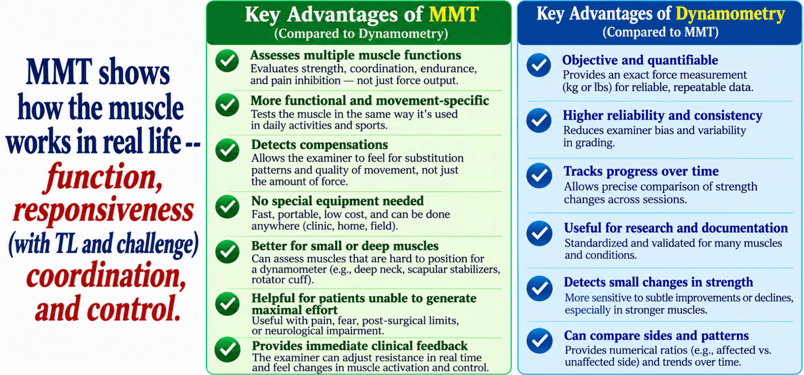
When precise force quantification is required, for research, high-level athletic testing, or detailed rehabilitation monitoring, hand-held or isokinetic dynamometry can add quantitative information. MMT and dynamometry are therefore best viewed as complementary tools.
Pelvic Stabilisation and the problem of substitution
One of the most important features of the gluteus maximus MMT is stabilisation of the pelvis. Without stabilisation, a patient may produce apparent hip extension partly through lumbar extension or pelvic rotation. The examiner must therefore control the pelvis sufficiently to make the test a meaningful assessment of hip-extensor performance.
Figure 8: Dynamometry vs MMT comparison of the Gluteus Maximus Muscle. Concurrent Validity of the Gluteus Maximus MMT
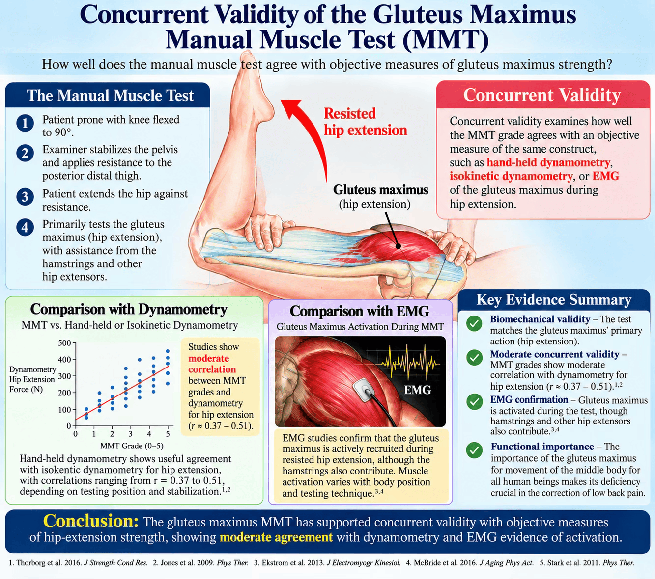
Clinical usefulness
- Minimal equipment - A Chiropractor needs an examination table, appropriate patient positioning, knowledge of anatomy and biomechanics, and trained hands.
- Rapid assessment - The test can be incorporated into a broader examination in a short period of time.
- Repeatability - The procedure can be repeated before and after an intervention or during rehabilitation if the same testing conditions are maintained.
- Side-to-side comparison - Comparing the right and left sides can identify clinically meaningful differences in force production.
- Functional relevance - Hip extension is fundamental to walking, acceleration, stair climbing, rising from a chair, jumping, running, lifting, and trunk control.
Current literature continues to investigate relationships between hip-extensor performance, gluteus maximus activation, and low back pain. A 2024 systematic review identified differences in hip muscle performance among people with low back pain, while a 2024 study using motion capture and electromyography investigated the clinical utility of prone hip extension testing for motor-control impairments associated with low back pain. (Ryan et al; Perry et al; Puzol et al, 2024) These studies support clinical investigation of hip muscle and its related joint function without establishing MMT as a stand-alone diagnosis of low back pain.
The neuromuscular significance of an inhibited gluteus maximus MMT
An ‘inhibited’ or weak’ gluteus maximus MMT indicates difficulty generating or maintaining force under the conditions of the test. Possible contributors include pain inhibition, local injury, postoperative weakness, disuse, altered motor control, neurological impairment, reduced conditioning, hip pathology, lumbopelvic dysfunction, or other neuromusculoskeletal factors. These are encompassed in the AK model of the ‘5-factors of the IVF’. (Cuthbert & Goodheart, 2007)
Accordingly, a weak gluteus maximus MMT is a finding, not a diagnosis. The result should guide additional examination rather than replace it. What distinguishes AK is its emphasis upon proprioceptive responses of the muscle rather than the absolute strength of the muscle itself. It essentially sees muscle function as a transcript of the central integrative state of the anterior horn motoneurons, summing all excitatory and inhibitory inputs from the totality of the patient connected to the muscle.
The appropriate sequences of testing, therapy localisation, and challenge, to correct the muscle inhibition found in the case of a gluteus maximus malfunction, to assess and to treat each tissue will become second nature for the experienced AK examiner.
In other words, the locus of dysfunction ultimately rests with the complete nervous system. A procedure called therapy localisation, strictly diagnostic, seeks a change of muscle strength when the patient’s hand is placed over an area of suspected involvement. Challenge defines a mechanism to test the body’s ability to cope with external stimuli, again assessed by muscle testing.
Challenge is a diagnostic procedure unique to AK that is used to determine the body’s ability to cope with external stimuli, which can be physical, chemical, or mental. Cranial challenge (for instance) has been described in the literature of this Journal previously. (Cuthbert) After an external stimulus is applied, muscle-testing procedures are done to determine a change in the muscle strength as a result of the stimulus. Through this approach, ineffective therapies that produced no improvements in muscle strength are rejected and only those that elicit a positive muscle response are used. This guides the treatment given to a patient.
Common errors in gluteus maximus MMT
(Schmitt, Cuthbert, 2008)
- Inadequate pelvic stabilisation, permitting lumbar extension or pelvic rotation.
- Testing with the knee extended, thereby changing the contribution of the hamstrings.
- Inconsistent resistance magnitude or speed.
- Changing the examiner’s contact point and therefore changing the lever arm.
- Inconsistent examiner position or mechanics.
- Testing a general movement rather than a standardised muscle-specific procedure.
- Over-interpreting a weak result as proof of a particular spinal, visceral, nutritional, or systemic disorder.
Finding remote factors of dysfunction and interaction with the gluteus maximus MMT
For example, if foot dysfunction is causing disorganisation to the gluteus maximus muscles they will usually test weak with manual muscle testing when the patient is standing. The primary effect of foot dysfunction is evidenced by the muscles again testing weak immediately after the patient walks. Applied kinesiology techniques are seldom employed in isolation but as part of a process designed to restore maximal pain free movement of articulations, restoration of postural balance, systemic functionality and facilitation of the self-regulatory mechanisms of the body.
Figures 9, 10: Assessing foot subluxations in cases of gluteus maximus muscle inhibition: the obvious importance
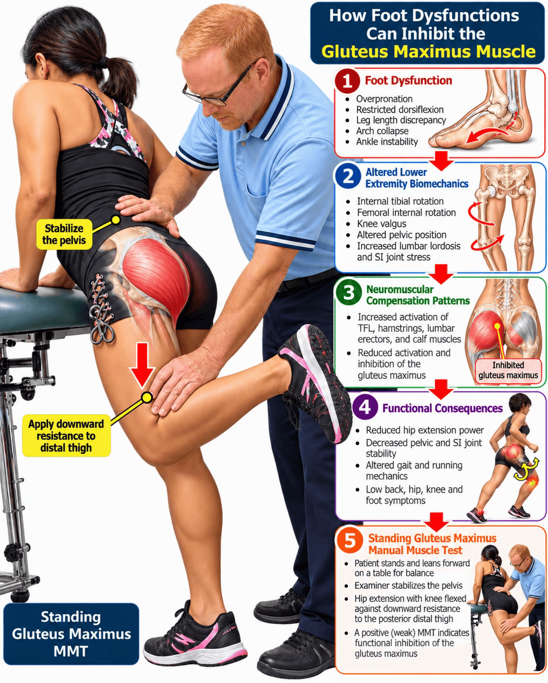
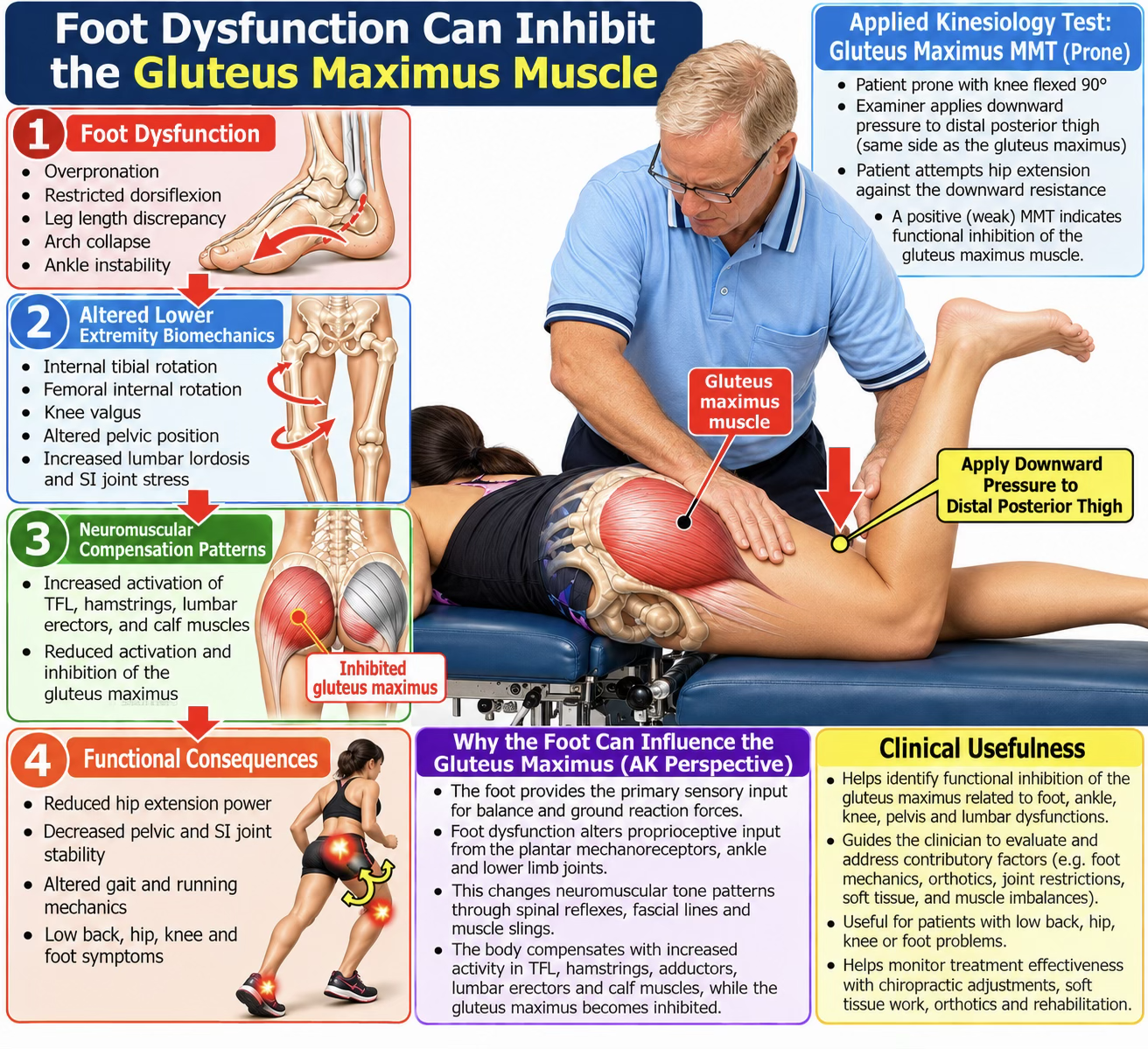
The standing gluteus maximus MMT: An alternative clinical approach
The variations of patient positioning for this test shown for far emphasises the diversity of approaches used in Applied Kinesiology and Chiropractic clinical assessment. Such variations may be clinically interesting, but they should not automatically be assumed to possess the same reliability and validity evidence as the standardised prone hip-extension test (Figure 2).
This distinction is especially important in Chiropractic education. The evidence supports teaching a clearly defined, standardised procedure and then separately teaching clinicians how to critically evaluate variations. A test should not acquire the evidence base of another test merely because both are described as ‘gluteus maximus MMT’.
Foot function and its effect on remote structure and function is an excellent example of the body’s integration, and how the entire body must be evaluated in order to find the basic underlying cause for many health problems. The major way the foot influences other areas is by way of the nervous system. Proprioceptors in the foot and ankle send organising impulses throughout the body. The role of these functional sensory receptors is to inform the central nervous system about ground reaction forces when the body stands and moves. The bottom of the low back is the feet, as many Chiropractors have declaimed.
Why this essential test belongs in Chiropractic education
(Ebrall, 2026)
A standardised gluteus maximus MMT integrates anatomy, biomechanics, neurological control, stabilisation, examination technique, and functional assessment. It provides a low-cost examination tool that can be incorporated into chiropractic education without requiring specialised equipment.
The educational objective should not be to teach students that MMT can explain every clinical problem. It should be to teach students that muscle performance is a clinically measurable component of the neuromusculoskeletal system and that the quality of the measurement depends on standardised procedure and disciplined interpretation.
The case for making gluteus maximus MMT a universal Chiropractic skill
Several lines of evidence converge on the clinical usefulness of standardised gluteus maximus MMT. First, inter-examiner studies have demonstrated meaningful agreement. Second, controlled research has demonstrated correspondence between manual hip-extensor grades and objectively measured torque. Third, the 2025 systematic review of AK-MMT identified standard MMT, including gluteus maximus testing as having potentially useful reliability when performed without non-musculoskeletal challenge procedures. Fourth, hip-extensor function has clear relevance to walking, propulsion, lifting, stair climbing, athletic activity, and lumbopelvic function. Finally, the procedure is accessible in virtually every clinical setting and obviously responsive to chiropractic treatments.
For these reasons, standardised gluteus maximus MMT can reasonably be considered a core Chiropractic examination competency. The strongest educational argument is not that MMT replaces other forms of examination, but that every Chiropractor should be capable of performing a reliable, reproducible functional strength assessment with their own hands.
What Chiropractors should - and should not - claim
The MMT can:
- assess force-producing capacity under standardised conditions;
- identify reproducible weakness;
- compare sides;
- contribute to neuromusculoskeletal examination;
- contribute to functional assessment;
- provide a baseline for reassessment; and
- assist rehabilitation monitoring.
The MMT cannot, by itself:
- identify the exact anatomical cause of weakness;
- prove a particular spinal subluxation;
- establish a visceral disorder;
- replace neurological examination;
- replace quantitative dynamometry when precise force measurement is required, or;
- diagnose a complex disorder from a single muscle response.
Conclusion
The gluteus maximus manual muscle test deserves a prominent position in Chiropractic education and clinical practice. The evidence does not support treating MMT as an infallible diagnostic instrument, nor does it support treating every procedure historically described as Applied Kinesiology as equally validated. Contemporary research instead supports a careful distinction between standardised musculoskeletal MMT and procedures involving additional non-musculoskeletal challenges.
What the evidence does support is a practical and scientifically defensible role for standardised gluteus maximus MMT. It can be reproducible, clinically meaningful, inexpensive, rapid, and relevant to hip-extensor function. Inter-examiner studies have demonstrated meaningful agreement; controlled research has demonstrated correspondence between manual grades and objectively measured hip-extension torque; and contemporary systematic-review evidence identifies gluteus maximus among muscles for which standard MMT may demonstrate favourable reliability.
Every Chiropractic student should therefore graduate knowing how to perform this test on the largest muscle in the human body correctly, recognise substitution patterns, document the result, understand what it measures and does not measure, and integrate the finding with the remainder of the patient’s neuromusculoskeletal examination.
That is not merely teaching ‘muscle testing’. It is teaching competent functional neuromuscular examination.
References
Cuthbert SC, Goodheart GJ Jr. On the reliability and validity of manual muscle testing: a literature review. Chiropr Osteopat. 2007;15:4. DOI 10.1186/1746-1340-15-4.
Schmitt WH Jr, Cuthbert SC. Common errors and clinical guidelines for manual muscle testing: “the arm test” and other inaccurate procedures. Chiropr Osteopat. 2008;16:16. DOI 10.1186/1746-1340-16-16.
Conroy et al. Kendall's Muscles: Testing and Function with Posture and Pain. Lippincott Williams & Wilkins; 2022.
Ebrall PS. Assessment of Muscle Change. In: Ebrall PS. The Chiropractic subluxation: A pragmatic conversation. XLibris. 2026. pp. 404-15.)
Lieberman et al. The human gluteus maximus and its role in running. J Exp Biol (2006) 209 (11): 2143–2155.
Pollard H, Calder D, Farrar L, Ford M, Melamet A, Cuthbert S. Inter examiner reliability of manual muscle testing of lower limb muscles without the ideomotor effect. Chiropr J Aust. 2011;41(1):23-30.
Perry J, Weiss WB, Burnfield JM, Gronley JK. The supine hip extensor manual muscle test: a reliability and validity study. Arch Phys Med Rehabil. 2004;85(8):1345-1350. DOI 10.1016/j.apmr.2003.09.019.
Soares JR, Stieven FF, Rocha CSDS, Miranda IF. Reliability of Manual Muscle Testing in Applied Kinesiology: A Systematic Review. J Manipulative Physiol Ther. 2025;48(6-9):862-870. DOI 10.1016/j.jmpt.2025.10.007.
Ryan N, Bruno P. The clinical utility of the prone hip extension test in the diagnosis of motor control impairments associated with low back pain: a cross-sectional study using motion capture and electromyography. Clin Biomech. 2024;118:106317.
Pizol GZ et al. Hip biomechanics in patients with low back pain, what do we know? A systematic review. BMC Musculoskelet Disord. 2024.
Perry J, Weiss WB, Burnfield JM, Gronley JK. The supine hip extensor manual muscle test: a reliability and validity study. Archives of Physical Medicine and Rehabilitation. 2004;85(8):1345–1350. DOI 10.1016/j.apmr.2003.09.019. PMID: 15295763.
Soares JR, Stieven FF, Rocha CSS, Miranda IF. Reliability of Manual Muscle Testing in Applied Kinesiology: A Systematic Review. Journal of Manipulative and Physiological Therapeutics. 2025;48(6–9):862–870. DOI 10.1016/j.jmpt.2025.10.007. PMID: 41236460.
Pollard H, Calder D, Farrar L, Ford M, Melamet A, Cuthbert S. Inter examiner reliability of manual muscle testing of lower limb muscles without the ideomotor effect. Chiropractic Journal of Australia. 2011;41(1):23–30.
Cuthbert SC, Goodheart GJ Jr. Common errors and clinical guidelines for manual muscle testing: 'the arm test' and other inaccurate procedures. Chiropractic & Manual Therapies. 2008;16:16. DOI 10.1186/1746-1340-16-16.
Avers D, Brown M. Daniels and Worthingham's Muscle Testing: Techniques of Manual Examination and Performance Testing. 10th ed. Elsevier; 2019.
Bohannon RW. Manual muscle testing: does it meet the standards of an adequate screening test? Clinical Rehabilitation. 2005. DOI 10.1191/0269215505cr873oa. PMID: 16180603.
Lyons K, Perry J, Gronley JK, Barnes L, Antonelli D. Timing and relative intensity of hip extensor and abductor muscle action during level and stair ambulation: an EMG study. Physical Therapy. 1983;63(10):1597–1605. PMID: 6622534.
Selkowitz DM, Beneck GJ, Powers CM. Which exercises target the gluteal muscles while minimizing activation of the tensor fascia lata? Electromyographic analysis of gluteus maximus, gluteus medius, and tensor fascia lata during rehabilitation exercises. Journal of Orthopaedic & Sports Physical Therapy. 2013.
Goodheart, G.J., Jr., “The fixation vertebral pattern,” Chiro Econ, Vol 16, No 1 (Jul/Aug 1973).
Triano J, Schultz A: Correlation of objective measure of trunk motion and muscle function with low-back disability ratings. Spine 1987,12:561-5.
Alston W, Carlson KE, Feldman DJ, Grimm Z, Gerontinos E. A quantitative study of muscle factors in the chronic low back syndrome. J Am Geriatr Soc. 1966 Oct;14(10):1041-7.
Thorstensson A, Arvidson A. Trunk muscle strength and low back pain. Scand J Rehabil Med. 1982;14(2):69-75.
Suzuki N, Endo S. A quantitative study of trunk muscle strength and fatigability in the low-back-pain syndrome. Spine. 1983 Jan-Feb;8(1):69-74.
Kang SY, Jeon HS, Kwon O, Cynn HS, Choi B. Activation of the gluteus maximus and hamstring muscles during prone hip extension with knee flexion in three hip abduction positions. Manual Therapy. 2013;18(4):303–307. DOI 10.1016/j.math.2012.11.006. PMID: 23312068.
Cuthbert S, et al. Applied Kinesiology Essentials: The Missing Piece in Healthcare. 2014. Available at: https://www.amazon.com/dp/0988745216?lv=shuf&channelId=500&plpRedirect=mhFallback
Clinical evidence as case reports
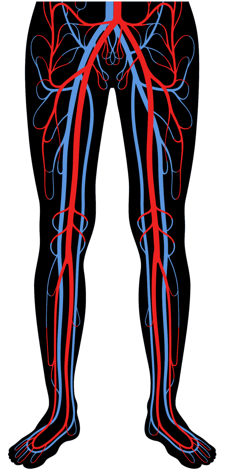
Ng Hannah Kwannoch
Critical limb ischemia can masquerade as lumbar radiculopathy and internal knee derangement in patients with type 2 diabetes, particularly when incidental degenerative imaging findings at the spine and knee provide a superficially plausible but ultimately incorrect explanation for severe, nocturnal leg pain.
First-contact musculoskeletal providers, including Chiropractors, are well positioned to recognise pain patterns disproportionate to imaging findings and to facilitate timely vascular referral, as illustrated by this patient’s complete symptom resolution within 48 hours of successful revascularisation.
AU: Ng Hannah Kwannoch MChiro. Chiropractor, New York Medical Group, Kowloon, Hong Kong. e: hannahng@nymg.com.hk
Narrative: Critical limb ischemia, now more broadly classified as chronic limb-threatening ischemia (CLTI), classically presents with rest pain, ischemic ulceration, or gangrene, but atypical presentations can closely mimic musculoskeletal and neurological disease, particularly in patients with type 2 diabetes mellitus (T2DM) who often lack classical claudication.
We report the case of a 64-year-old woman with a 10-year history of T2DM who presented to a Chiropractic clinic with two months of severe, nocturnal right knee and leg pain radiating to the foot, accompanied by sensory disturbance. Lumbar spine and knee magnetic resonance imaging (MRI) demonstrated multilevel degenerative disc disease with mild right L5 nerve root impingement and partial retinacular and meniscal tears, respectively, and the patient was initially managed for presumed lumbar radiculopathy and internal knee derangement.
Clinical examination was notable for warm, non-tender lower limbs without classical ischemic signs, and pain that was disproportionate to the modest imaging abnormalities. A concurrent diagnosis of diabetic nephropathy prompted the treating Chiropractor to suspect underlying peripheral arterial disease; computed tomography angiography and subsequent digital subtraction angiography confirmed a 90% flow-limiting stenosis of the right superficial femoral artery, consistent with CLTI. Percutaneous transluminal angioplasty with drug-eluting stent placement produced complete resolution of the nocturnal rest pain within 48 hours, confirming the vascular etiology. This case demonstrates how coexisting incidental degenerative spine and knee imaging findings can anchor clinicians to musculoskeletal diagnoses and delay recognition of limb-threatening vascular disease, and highlights diabetic nephropathy as a sentinel marker of comorbid peripheral arterial disease.
Clinicians, including chiropractors and other first-contact musculoskeletal providers, should maintain a high index of suspicion for vascular pathology when nocturnal rest pain is disproportionate to imaging findings, particularly in diabetic patients with microvascular comorbidities.
Indexing Terms: critical limb ischemia; chronic limb-threatening ischemia; peripheral artery disease; lumbar radiculopathy; knee internal derangement; diabetic nephropathy; type 2 diabetes mellitus; Chiropractic red flags.
Cite: Ng HK. Critical limb ischemia masquerading as lumbar radiculopathy and internal knee derangement in a diabetic patient: A Case Report and Literature Review. Asia-Pac Chiropr J. 2026;7.3.https://www.apcj.site/NgLimbIschaemia.pdf
Introduction
Critical limb ischemia, increasingly termed chronic limb-threatening ischemia (CLTI) to reflect its natural history of pain, tissue loss, and limb-threatening potential without revascularisation, represents the most advanced clinical stage of peripheral artery disease (PAD). (1)
PAD disproportionately affects patients with type 2 diabetes mellitus (T2DM): systematic reviews of randomised controlled trials estimate a comorbid PAD prevalence of 12.5% to 22% among people with T2DM, substantially higher than in the general population. (2) Diabetes accelerates atherosclerosis through a combination of endothelial dysfunction, chronic hyperglycaemia, and microvascular injury, and several of its microvascular complications independently predict macrovascular PAD risk. Diabetic nephropathy, in particular, has been shown to be an independent risk factor for PAD in patients with T2DM (3), and additional microvascular complications, including diabetic peripheral neuropathy and retinopathy, further compound this risk. (4, 5) These associations position renal and neuropathic complications as potential sentinel markers that should prompt vascular risk stratification even in the absence of classical claudication.
Distinguishing PAD-related leg pain from neurogenic or musculoskeletal causes is diagnostically challenging because all three conditions can produce radiating leg pain, sensory disturbance, and functional limitation. Classical intermittent claudication, cramping pain in the calf, thigh, or buttock provoked by exertion and relieved by rest, is a comparatively reliable clinical clue, but many patients, particularly those with diabetic neuropathy, present atypically with rest pain, nocturnal pain, or non-exertional symptoms that closely resemble radiculopathy or arthropathy. (6)
The ankle-brachial index remains a simple, accurate bedside tool for confirming or excluding PAD in this setting and should be considered whenever the diagnosis is entertained. (7) Several published cases illustrate how advanced PAD can masquerade as lumbar radiculopathy or sciatica: Lin et al. described three patients with aortoiliac PAD initially treated for radiculopathy after their absent pulses were overlooked, (8) and Feller et al. reported a similar case identified only after a physiotherapist recognised red-flag features and screened for vascular disease. (9) More broadly, musculoskeletal and vascular mimickers of lumbar radiculopathy are well recognised in the orthopedic literature, underscoring the need for a systematic differential diagnosis whenever radicular-appearing leg pain does not fit the expected clinical or imaging pattern. (10)
A further diagnostic pitfall arises when incidental degenerative findings on lumbar spine or knee imaging are mistakenly assumed to be causal. Meniscal tears and other internal derangements are frequently identified on knee MRI in asymptomatic middle-aged and older adults, (11) and knee osteoarthritis features are similarly common in uninjured, asymptomatic populations. (12) Analogous data from the spine literature show that degenerative changes, including disc bulges, protrusions, and annular tears, are highly prevalent on MRI in asymptomatic individuals and increase steadily with age. (13)Because patients frequently present to Chiropractors and other first-contact musculoskeletal providers before a vascular or systemic diagnosis is considered, these clinicians are well positioned to recognise red-flag features, such as pain disproportionate to imaging findings, that warrant referral for further systemic workup, a role illustrated in previous chiropractic case reports describing serious pathology, including malignancy and nerve tumors, masquerading as spinal pain. (14, 15, 16, 17)
Here we report a case in which a diabetic patient’s severe nocturnal leg pain was initially attributed to concurrent lumbar radiculopathy and internal knee derangement identified on MRI, but was ultimately found to represent critical limb ischemia, illustrating how compound incidental musculoskeletal findings can obscure a limb-threatening vascular diagnosis.
Case presentation
A 64-year-old retired woman presented to a Chiropractic clinic with severe right knee pain of approximately two months’ duration, on a background of a similar but milder episode two years earlier that had resulted in hospital admission and discharge with analgesics alone. She described the pain as sudden, sharp, and radiating from the right knee down to the foot, accompanied by numbness in the same distribution. The most distressing feature was its nocturnal nature: the pain consistently worsened before sleep and frequently woke her in the early morning. Attacks were unpredictable and rated 8-9/10 in severity, with non-steroidal anti-inflammatory drugs providing only partial relief. She also reported intermittent bilateral hand numbness that she considered less bothersome.
She had previously been treated with physiotherapy and traditional Chinese medicine, achieving only short-term benefit. Her medical history included a 10-year history of T2DM managed with oral hypoglycemic agents. During the course of Chiropractic treatment she was newly diagnosed with diabetic nephropathy, with an estimated glomerular filtration rate indicating 50% of expected renal function. There was no known history of intermittent claudication.
On examination, there was no swelling, erythema, or bruising around either knee, and both lower legs were warm to touch. Palpation of the right knee joint itself did not reproduce her pain. Mild tenderness was noted over the right sacroiliac joint, right quadratus lumborum, and right gluteal muscles, and lumbar spine range of motion was limited but not painful. Knee active, passive, and resisted ranges of motion were full and pain-free. Neurological examination revealed diminished light touch and sharp/dull discrimination in the right leg below the knee. Patellar and Achilles reflexes were 1+ bilaterally, and muscle strength was 5/5 throughout the lower limbs. Straight leg raise was positive on the right, as was the sacroiliac joint spring test. All knee-specific orthopedic tests, including McMurray, valgus/varus stress, and anterior/posterior drawer, were negative.
Investigations
Lumbar spine radiographs showed multilevel degenerative changes, and knee radiographs demonstrated mild osteoarthritis. Given persistent symptoms, the Chiropractor ordered MRI of the right knee and lumbar spine. Knee MRI revealed partial tears of the lateral and medial patellar retinaculum, tendinosis of the semimembranosus tendon, a stress response at the medial collateral ligament, and tiny tears of the posterior horn of the medial meniscus (Figures 1, 2). Lumbar spine MRI showed posterior disc prolapses from L1/2 to L5/S1 with mild bilateral foraminal narrowing, slight impingement of the right L5 nerve root, and a tiny anterior annular tear at L4/5 (Figure 3). Despite these findings, the patient continued to experience severe, nocturnal pain that responded poorly to treatment.
After her nephropathy was diagnosed, the treating Chiropractor raised concern for peripheral vascular disease, and a computed tomography angiogram of the lower limbs was arranged. This demonstrated a significant, flow-limiting stenosis in the right superficial femoral artery, consistent with critical limb ischemia.
Figure 1: Partial tear of lateral patellar retinaculum (LPR) B) Tendinosis changes at semimembranosus tendon (SM)
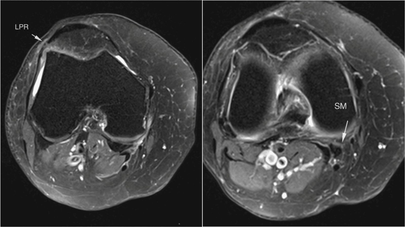
Figure 2: Tiny tears at posterior horn (post) of medial meniscus (MM) B) Stress response at the medial cruciate ligament (MCL)
Figure 3: Posterior prolapsed disc from L1/2 to L5/S1, slightly narrowing bilateral intervertebral foramina.
Differential diagnosis
The compound diagnostic picture, comprising imaging abnormalities at both the lumbar spine and knee alongside sensory and vascular risk features, generated a broad differential diagnosis, summarised in Table 1.
Treatment
The patient was urgently referred to the vascular surgery service. Diagnostic subtraction angiography confirmed a 90% stenosis of the right superficial femoral artery. Successful percutaneous transluminal balloon angioplasty with deployment of a drug-eluting stent was performed. She was commenced on dual anti-platelet therapy (aspirin and clopidogrel) and a high-intensity statin, and her diabetes and blood pressure were optimised.
She also received structured smoking cessation advice in view of a remote smoking history. Chiropractic care, comprising spinal and pelvic adjustments, soft-tissue mobilisation, electrical muscle stimulation, and short-wave therapy over 16 sessions, was paused during the acute vascular intervention and subsequently resumed post-operatively as low-grade supportive care for residual axial lumbar discomfort only; no further manual therapy was directed at the right leg.
Outcome and follow-up
Within 48 hours of revascularisation, the patient’s nocturnal rest pain resolved completely, and for the first time in months she slept through the night without waking; the radiating pain and numbness in the leg also disappeared.
At two-week post-operative follow-up, she was walking independently without analgesics, and her diabetic nephropathy remained stable under nephrology follow-up. At three months, she was pain-free and had resumed all activities of daily living. Mild residual lumbar discomfort and bilateral hand numbness persisted, consistent with pre-existing degenerative spine disease and mild diabetic neuropathy, but these were managed conservatively.
She remained under vascular surveillance with regular ankle-brachial index measurements (7) and continued monthly chiropractic care for wellness and gentle spinal mobilisation, though her primary disabling pain had been eliminated entirely by the vascular procedure.
Discussion
This case highlights how coexisting incidental imaging findings at two anatomically unrelated sites, the lumbar spine and the knee, can compound diagnostic confusion and delay recognition of a limb-threatening vascular process. Meniscal and retinacular abnormalities of the kind seen on this patient's knee MRI are common incidental findings in asymptomatic middle-aged and older adults, (11) and radiographic knee osteoarthritis is similarly prevalent in uninjured, pain-free populations. (12)
Degenerative lumbar spine findings, including disc protrusions, foraminal narrowing, and annular tears, are likewise near-ubiquitous on MRI in asymptomatic adults and increase with age, such that their presence does not establish causation for a given patient's pain. (13) In this patient, the mild right L5 nerve root impingement and the minor knee structural findings were plausible but ultimately incidental explanations that anchored clinicians to musculoskeletal diagnoses, delaying recognition that her pain pattern, severe, nocturnal, and disproportionate to imaging severity, was in fact vascular in origin.
A striking feature of this case is the absence of classical ischemic signs: both lower limbs were warm to touch, and there was no history of intermittent claudication, findings that would ordinarily lower clinical suspicion for PAD. (6).However, the emergence of diabetic nephropathy proved to be the pivotal clue. Diabetic nephropathy is independently associated with an increased risk of PAD in patients with T2DM, (3) and diabetic microvascular disease more broadly, including neuropathy and retinopathy, predicts a substantially higher risk of PAD, foot ulceration, and amputation. (4, 5) This patient’s new nephropathy diagnosis, combined with pain that was disproportionate to her imaging findings, was the trigger that prompted vascular referral.
Clinicians managing diabetic patients with atypical leg pain should therefore treat a new or worsening microvascular complication as a sentinel marker warranting vascular risk stratification, even when the limb appears clinically warm and pulses are not formally documented as diminished.
This case also underscores the role that Chiropractors and other first-contact musculoskeletal providers can play as red-flag screeners for systemic and vascular disease. (14 - 18) Similar to previously reported cases in which chiropractic clinicians identified occult malignancy or nerve tumors masquerading as spinal pain, (19 - 32) the treating chiropractor’s recognition that this patient's nocturnal pain pattern and new nephropathy diagnosis did not fit a purely musculoskeletal picture led directly to the correct diagnosis and timely revascularisation. (33, 34, 35) The dramatic and complete resolution of nocturnal rest pain within 48 hours of angioplasty, an interval far too short to reflect natural resolution of lumbar radiculopathy or knee derangement, provides strong clinical evidence that critical limb ischemia, rather than the incidental musculoskeletal findings, was the true driver of her symptoms. This temporal relationship, together with confirmatory angiographic findings, illustrates the value of a definitive vascular intervention as both treatment and diagnostic confirmation, and supports incorporating simple bedside tools such as the ankle-brachial index (7) into the assessment of diabetic patients presenting with atypical leg pain in musculoskeletal practice settings.
Conclusions
Critical limb ischemia can masquerade as lumbar radiculopathy and internal knee derangement in patients with type 2 diabetes, particularly when incidental degenerative imaging findings at the spine and knee provide a superficially plausible but ultimately incorrect explanation for severe, nocturnal leg pain.
A new diagnosis of diabetic nephropathy should prompt clinicians to consider peripheral artery disease as a sentinel vascular marker, even in the absence of classical claudication or overt ischemic signs.
First-contact musculoskeletal providers, including Chiropractors, are well positioned to recognise pain patterns disproportionate to imaging findings and to facilitate timely vascular referral, as illustrated by this patient’s complete symptom resolution within 48 hours of successful revascularisation.
References
- Conte MS, Bradbury AW, Kolh P, et al. Global vascular guidelines on the management of chronic limb-threatening ischemia. J Vasc Surg. 2019;69(6S):3S-125S.e40. PMID: 31159978.
- Verma S, Leiter LA, Mangla KK, Nielsen NF, Hansen Y, Bonaca MP. Epidemiology and burden of peripheral artery disease in people with type 2 diabetes: a systematic literature review. Diabetes Ther. 2024;15(9):1893-1961. PMID: 39023686.
- Hsieh MC, Tien KJ, Perng DS, et al. Diabetic nephropathy and risk factors for peripheral artery disease in Chinese with type 2 diabetes mellitus. Metabolism. 2009;58(4):504-509. PMID: 19303971.
- Chen T, Xiao S, Chen Z, Yang Y, Yang B, Liu N. Risk factors for peripheral artery disease and diabetic peripheral neuropathy among patients with type 2 diabetes. Diabetes Res Clin Pract. 2024;207:111079. PMID: 38154538.
- Yen FS, Yen YH, Hung YM, et al. Diabetic microvascular disease and risk of peripheral artery disease, foot ulcer, leg infection, and amputation. Thromb Haemost. 2025 Aug 5. Online ahead of print. PMID: 40763784.
- Lyden SP, Joseph D. The clinical presentation of peripheral arterial disease and guidance for early recognition. Cleve Clin J Med. 2006;73(Suppl 4):S15-S21. PMID: 17385387.
- Patel Y, Shah A, Patel P, et al. Diagnostic accuracy of ankle-brachial index in patients with peripheral arterial disease: a meta-analysis. Cardiol Rev. 2025 Oct 9. Online ahead of print. PMID: 41297000.
- Lin JD, Morrissey NJ, Levine WN. Don't forget the pulses! Aortoiliac peripheral artery disease masquerading as lumbar radiculopathy-a report of 3 cases. Am J Orthop (Belle Mead NJ). 2016;45(5):314-318. PMID: 27552456.
- Feller D, Giudice A, Maritati G, et al. Physiotherapy screening for referral of a patient with peripheral arterial disease masquerading as sciatica: a case report. Healthcare (Basel). 2023;11(11):1527. PMID: 37297667.
- Grimm BD, Blessinger BJ, Darden BV 2nd, Brigham CD, Kneisl JS, Laxer EB. Mimickers of lumbar radiculopathy. J Am Acad Orthop Surg. 2015;23(1):7-17. PMID: 25538126.
- Englund M, Guermazi A, Gale D, Hunter DJ, Aliabadi P, Clancy M, Felson DT. Incidental meniscal findings on knee MRI in middle-aged and elderly persons. N Engl J Med. 2008;359(11):1108-1115. PMID: 18784100.
- Culvenor AG, Øiestad BE, Hart HF, Stefanik JJ, Guermazi A, Crossley KM. Prevalence of knee osteoarthritis features on magnetic resonance imaging in asymptomatic uninjured adults: a systematic review and meta-analysis. Br J Sports Med. 2019;53(20):1268-1278. PMID: 29886437.
- Brinjikji W, Luetmer PH, Comstock B, et al. Systematic literature review of imaging features of spinal degeneration in asymptomatic populations. AJNR Am J Neuroradiol. 2015;36(4):811-816. PMID: 25430861.
- Lin AFC, Chu EC, Chu VK, et al. Recognizing Hong Kong Chiropractors' Sick Leave Authority: Valuing a Conservative Approach to Workers' Compensation. Cureus. 2023;15(3):e36879. Published 2023 Mar 29. DOI 10.7759/cureus.36879
- Lee WT, Chu EC, Lam K, et al. Exploring Chiropractic Healthcare in Hong Kong: Sick Leave Certification Dilemma. Cureus. 2024;16(1):e52957. Published 2024 Jan 25. DOI 10.7759/cureus.52957
- Chow CLJ, Shum JS, Hui KTP, Lin AFC, Chu EC. Optimizing Primary Healthcare in Hong Kong: Strategies for the Successful Integration of Radiology Services. Cureus. 2023;15(4):e37022. Published 2023 Apr 2. DOI 10.7759/cureus.37022
- Ng L, Chu V, Lam KK, et al. Meeting Future Demand for Chiropractic Services in Hong Kong: A Strategic Manpower Planning Approach. Cureus. 2023;15(4):e37481. Published 2023 Apr 12. DOI 10.7759/cureus.37481
- Lin AFC, Cunliffe C, Chu VK, et al. Prevention-Focused Care: The Potential Role of Chiropractors in Hong Kong's Primary Healthcare Transformation. Cureus. 2023;15(3):e36950. Published 2023 Mar 31. DOI 10.7759/cureus.36950
- Chu EC, Tao C, Cheung G. Langerhans Cell Histiocytosis Presenting as a Limp in a Child: A Case Report. Cureus. 2025;17(4):e83149. Published 2025 Apr 28. DOI 10.7759/cureus.83149
- Chu EC, Yun SMH. Metastatic Breast Cancer Presenting As Neck Pain in a Pregnant Patient. Cureus. 2024;16(1):e51444. Published 2024 Jan 1. DOI 10.7759/cureus.51444
- 2Chu EC, Piong SZ, Tao C. Chiropractic Management of Lumbar Disc Herniation in a Patient With Co-existing Liver Cancer: A Case Report. Cureus. 2024;16(1):e51445. Published 2024 Jan 1. DOI 10.7759/cureus.51445
- Mok S, Chu EC. The Importance of Early Detection of Spinal Tumors Through Magnetic Resonance Imaging in Chiropractic Practices. Cureus. 2024;16(1):e51440. Published 2024 Jan 1. DOI 10.7759/cureus.51440
- Chu EC, Trager RJ. Prevalence of serious pathology among adults with low back pain presenting for chiropractic care: a retrospective chart review of integrated clinics in Hong Kong. Med Sci Monit. 2022;28:e938042. PMID: 36164262.
- Chu EC, Sabourdy E, Cheong B. Secondary Holocord Syringomyelia Associated With Spinal Hemangioblastoma in a 29-Year-Old Female. Cureus. 2023;15(6):e40022. Published 2023 Jun 6. DOI 10.7759/cureus.4002
- Chan AK, Ng GSN, Cheong BKC, Ng KKP, Chu EC. Sacral Chordoma Presenting as Back Pain in the Chiropractic Clinic: A Case Report. Cureus. 2023;15(5):e39810. Published 2023 May 31. DOI 10.7759/cureus.39810
- Chu EC, Trager RJ, Lee WT, Tam DMY, Kwok R. Spinal Metastasis Causing Dropped Head Syndrome in the Chiropractic Office: A Case Report. Cureus. 2023;15(2):e34796. Published 2023 Feb 9. DOI 10.7759/cureus.34796
- Chu EC, Sabourdy E. Sporadic Desmoid Tumor Mimicking Myofascial Pain Syndrome in a Chiropractic Clinic. Cureus. 2023;15(9):e44653. Published 2023 Sep 4. DOI 10.7759/cureus.44653
- Chu EC, Sabourdy E. Dorsal Scapular Osteochondroma in the Chiropractic Clinic: A Case Report and Literature Review. Cureus. 2023;15(7):e42007. Published 2023 Jul 17. DOI 10.7759/cureus.42007
- Chu EC, Trager RJ, Shum JS, Ng KK. Sacral Giant Cell Tumor Presenting as Low Back Pain in the Chiropractic Office: A Case Report. Cureus. 2023;15(1):e33262. Published 2023 Jan 2. DOI 10.7759/cureus.33262
- Chu EC, Yau K, Yun SMH. Spondylodiscitis in a Patient Undergoing Chronic Peritoneal Dialysis Presenting to a Chiropractor: Case Report and a Review of the Literature. Cureus. 2023;15(8):e44312. Published 2023 Aug 29. DOI 10.7759/cureus.44312
- Chu EC, Lee WT. Prostate Cancer Presenting as Hip Pain at the Chiropractic Office: A Case Report and Literature Review. Cureus. 2023;15(1):e34049. Published 2023 Jan 21. DOI 10.7759/cureus.34049
- Chu EC, Trager RJ, Lee WT, Cheong BKC, Hei SYM. Lung Cancer With Vertebral Metastases Presenting as Low Back Pain in the Chiropractic Office: A Case Report. Cureus. 2023;15(2):e34821. Published 2023 Feb 9. DOI 10.7759/cureus.34821
- Chu EC. Idiopathic Avascular Necrosis of the Femoral Head in an Older Adult: A Case Report. Cureus. 2026;18(7):e112084. Published 2026 Jul 5. DOI 10.7759/cureus.112084
- Chu EC, Chiang R. Giant cell arteritis: a cause of temporomandibular joint claudication. J Med Life. 2022;15(10):1322-1326. DOI 10.25122/jml-2022-0076
- Chu EC, Lai C, Huang KS, Lin AFC. Bilateral Cellulitis Presented As Lower-Extremity Pain Reported in a Chiropractic Clinic: A Case Report. Cureus. 2023;15(2):e35470. Published 2023 Feb 25. DOI 10.7759/cureus.35470

Shaan Rai
At presentation, the patient’s goals were improved sleep quality, more consistent tolerance of food, and general improvement in wellbeing; he described feeling frustrated by the persistence of his symptoms and motivated to address them.
AU: Shaan Rai MChirop (AECC). Private practice of Chiropractic. Singapore. e: help@vitalitychiropracticcentres.com
Background: A 39-year-old male IT engineer presented with a five-year history of gastro-oesophageal reflux, gastritis, and bloating, alongside a co-existing medical diagnosis of cardiac ectopy, despite prior medical investigation and treatment including endoscopy, antacid therapy, and dietary modification. Examination revealed postural asymmetry, restricted cervical range of motion, a weight-bearing asymmetry, a functionally short leg that corrected with cervical rotation, and position-specific upper limb weakness.
Intervention: The patient underwent a course of Knee Chest Upper Cervical (KCUC) Chiropractic care, with correction of a C1-right listing identified using postural, weight-bearing, and thermographic assessment and confirmed radiographically.
Outcomes: Serial reassessment showed normalisation of postural, weight-bearing, and cervical range-of-motion asymmetries, alongside a reduction in paraspinal thermographic asymmetry. The patient reported substantial improvement in bloating, digestion, and general wellbeing, corroborated by a recorded patient testimonial. His co-existing cardiac ectopy was not reassessed as part of this report.
Conclusion: This case adds to a small body of literature linking upper cervical and vagal function to gastrointestinal symptoms, and supports further research into upper cervical chiropractic care as a component of management for treatment-resistant gastrointestinal presentations.
Indexing Terms: Chiropractic; Subluxation; gastro-oesophageal reflux; Knee Chest Upper Cervical; vagus nerve.
Cite: Rai S. Improvement in gastrointestinal symptoms concomitant with Knee Chest Upper Cervical Chiropractic care: A Case Report. Asia-Pac Chiropr J. 2026;7.3. https://www.apcj.site/RaiGIKneeChest.pdf
Take-away: ' ... this case provides a rationale for further research into upper cervical Chiropractic care as part of the management of treatment-resistant gastrointestinal symptoms …’
Background
Gastro-oesophageal reflux disease (GERD) and functional dyspepsia are highly prevalent gastrointestinal conditions, and a substantial proportion of affected patients continue to report bloating, postprandial discomfort, and sleep disturbance despite acid-suppressive therapy and dietary modification. The vagus nerve is the principal parasympathetic conduit of the gut-brain axis, modulating gastric motility, secretion, and visceral sensation, and disruption of vago-vagal reflex signalling has been implicated in functional gastrointestinal disorders such as functional dyspepsia, in which patients demonstrate altered vagal afferent processing of gastric distension and impaired gastric accommodation. (1, 2)
Anatomically, the vagus nerve exits the jugular foramen and travels through the neck in close proximity to the upper cervical spine before descending through the thorax to the abdominal viscera, and vagal outflow originates from brainstem nuclei situated immediately adjacent to the atlanto-occipital junction. In an experimental model of functional dyspepsia, altered vagal afferent signalling from the stomach was shown to drive both visceral hypersensitivity and associated affective symptoms, implicating vagal dysregulation as a contributor to functional gastrointestinal symptoms. (2) This anatomical and physiological relationship raises the possibility that upper cervical dysfunction, situated close to the upper spinal cord and brainstem origin of the vagus nerve, could plausibly influence gastrointestinal symptoms in some patients, though this remains a hypothesis rather than an established mechanism.
A separate body of chiropractic literature has examined the relationship between cervical adjustment and autonomic nervous system activity. Adjustment of the cervical spine has been associated with a measurable parasympathetic shift in healthy volunteers, attributed to the proximity of the upper cervical segments to brainstem autonomic centres. (3) Case reports have also described improvement in gastrointestinal symptoms, including cyclic vomiting and gastro-oesophageal reflux, concomitant with upper cervical or cervical chiropractic care, though this literature remains limited to isolated cases. (4, 5) We report a case of longstanding, treatment-resistant gastro-oesophageal reflux and gastritis, in a patient with a co-existing diagnosis of cardiac ectopy, who underwent a course of KCUC chiropractic care. Written informed consent was obtained from the patient for publication of this case report, including a recorded video testimonial quoted herein.
Patient information
A 39-year-old male information technology engineer presented for a Chiropractic consultation in September 2020 with a five-year history of gastro-oesophageal reflux and associated symptoms.
History of presenting complaint
The patient’s symptoms began approximately five years prior to presentation with gastro-oesophageal reflux. The condition was initially marked by an episode of blood in the stool, prompting endoscopic investigation and a course of medication; rectal bleeding resolved, but the patient continued to experience torso pain, for which antacid therapy was recommended. Bloating and abdominal pain persisted despite this management. Symptoms were reported as somewhat better with salad-based meals, self-massage of the abdomen, and sleeping on a firm mattress, and worse with rapid ingestion of large volumes of water, at bedtime, during periods of anxiety, with poor posture or prolonged desk-based work, and when lying supine; specific food triggers were inconsistent and difficult for the patient to identify.
Sleep was affected by abdominal discomfort that was worse at bedtime and in the supine position, with symptoms improving only after approximately eight hours of sleep. The patient also carried a separate medical diagnosis of cardiac ectopy (premature ventricular contractions), for which he attended regular cardiology follow-up and had previously used an ambulatory cardiac monitor; this diagnosis is reported here as relevant medical history and was not formally reassessed as part of chiropractic care. Gastrointestinal symptoms were reported to respond partially to garlic and ginger supplementation.
At presentation, the patient’s goals were improved sleep quality, more consistent tolerance of food, and general improvement in wellbeing; he described feeling frustrated by the persistence of his symptoms and motivated to address them.
The patient’s working medical diagnoses, established by his treating gastroenterologist and cardiologist respectively, were gastro-oesophageal reflux disease, gastritis, and cardiac ectopy. He reported no relevant prior surgery, and no current medication (previously prescribed antacid therapy had been discontinued). Prior management consisted of medication, dietary elimination, and self-massage of the abdomen, with only partial and inconsistent relief. No relevant familial or hereditary conditions were reported. The patient denied tobacco, alcohol, or substance use. He was physically active, weight training and cycling three times per week.
Physical examination
Postural assessment revealed a low right shoulder and anterior head carriage. Cervical range of motion was restricted in left lateral flexion, extension, and left rotation, without associated pain. Cervical distraction and cervical compression testing were both negative. Static bilateral weight-bearing assessment showed a 4.1 kg side-to-side discrepancy (39.8 kg left, 35.7 kg right). Blood pressure was 128/100 mmHg with a pulse of 83 beats per minute. Prone leg length assessment showed a functionally short left leg of approximately 1.5 cm that normalised with right cervical rotation, suggesting a cervical rather than structural origin.
Upper cervical manual muscle testing demonstrated weakness on shoulder abduction specifically when the head was turned right in combined rotation and extension (looking up), with no weakness elicited in other head positions; this position-specific pattern, together with the corrected leg length and absence of pain on orthopaedic provocation testing, comprised the objective examination findings, which were assessed and recorded as a single grouped set of clinical indicators. No focal neurological deficit was identified.
Diagnostic imaging
Upper cervical radiographs (lateral cervical, base posterior, and anteroposterior open-mouth views in neutral and left/right lateral flexion) were obtained on 23 September 2020 (Figures 1 - 5). Radiologist interpretation reported loss of the normal cervical lordosis with straightening of the cervical spine and mild osteophytosis of the vertebral bodies, without loss of vertebral height or significant narrowing of the disc spaces. Computerised infrared paraspinal thermographic scanning was also performed at baseline as part of standard KCUC workup (Figure 6).
Thermographic findings showed asymmetric thermal patterns in the upper cervical paraspinal region, consistent with underlying nerve irritation; together with the postural, weight-bearing, and leg length findings described above, these clinical indicators identified the upper cervical listing, which was subsequently confirmed by radiographic analysis (C1 on the right). No magnetic resonance or computed tomography imaging was obtained; the patient's gastrointestinal and cardiac symptoms had already been medically investigated and organically excluded by his treating gastroenterologist (via endoscopy) and cardiologist (via ambulatory cardiac monitoring) prior to chiropractic consultation, and no red-flag features were present to warrant central nervous system imaging at the time.
Figure 1: Lateral cervical radiograph showing loss of the normal cervical lordosis with straightening of the cervical spine. Date of imaging: 23 September 2020.
Figure 2: Base posterior radiograph of the upper cervical spine. Date of imaging: 23 September 2020.
Figure 3: Anteroposterior open-mouth (APOM) radiograph, neutral position. Date of imaging: 23 September 2020.
Figure 4: Anteroposterior open-mouth (APOM) radiograph, right lateral flexion. Date of imaging: 23 September 2020.
Figure 5: Anteroposterior open-mouth (APOM) radiograph, left lateral flexion. Date of imaging: 23 September 2020.
Figure 6: Baseline computerised infrared paraspinal thermographic scan sequence from the initial visit (23 September 2020), showing pre- and post-adjustment DELTA-T (left-right paraspinal thermal differential) tracings recorded during progressive listing correction.
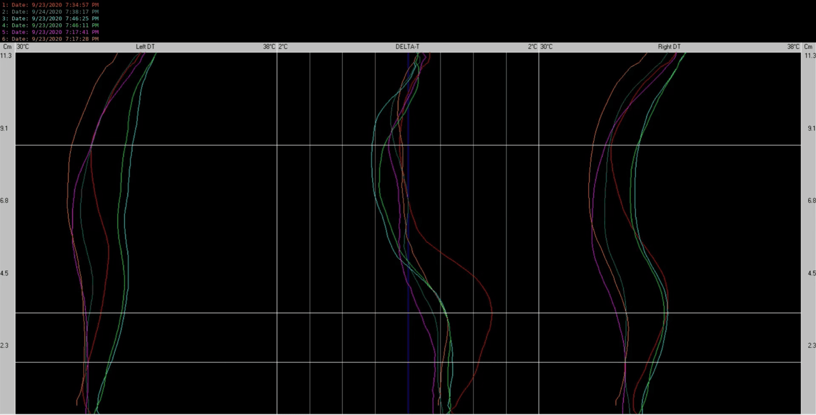
Intervention
Based on the history, examination, and imaging findings, the Chiropractic working diagnosis was upper cervical dysfunction, in a patient carrying separate medical diagnoses of gastro-oesophageal reflux disease, gastritis, and cardiac ectopy made by his treating gastroenterologist and cardiologist respectively.
This Chiropractic impression was reached by a process of elimination: the patient had already pursued standard medical management for his gastrointestinal and cardiac symptoms without full resolution. The examination findings, including position-dependent upper limb weakness specific to combined right cervical rotation and extension, a functionally short leg that normalised with cervical rotation, and a weight-bearing asymmetry, were judged consistent with a cervical contribution to autonomic function. This impression was informed by the anatomical proximity of the upper cervical spine to the upper spinal cord and brainstem structures that give rise to the vagus nerve, the principal parasympathetic pathway influencing gastrointestinal motility and secretion. (1) This relationship is discussed further in the Conclusion below.
The Knee Chest Upper Cervical (KCUC) technique is a high-velocity, low-amplitude thrust adjustment used to correct upper cervical (atlanto-occipital and atlanto-axial) misalignment, delivered with the patient prone in a knee-chest position with the head rotated to one side. The patient underwent care using this technique, performed as per standard clinical procedure with no variation. The specific contact point (listing) corrected was C1 on the right, identified using postural, weight-bearing, and thermographic findings and confirmed by the initial upper cervical radiographic series, in accordance with standard KCUC protocol.Visit frequency began at twice weekly, reducing to once weekly as reassessment findings indicated stabilisation. The patient completed 24 visits during the initial active care plan over approximately five months, followed by a further period of maintenance checkups (six visits over an additional five months). The patient subsequently continued checkups at intervals of approximately three weeks until 10 June 2021, after which he ceased attending; continued periodic checkups had been advocated by the clinic throughout this period.
Outcomes
The Bournemouth Questionnaire (BQ), a validated seven-item patient-reported outcome measure assessing pain, disability, and cognitive/affective dimensions, was used to record baseline status. (6) Baseline BQ score at the initial consultation (23 September 2020) was 24/70. A discharge BQ score was not recorded; this is addressed as a limitation below.
First reassessment (29 October 2020)
Postural asymmetry persisted, with uneven shoulders still present. Weight-bearing scale readings were 40 kg on the left and 34 kg on the right (6 kg discrepancy). Arm and shoulder strength was now balanced, with no weak positions identified on manual muscle testing. Leg length was even. Cervical range of motion was normal except for persistent stiffness on left lateral flexion. Comparative thermographic scanning at this visit (Figure 7) showed the paraspinal DELTA-T tracing running closer to the zero-differential midline in the upper cervical region than at baseline, consistent with reduced thermal asymmetry. The patient reported improved digestion and that his neck moved better, despite having initially reported no neck complaints. He described his stomach as feeling better and less bloated, with improvement noted from around the time of his second adjustment.
Figure 7:Comparative paraspinal thermographic scan: baseline (23 September 2020, red) versus first reassessment (29 October 2020, cyan).
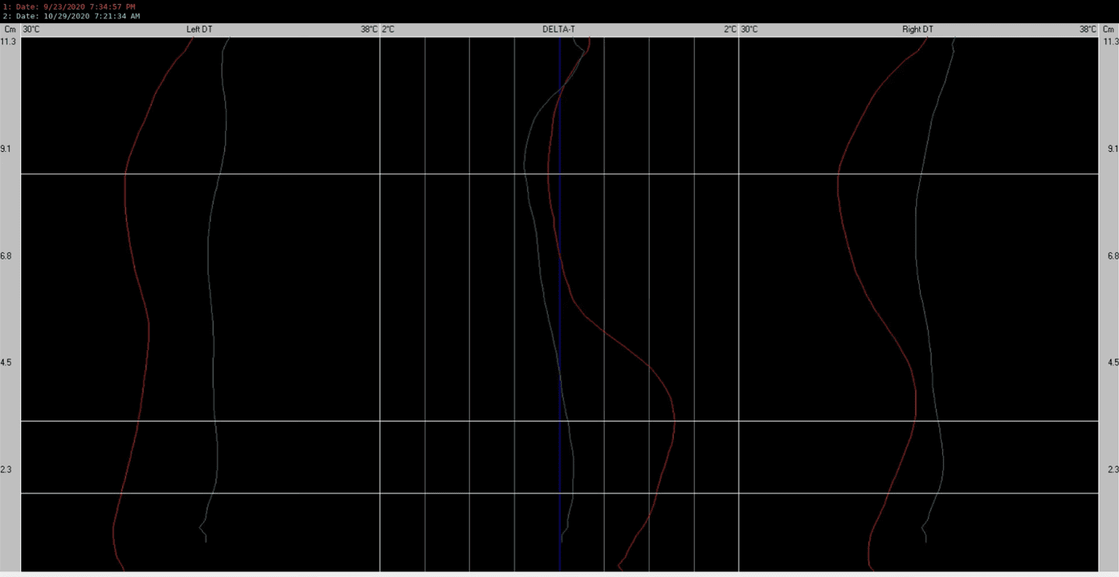
Second reassessment (4 February 2021)
Shoulder posture was even. Weight-bearing scale readings were 35.3 kg on the left and 32.2 kg on the right (3.1 kg discrepancy). Arm and shoulder strength remained balanced with no weak positions. Leg length was even. Cervical range of motion was normal. Comparative thermographic scanning at this visit (Figure 8) continued to show the paraspinal DELTA-T tracing running close to the zero-differential midline, consistent with the postural and range-of-motion findings above. The patient reported that his stomach was doing well, describing himself as ’80% better’, with improved general wellbeing, better digestion and overall health, and a slight improvement in mood. He reported being able to start exercising more and was considering joining a gym.
Figure 8:Comparative paraspinal thermographic scan: baseline (23 September 2020, red) versus first reassessment (29 October 2020, cyan).
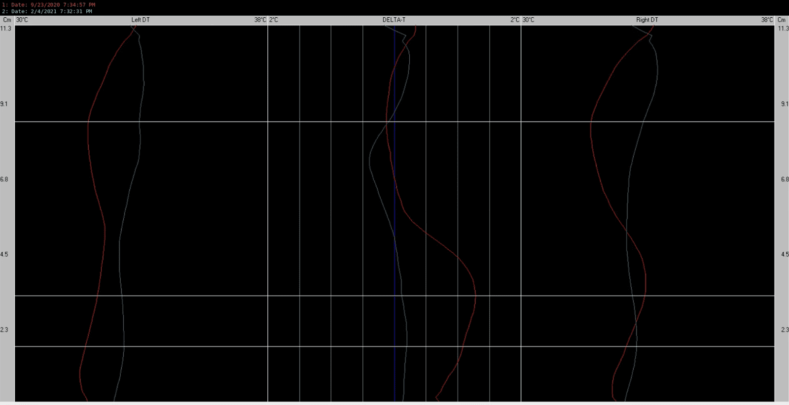
Patient-reported experience
In addition to the reassessment findings above, the patient recorded a video testimonial partway through his care, in which he stated:
‘I am halfway through and I’m already seeing such great improvement’.
Later in the same recording, he described his bloating specifically:
‘I used to get a lot of bloating in my stomach as well, so that’s more or less gone, right. So it’s almost like a complete 100% recovery on the bloating side’.
He went on to say:
‘And generally, I feel like my stomach itself feels better. And something that I couldn’t get in any other kind of treatment, right. It’s only Chiropractic that has given me so much improvement’.
He further noted the absence of any concurrent medication:
‘And this is happening without any kind of medication, that is very important for me’.
Adverse events
No adverse reactions or side effects related to Chiropractic care were reported at any point during the course of treatment.
Discharge and follow-up
No referrals were made during the course of Chiropractic care, as the patient had already been evaluated by his medical doctors prior to presentation. The patient continued periodic checkups, spaced progressively further apart, until 10 June 2021, at which point he ceased attending; the clinic continued to advocate for ongoing checkups throughout this period. The reason for discontinuation was not formally documented, though the patient may have relocated overseas.
Conclusion
This case describes significant improvement in gastrointestinal symptoms, bloating, digestion, and general wellbeing, concomitant with a course of KCUC Chiropractic care, alongside normalisation of postural, weight-bearing, and cervical range-of-motion asymmetries, in a patient with a five-year history of treatment-resistant gastro-oesophageal reflux, gastritis, and separately diagnosed cardiac ectopy. The patient’s own account, corroborated by a recorded testimonial, described near-complete resolution of bloating and marked improvement in digestion without concurrent medication, alongside objective examination findings that moved in the same direction over the same period.
The vagus nerve is the principal parasympathetic mediator of the gut-brain axis, modulating gastric motility, secretion, and visceral sensation, and it exits the skull and travels through the neck in close anatomical proximity to the upper cervical spine before descending to the abdominal viscera. (1)
Experimental models of functional dyspepsia have demonstrated that altered vagal afferent signalling from the stomach can drive both visceral hypersensitivity and associated affective symptoms, implicating vagal dysregulation as a contributor to functional gastrointestinal symptoms of the kind described by this patient. (2) This offers a plausible rationale for why gastrointestinal symptoms improved concomitant with an intervention directed at upper cervical function. The patient’s separately diagnosed cardiac ectopy is noted here as relevant medical background only: his cardiac symptoms were not formally reassessed during Chiropractic care, and no claim is made regarding any effect of care on his cardiac status.
This is consistent with a small body of Chiropractic literature suggesting that cervical spinal adjustment can produce measurable shifts in autonomic nervous system activity, with cervical adjustment associated with a parasympathetic-dominant response in healthy volunteers, attributed to the proximity of the upper cervical segments to brainstem autonomic centres. (3) Isolated case reports have also described improvement in gastrointestinal symptoms concomitant with upper cervical or cervical chiropractic care, including cessation of cyclic vomiting in a paediatric patient following upper cervical adjustment (4) and resolution of gastro-oesophageal reflux disease following correction of postural dysfunction in an adult patient. (5) This case adds to that literature.
An important feature of this case is that the patient's improvement occurred in a context where conventional medical management, endoscopy-guided treatment, antacid therapy, and dietary elimination, had already been pursued without full resolution of symptoms, and where Chiropractic care was undertaken without concurrent medication. This sequencing, together with the convergence of the patient’s subjective report and the objective examination findings, supports a genuine treatment-related effect rather than a purely coincidental or natural-history explanation for the improvement.
Practitioners seeing patients with treatment-resistant gastrointestinal symptoms should consider an upper cervical structural and functional assessment as part of a broader, medically co-managed approach.
This is a single case report, and the proposed vagal mechanism was not directly tested in this patient (for example, by heart rate variability testing); the examination findings described are consistent with, but not diagnostic of, this mechanism. A discharge Bournemouth Questionnaire score was not recorded, so outcome assessment in this report relies on serial clinical reassessment findings and the patient’s own reported experience rather than a validated discharge score; the BQ itself was not developed or validated specifically for gastrointestinal symptoms.
Thermography was used as an adjunct clinical assessment tool to help identify and reassess the upper cervical listing; the trend toward reduced paraspinal thermal asymmetry on serial scanning (Figures 6 - 8) is reported descriptively rather than as evidence of clinical change in its own right. The patient’s separately diagnosed cardiac ectopy was not formally reassessed during care, and no claim is made regarding any effect of chiropractic care on cardiac status. Central nervous system imaging (MRI/CT) was not obtained, as the patient’s presentations had already been medically investigated and organic pathology addressed prior to chiropractic consultation.What this report adds
This case report describes significant, sustained improvement in gastrointestinal symptoms and general wellbeing, alongside normalisation of objective upper cervical examination findings, in a patient with treatment-resistant, medically diagnosed gastro-oesophageal reflux disease and gastritis, concomitant with a course of Knee Chest Upper Cervical Chiropractic care undertaken without concurrent medication. Given the anatomical and physiological links between the upper cervical spine, the vagus nerve, and gastrointestinal function, this case report provides a rationale for further research into upper cervical Chiropractic care as part of the management of treatment-resistant gastrointestinal symptoms.
Consent
Written informed consent for publication of this case report, including clinical images and a recorded patient testimonial, was obtained from the patient using the clinic’s in-house consent process.
Conflicts of interest
The author declares no conflicts of interest and received no funding for this work.
References
- Breit S, Kupferberg A, Rogler G, Hasler G. Vagus nerve as modulator of the brain-gut axis in psychiatric and inflammatory disorders. Front Psychiatry. 2018;9:44. DOI 10.3389/fpsyt.2018.00044
- Cordner ZA, Li Q, Liu L, Tamashiro KL, Bhargava A, Moran TH, Pasricha PJ. Vagal gut-brain signaling mediates amygdaloid plasticity, affect, and pain in a functional dyspepsia model. JCI Insight. 2021;6:e144046. DOI 10.1172/jci.insight.144046
- Welch A, Boone R. Sympathetic and parasympathetic responses to specific diversified adjustments to chiropractic vertebral subluxations of the cervical and thoracic spine. J Chiropr Med. 2008;7:86-93. DOI 10.1016/j.jcm.2008.04.001
- Hubbard TA, Crisp CA. Cessation of cyclic vomiting in a 7-year-old girl after upper cervical chiropractic care: a case report. J Chiropr Med. 2010;9:179-183. DOI 10.1016/j.jcm.2010.07.006
- Chu EC-P, Butler KR. Resolution of gastroesophageal reflux disease following correction for upper cross syndrome—a case study and brief review. Clin Pract. 2021;11:322-326. DOI 10.3390/clinpract11020045
- Bolton JE, Breen AC. The Bournemouth Questionnaire: a short-form comprehensive outcome measure. I. Psychometric properties in back pain patients. J Manipulative Physiol Ther. 1999;22:503-510. DOI 10.1016/S0161-4754(99)70001-1

Jessey Wong and Shaan Rai
• A 38-year-old female instructor with a 7-year history of chronic gastrointestinal symptoms and comorbid generalised anxiety disorder (GAD), alongside dizziness, tinnitus, palpitation, headache and jaw tightness.
• Knee Chest Upper Cervical (KCUC) care was directed at a working chiropractic finding of vertebral subluxation complex at C1.
• By the eleventh follow-up visit (16 August 2024) the patient reported approximately 80% improvement in gastrointestinal symptoms, discontinuation of esomeprazole after roughly two years of use, resolution of headache, jaw tightness and finger paraesthesia, and improved sleep.
• At a follow-up visit on 31 July 2026 the patient's Bournemouth Questionnaire (BQ) score had fallen from a baseline of 70/70 to 17/70 (a 75.7% reduction), and she is now managed on a maintenance/check-up schedule.
• The patient paused care after 33 of a planned 36 visits without a formal discharge assessment before this follow-up; the interval between assessments and current maintenance status are discussed as limitations.
AU1: Jessey Wong MChiro (AECC). Private practice of Chiropractic, Singapore. Vitality Chiropractic Centres. e: jesseyuxin@gmail.com
AU2: Shaan Rai MChirop (AECC). Private practice of Chiropractic. Singapore
Background: A 38-year-old female instructor presented with a 7-year history of chronic gastrointestinal symptoms (nausea, oesophageal and upper-abdominal pain, bloating, and a sensation of reflux rising into the throat), alongside neck and shoulder stiffness, bilateral finger paraesthesia, tinnitus and ear fullness, dizziness, palpitation, bilateral temporal and occipital headache, and jaw tightness. The patient carried a prior medical diagnosis of generalised anxiety disorder (GAD), for which she was taking clonazepam, and was concurrently managed with esomeprazole and domperidone for gastrointestinal symptoms.
Intervention: Knee Chest Upper Cervical (KCUC) Chiropractic care was directed at a working finding of vertebral subluxation complex at C1, identified through postural, orthopaedic and thermographic assessment and confirmed radiographically. The correction was delivered in the standard prone knee-chest position; the patient was rested seated, rather than supine, in the recovery room following each adjustment because of her reflux symptoms. Care was delivered twice weekly, tapering to weekly.
Outcomes: Baseline Bournemouth Questionnaire (BQ) score was 70/70. By the eleventh follow-up visit (16 August 2024) the patient reported roughly 80% improvement in her gastrointestinal symptoms, had discontinued esomeprazole after approximately two years of use, and reported complete resolution of headache, jaw tightness and finger paraesthesia, and improved sleep. The patient paused care after 33 of a planned 36 visits without a formal discharge assessment; she subsequently returned to the practice, and at a follow-up visit on 31 July 2026 her BQ score had fallen to 17/70, a 75.7% reduction from baseline. She is now managed on a maintenance/check-up schedule. No adverse events were reported during the course of care.
Conclusion: This case describes a temporal association between KCUC care and marked, sustained improvement in chronic, multisystem gastrointestinal and autonomic symptoms in a patient with comorbid GAD, with a 75.7% reduction in BQ score (70/70 to 17/70) documented at follow-up despite a pause in active care. As a single, uncontrolled case, causation cannot be inferred; the report is offered as hypothesis-generating and as a contribution to the case-report evidence base for upper cervical Chiropractic and visceral/autonomic presentations.
Indexing Terms: Chiropractic; Subluxation; Knee Chest Upper Cervical; gastrointestinal dysfunction; generalised anxiety disorder; autonomic nervous system.
Cite: Wong J, Rai S. Improvement in chronic Gastrointestinal and multi-system Autonomic symptoms in a 38-year-old female concomitant with Knee Chest Upper Cervical Chiropractic care: A Case Report. Asia-Pac Chiropr J. 2026;7.3. https://www.apcj.site/WongRaiKCDysautonomia.pdf
Take-away: ' … at a follow-up visit on 31 July 2026 a repeat BQ was completed, scoring 17/70, a reduction of 53 points, or 75.7%, from baseline …’
Background
Upper cervical Chiropractic techniques share a common theoretical premise: that the craniocervical junction, given its proximity to the brainstem and its role in postural and proprioceptive regulation, is a clinically significant site for the assessment and correction of vertebral subluxation. The Knee Chest Upper Cervical (KCUC) procedure is one of several articular-model upper cervical protocols descended from the work of BJ Palmer, using a knee-chest patient position and radiographic analysis to guide a precise, low-force correction of the atlas.
Chronic functional gastrointestinal symptoms frequently present alongside anxiety and other autonomic complaints, consistent with the recognised bidirectional relationship between the gut and the central nervous system, the ‘gut-brain axis’. This case is reported to add to the case-based literature examining the relationship between upper cervical chiropractic care and multisystem, autonomically-mediated presentations.
Patient history
A 38-year-old female, employed as an instructor, presented with a seven-year history of chronic gastrointestinal symptoms comprising nausea, pain localised to the oesophagus and upper abdomen, bloating, and a sensation of something rising from the stomach into the throat. Onset of these symptoms coincided with a diagnosis of generalised anxiety disorder (GAD) seven years earlier, made by the patient’s medical team. She additionally reported neck and shoulder stiffness, bilateral finger numbness and tingling, tinnitus and ear fullness, dizziness and vertigo of eight months' duration, heart palpitation, bilateral temporal and occipital headache, and jaw tightness.
At presentation the patient was taking esomeprazole (two years) for gastrointestinal symptoms, domperidone for nausea, and clonazepam (nine months) for anxiety. She had previously trialled homeopathy without documented benefit, and a three-month course of physiotherapy that she reported helped her dizziness. Family history was notable for type 2 diabetes mellitus in her father. She reported no smoking, alcohol or substance use, an active lifestyle including regular Pilates practice, and approximately two hours of daily screen use.
Clinical findings
Postural observation identified a left head tilt. Cervical range of motion was unremarkable. Static weight-bearing analysis showed asymmetric distribution of 33.25 kg on the left and 28.5 kg on the right (a 4.75 kg leftward lean), with a left short leg on prone leg check, which was accentuated on left head rotation. Upper-limb manual muscle testing, cranial nerve screening, cervical distraction and compression testing, deep tendon reflexes, and Romberg testing were all unremarkable.
Diagnostic imaging
A standard five-view cervical radiographic series (lateral cervical, base posterior, and APOM neutral, left and right lateral flexion) was obtained (Figures 1 - 5). Radiological findings showed loss of the normal cervical lordosis with straightening of the cervical spine, moderate narrowing of the C5/6 disc space with mild osteophytosis at C5 and C6, and mild osteophytic lipping at C4, without loss of vertebral height.
The atlanto-axial articulation was unremarkable and the lateral atlanto-dental intervals were symmetrical bilaterally. An incidental finding of left-sided sinusitis with a narrowed nasal septum was also noted on imaging.
Figure 1: Lateral cervical radiograph.
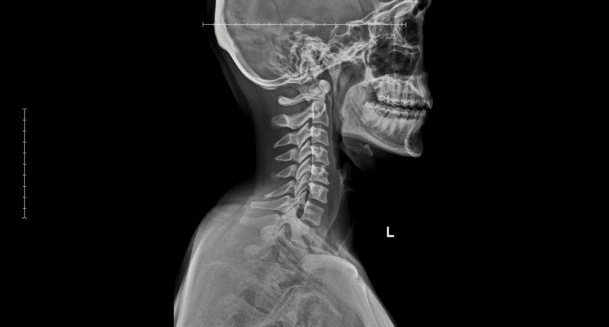
Figure 2: Base posterior radiograph.
Figure 3: APOM (anterior-posterior open mouth) neutral radiograph.
Figure 4: APOM right lateral flexion radiograph.
Figure 5: APOM left lateral flexion radiograph.
Serial computed paraspinal thermography was performed as part of this practice's standard pre-adjustment assessment protocol, with a baseline scan at the initial visit (4 July 2024; Figure 6) and a further re-scan approximately three months into care (3 October 2024; Figure 7). As in the authors’ other published case reports, thermography was used as an adjunct clinical assessment of paraspinal thermal asymmetry, employed to support, rather than independently establish, the presence of subluxation; it does not itself constitute a validated diagnostic or outcome measure, and radiographic confirmation was used as the reference standard for the subluxation finding reported below.
Qualitatively, the baseline scan showed a marked bilateral thermal asymmetry pattern, most pronounced in the upper cervical/occipital region, while the three-month re-scan showed a visibly reduced deviation from thermal symmetry through most of the cervical region compared with baseline.
Figure 6: APOM left lateral flexion radiograph. Baseline paraspinal thermography, 4 July 2024 (initial consultation; pre- and serial post-adjustment scans). Free-text clinical comments have been redacted from the scan header; dates and reference values are preserved.
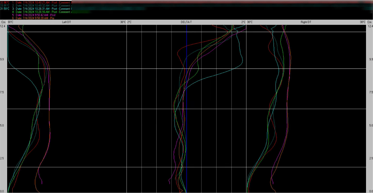
Figure 7: Paraspinal thermography at approximately three months of care, 3 October 2024 (red = baseline, cyan = follow-up). Free-text clinical comments have been redacted from the scan header; dates and reference values are preserved.
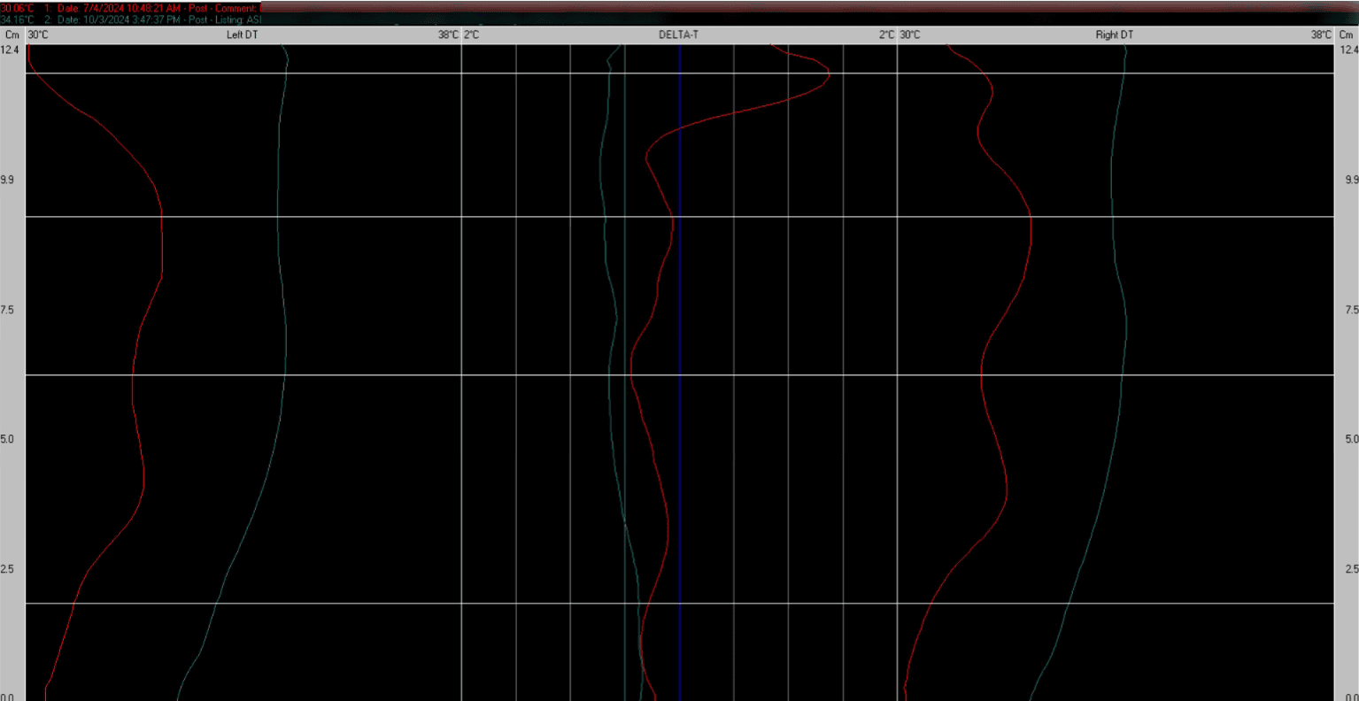
Diagnosis
On the basis of postural, orthopaedic and radiographic findings, a working Chiropractic diagnosis of vertebral subluxation complex at C1 was made. The patient’s generalised anxiety disorder is recorded here as the working medical diagnosis established by her treating medical team; it was not independently confirmed by the authors and is not presented as a Chiropractic diagnosis.
Given the multisystem, partly functional nature of the presenting symptoms, differentials considered by the referring medical team reportedly included primary anxiety-related and functional gastrointestinal disorders; central or vestibular contributions to the dizziness were not radiologically excluded, as no MRI or CT had been obtained at the time of writing.
Intervention
KCUC care
KCUC care was delivered following standard procedure, with the patient positioned prone in the standard knee-chest posture for the correction itself. The one variation from standard procedure occurred in the recovery room; the patient rested seated, rather than supine, following each adjustment, because of her acid-reflux symptoms. The correction followed the KCUC model of a precise, low-force contact to the atlas, with vector and contact point determined by the radiographic analysis described above.
Care plan
Visit frequency was set at twice weekly initially, gradually reducing to once weekly, over a planned corrective course of 36 visits across an anticipated 4 to 6 months, with no changes made to the treatment plan during that phase.
The patient attended 33 of the 36 planned visits before a pause in care, without a formal discharge assessment. She has since returned to the practice and is currently receiving maintenance care.
Outcomes
Objective and patient-reported
Baseline Bournemouth Questionnaire (BQ) score, taken on 4 July 2024, was 70/70, the maximum possible score on this instrument. At the 11th (eleventh) follow-up visit (the 12th visit overall counting the initial consultation as visit one), on 16 August 2024, the patient reported marginal improvement in her gastrointestinal symptoms at that point in care and stated she had discontinued esomeprazole (Nexium), which she had previously used for approximately two years, together with complete resolution of headache and jaw tightness, resolution of finger numbness and tingling, and improved sleep quality. By the same milestone she described roughly an 80% improvement in her gastrointestinal symptoms overall.
The patient paused care after 33 of the planned 36 visits without a formal discharge assessment. She subsequently returned to the practice, and at a follow-up visit on 31 July 2026 a repeat BQ was completed, scoring 17/70, a reduction of 53 points, or 75.7%, from baseline.
She is now managed on a maintenance/check-up schedule rather than active corrective care. The table below summarises the reported timeline.
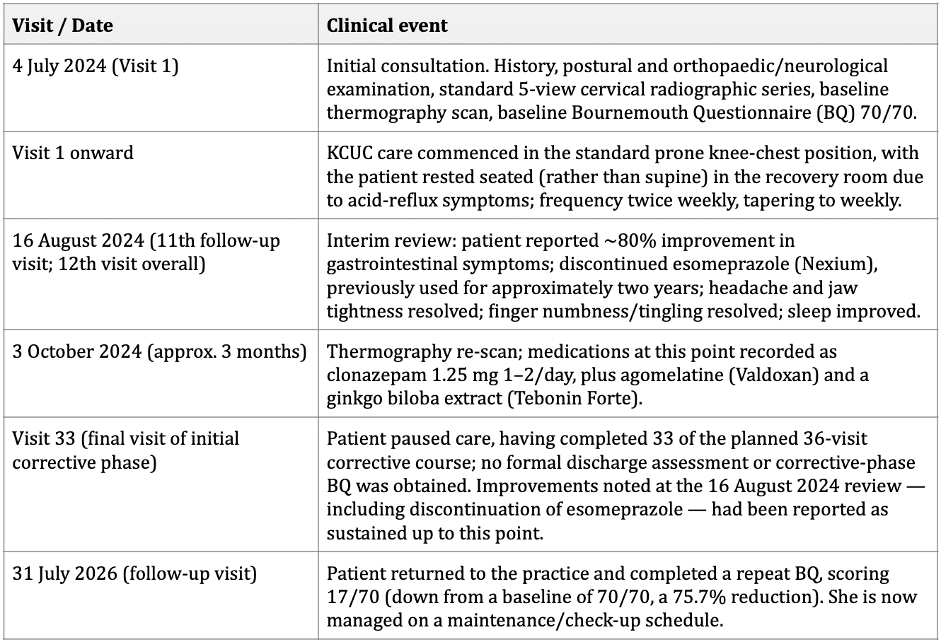
Patient perspective
‘Jessey has helped me get through my acid reflux issues. I started feeling better starting session 4. As of session 13 I have 90% relief from my symptoms. Thank you Jessey, this has given me a better quality of life. I am so thankful to Jessey. The Vitality team is amazing; they are very helpful in every step of the way. Wonderful experience at Vitality’.
This account, given in the patient’s own words, is broadly consistent with the clinical record; she describes symptomatic improvement beginning around the 4th visit and roughly 90% relief by the 13th visit, compared with the approximately 80% improvement in gastrointestinal symptoms, and discontinuation of esomeprazole, documented in clinical notes at the 16 August 2024 (11th follow-up) review. The two figures are close enough to be considered concordant, with the small difference plausibly reflecting one additional visit and the retrospective, whole-experience framing typical of patient testimonials versus a same-visit clinical rating.
Adverse events
No adverse reactions or side effects were reported during the course of care.
Discussion
This case is of interest for the co-occurrence of chronic, treatment-resistant gastrointestinal symptoms with GAD and a constellation of other autonomically-mediated complaints (palpitation, dizziness, tinnitus), in a patient whose cervical radiographs demonstrated loss of normal lordosis and a working finding of upper cervical subluxation.
The reported improvement in gastrointestinal and associated symptoms following the commencement of KCUC care is, at this stage, a temporal association only, and no causal or mechanistic claim is made.
A proposed, explicitly hypothetical, mechanism
One hypothesis under active discussion in the Chiropractic literature is that correction of upper cervical subluxation may influence autonomic balance, given the proximity of the craniocervical junction to brainstem centres governing cardiovascular, respiratory and gastrointestinal regulation.
Small studies have reported shifts toward parasympathetic activity following cervical adjustment, and a proposed visceral link between upper cervical/craniocervical dysfunction and gastrointestinal symptoms (framed by Rome & Waterhouse as a ‘vertebrogenic dyspepsia syndrome’) has been discussed in the Chiropractic literature.
This remains a hypothesis; the mechanism by which any change in autonomic tone might translate into the specific symptom pattern reported here (nausea, reflux sensation, bloating) has not been established by controlled research, and the patient’s separately diagnosed and pharmacologically managed GAD is itself a well-recognised driver of functional gastrointestinal symptoms via the gut-brain axis.
Limitations
- This is a single, uncontrolled case, and no inference of efficacy or causation can be drawn from a temporal association alone.
- The patient did not complete a formal discharge assessment at the end of the initial corrective phase (33 of 36 planned visits); a repeat BQ was instead obtained roughly two years after baseline, at a follow-up visit that also marked the start of a maintenance/check-up phase, so no interim outcome measures document her status during the gap between these two assessments.
- The follow-up BQ score was taken at what the patient and practice now regard as a maintenance/check-up stage of care rather than at a defined, pre-planned discharge point, which limits direct comparison with case reports using a fixed corrective-phase endpoint.
- Gastrointestinal and autonomic symptom improvement is based on patient self-report rather than objective, validated gastrointestinal outcome measures.
- The patient’s GAD and its pharmacological management (clonazepam, domperidone, and esomeprazole, the latter discontinued during the corrective phase of care) are important potential confounders that were not controlled for.
- Central or vestibular contributions to the patient’s dizziness were not excluded by cross-sectional imaging (MRI/CT), as none had been obtained.
- Thermography findings from the baseline scan were not fully available to the authors at the time of writing and are noted as a documentation gap to be resolved before submission.
Conclusion
In this patient, a temporal association was observed between the commencement of Knee Chest Upper Cervical care, directed at a working finding of C1 subluxation, and marked improvement in a longstanding, multisystem pattern of gastrointestinal and autonomic symptoms occurring alongside generalised anxiety disorder.
Gains reported during the initial corrective phase were reflected at a follow-up visit roughly two years after baseline, at which the patient's BQ score had fallen from 70/70 to 17/70 (a 75.7% reduction); she is now managed on a maintenance/check-up schedule.
Given the gap between the initial corrective phase and this follow-up assessment, the absence of interim outcome measures, and the case's single-subject, uncontrolled design, these findings are offered as hypothesis-generating rather than confirmatory. They add to a small body of chiropractic case literature exploring links between upper cervical care and visceral/autonomic presentations, and support further, more rigorously controlled inquiry into this relationship.
Informed consent
Written informed consent for Chiropractic care and for the publication of anonymised clinical details, including the patient’s own words, was obtained from the patient using Vitality Chiropractic Centres’ standard in-house consent documentation, which covers both clinical care and the publication of case material. This is the same consent form and process previously accepted for a published case report by Cureus. No identifying images or personal details are included in this report.
Bibliography
Bolton JE, Breen AC. The Bournemouth Questionnaire: a short-form comprehensive outcome measure. I. Psychometric properties in back pain patients. J Manipulative Physiol Ther. 1999;22(8):503-510. DOI 10.1016/S0161-4754(99)70001-1
Mayer EA. Gut feelings: the emerging biology of gut-brain communication. Nat Rev Neurosci. 2011;12(8):453-466.
Riley DS, Barber MS, Kienle GS, et al. CARE guidelines for case reports: explanation and elaboration document. J Clin Epidemiol. 2017;89:218-235.
Rome P. Waterhouse JD. A neurological evidence base for the Vertebrogenic Dyspepsia Syndrome: A somatosensory link to visceral dysfunction. Asia-Pacific Chiropr J. 2022;2.6. apcj.net/papers-issue-2-6/#RomeWaterhouseDyspepsia
Welch A, Boone R. Sympathetic and parasympathetic responses to specific diversified adjustments to chiropractic vertebral subluxations of the cervical and thoracic spine. J Chiropr Med. 2008;7(3):86-93.
Woodfield HC III, York C, Rochester RP, Bales S, Beebe M, Salminen B, Scholten JN. Craniocervical chiropractic procedures – a précis of upper cervical chiropractic. J Can Chiropr Assoc. 2015;59(2):173-192.
ASRF Case Report project
Two Case Reports in this issue are part of the ASRF Case Report Project, a project designed to gather client studies from chiropractors and transform them into much-needed case reports, focused on the effects of Chiropractic care on clinical presentations highly relevant to Chiropractic, such as stress, immunity, adaptability, and Quality of Life.
This valuable project is made possible by the generous fundraising and contributions of ASRF supporters.
Here are the two latest:
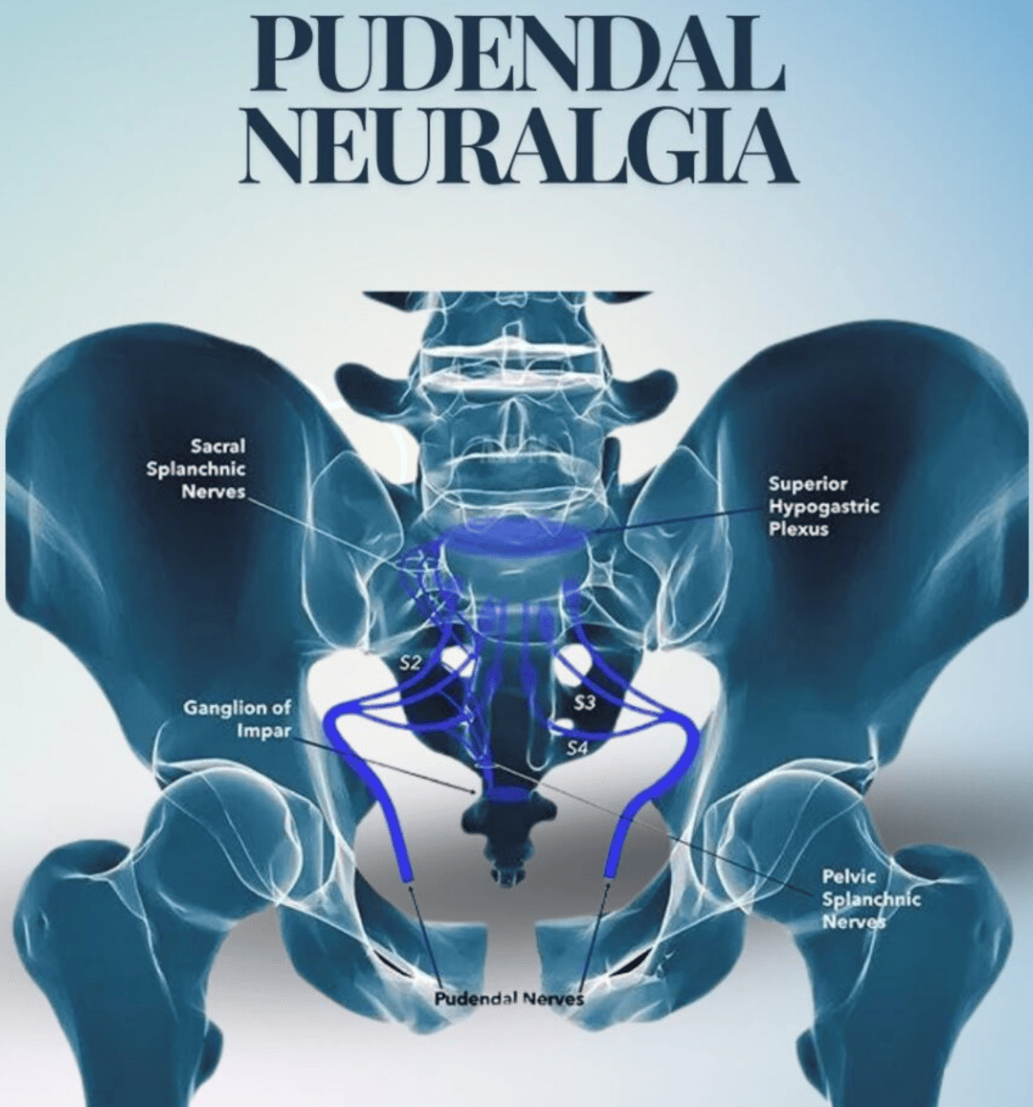
Jessey Wong, Ruth Postlethwaite, & Clare McIvor
A 32-year-old female business owner with a 15-month history of fibromyalgia and an existing diagnosis of pudendal neuralgia presented for chiropractic care with primary concerns relating to widespread body pain, chronic fatigue, brain fog and depression.
Pudendal neuralgia describes neuropathic pain within the territory of the pudendal nerve, which supplies the perineal, genital and anorectal regions.
In terms of her daily activities her Quality of Life is significantly improved as the widespread body discomfort and depression are reduced. She was able to enjoy life, spend quality time with her family and friends, eat the food she likes as well as travel again. Prior to engaging with care, she found these aspects of life to be impacted due to her pain and disability.

Brian Lanoue, Thea Lanoue, Ruth Postlethwaite & Clare McIvor
A 29-year-male flight attendant presented for Chiropractic care with primary concerns related to spinal alignment and general health. He had existing diagnoses of scoliosis, ADHD and depression. Additional concerns included shoulder crepitus, back pain when bending or standing, and poor posture.
The patient suffered a substance use relapse during his period of care, from which he reported recovering more quickly than usual.
This case exemplifies how Chiropractic care can serve as a meaningful entry point for individuals to re-engage with their health, physically, mentally, and emotionally. It often sparks awareness, routine, and a reconnection with the body that can inspire further lifestyle changes, even beyond the adjustment itself.

About case reports as evidence
Case reports are observational in design and thus limited by lacking controls; the effect of potential confounding factors, including comorbidities, cannot be excluded. The findings could support a clinically relevant hypothesis that the identification and correction of spinal subluxation and associated functional dysfunction are modifiable contributors to the effective management and resolution of the specific topic of the case report. In other words, they have meaning.
We must appreciate that case reports are eligible for inclusion as ‘expertise’ bringing clinical insights into the JBI FAME evidential ring (JBI Manual for Evidence Synthesis; 2024) to inform evidence-based healthcare in general and the science of Chiropractic in particular. This Journal specifically recognises that subluxation identification and correction is the art of the individual Chiropractor.
Book review
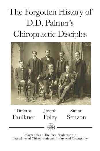
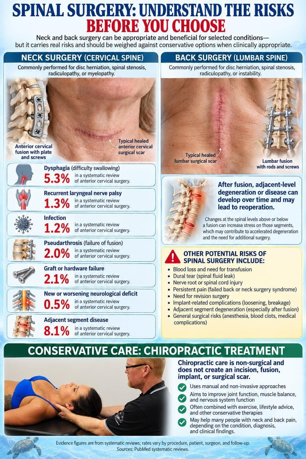
Quick-take
On-line exclusive
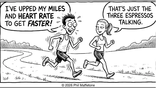
The Caffeine connection: An endurance drug
Does it really give us energy? Dr. Phil Maffetone
Like any respectable psychoactive drug, caffeine helps us feel good, be sharp, and demand another cup. From mental focus to physical vigor, fat burning to athletic output, caffeine orchestrates performance improvements starting straight in the central nervous system.
Caffeine occurs naturally in over 60 plants, with coffee claiming the lion's share, while tea and cacao settle for lesser roles. Having evolved millions of years ago as a natural chemical pesticide to ward off hungry bugs, humans have willingly consumed it at least since the dawn of recorded history.
Today, industrial plants synthesise massive quantities of caffeine from a base of urea and chloroacetic acid, fuelling soft drinks, 'energy' concoctions, supplements and pharmaceuticals. Globally, more than half of all consumed caffeine is synthetic, born in a chemical reactor rather than a bean.
As the planet’s favorite legal drug, caffeine remains the most widely consumed psychoactive substance. Its ergogenic profile includes:
- Enhanced Cognitive & Creative Drive: Sharper attention, heightened vigilance, and improved focus.
- Elevated Physical Output: Gains in muscular endurance, strength, and velocity—boosting aerobic performance by 2–4%, with lesser effects in anaerobic power sports.
- Precision Dosing: Target doses range from 3–6 mg/kg of body mass. For a 150-pound individual, that translates to 225–450 mg (roughly 1.5 to 3 large mugs of strong coffee). More subtle performance benefits begin around 2 mg/kg.
- The Law of Diminishing Returns: Escalating doses yield increased side effects without additional performance gains—more is emphatically not better. (NCAA and elite athletes must keep urinary concentrations below equivalent thresholds, typically capped around 10 mg/kg.)
- Kinetics: While metabolic changes kick off at the very first sip, noticeable effects take roughly 30 minutes or more to peak. Its half-life ranges from 2.5 to 5 hours. In the brain, caffeine acts as an adenosine antagonist, blocking the central nervous system's natural depressant and reducing the feeling of fatigue. Concurrently, it upregulates dopamine and related neurotransmitters, unleashing that familiar “feel-good” cascade that elevates motivation, creativity, and memory.
The Fat Connection
While the neurological sparkle begins in the brain, caffeine simultaneously recruits systemic metabolism to accelerate fat burning. Mobilizing stored body fat provides a sustained energy substrate while elevating circulating blood ketones for even more. Paired with a low-carbohydrate, nutrient-dense diet, caffeine acts as a metabolic catalyst for optimal physical and mental stamina.
Dr. Phil’s Fat-Burning Coffee
Yes, I’m sipping this exact concoction as these words hit the page, my daily morning ritual. Here is the video recipe showing how I brew it. (Today's variation: whole cacao substituted for coconut oil)
Hold the Sugar
Added sugars, including non-caloric artificial sweeteners and refined carbohydrates, disrupts metabolism, suppress fat burning, and directly counteract caffeine’s ergogenic potential. The cultural myth of the “sugar rush” is physiologically false; sugar consumption consistently lowers alertness, induces fatigue, and degrades mood. Despite deceptive marketing pushing caffeine-sugar cocktails as energy boosters, sweetened coffee, tea, and cacao remain mainstay vehicles for the junk-food enterprise.
Sugar and other refined carbohydrates trigger rapid blood glucose spikes and insulin surges, driving subsequent hypoglycemia, fat storage, fatigue, and intense cravings.
If you must sweeten your brew, you are likely dealing with two issues:
- Bitterness Management: Caffeine is naturally bitter. To optimize flavor without sugar, grind whole beans longer and finely while adjusting extraction times. Avoid over-steeping tea, too.
- Desensitized Palate: Sugar habituation alters taste perception rendering natural bitter compounds unpalatable (the same reason children reject cruciferous vegetables). See my article on Sugar Addiction.
Finally, respect the physiology: caffeine stimulates the sympathetic nervous system and raises stress hormones like cortisol. Excess intake triggers elevated heart rate and blood pressure, anxiety, sleep disruption, and gastrointestinal distress. (Due to their liver and brain metabolism, some people respond much less to caffeine.)
While caffeine does not directly supply calories or energy, it acts as a powerful metabolic lever—enhancing energy-producing fat burning, elevating mental clarity, and powering human performance.
Philip Maffetone DC. Retired practitioner, health-fitness writer, researcher, songwriter at PhilMaffetone.com
These pages are under construction ...